
Article Figures & Data
Figures
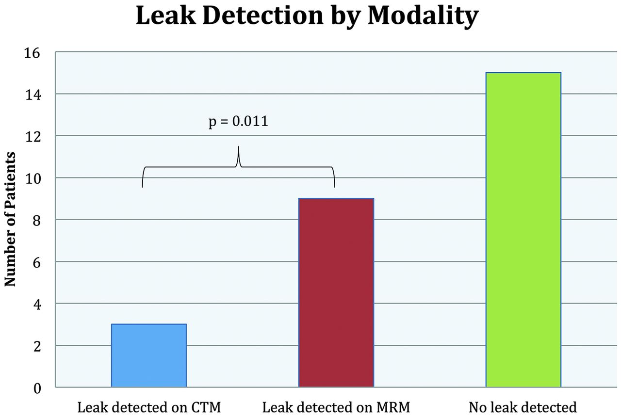
- Fig 1.
Bar graph illustrating the rate of leak detection by CTM and MRM. A statistically significant difference was observed between CTM and MRM by a 2-tailed paired t test (P = .011).
- Fig 2.
Patient 4. A 52-year-old former fire fighter with a 1-year history of postural headaches. Axial CTM (A and C) and axial T1 fat-suppressed images from MRM (B and D) following intrathecal administration of iodinated contrast and gadolinium. Multiple spinal diverticula are seen along with an extradural contrast collection (arrows, B) not evident on a concurrent CTM examination.
- Fig 3.
Patient 2. A 62-year-old man with headache and recurrent subdural hemorrhage following evacuation, found to have imaging findings of SIH. Axial CTM (A) and MRM (B) images at the T9–T10 level. Spinal diverticula are evident on both examinations. The MRM demonstrates an extra-arachnoid contrast collection and ill-defined increased T1 signal surrounding the enlarged right spinal diverticula. Subsequent CT performed for epidural blood patch planning with a localization grid in place (C) shows the spinal diverticula; the extra-arachnoid contrast collection is not evident. The patient reported symptomatic relief following directed blood patch and was without headache as of the 2-month follow-up note.
- Fig 4.
Patient 7. A 55-year-old man with a history of lethargy, fatigue, and hearing loss. Axial CTM (A and C) and axial MRM (B and D) images. CT shows bilateral spinal diverticula. MR image demonstrates prominent contrast leakage from the right-sided T5–T6 (B) and left T10–T11 (D) diverticula, with gadolinium extending into the adjacent paraspinal musculature. The patient underwent targeted epidural blood patches of both leaking spinal diverticula.
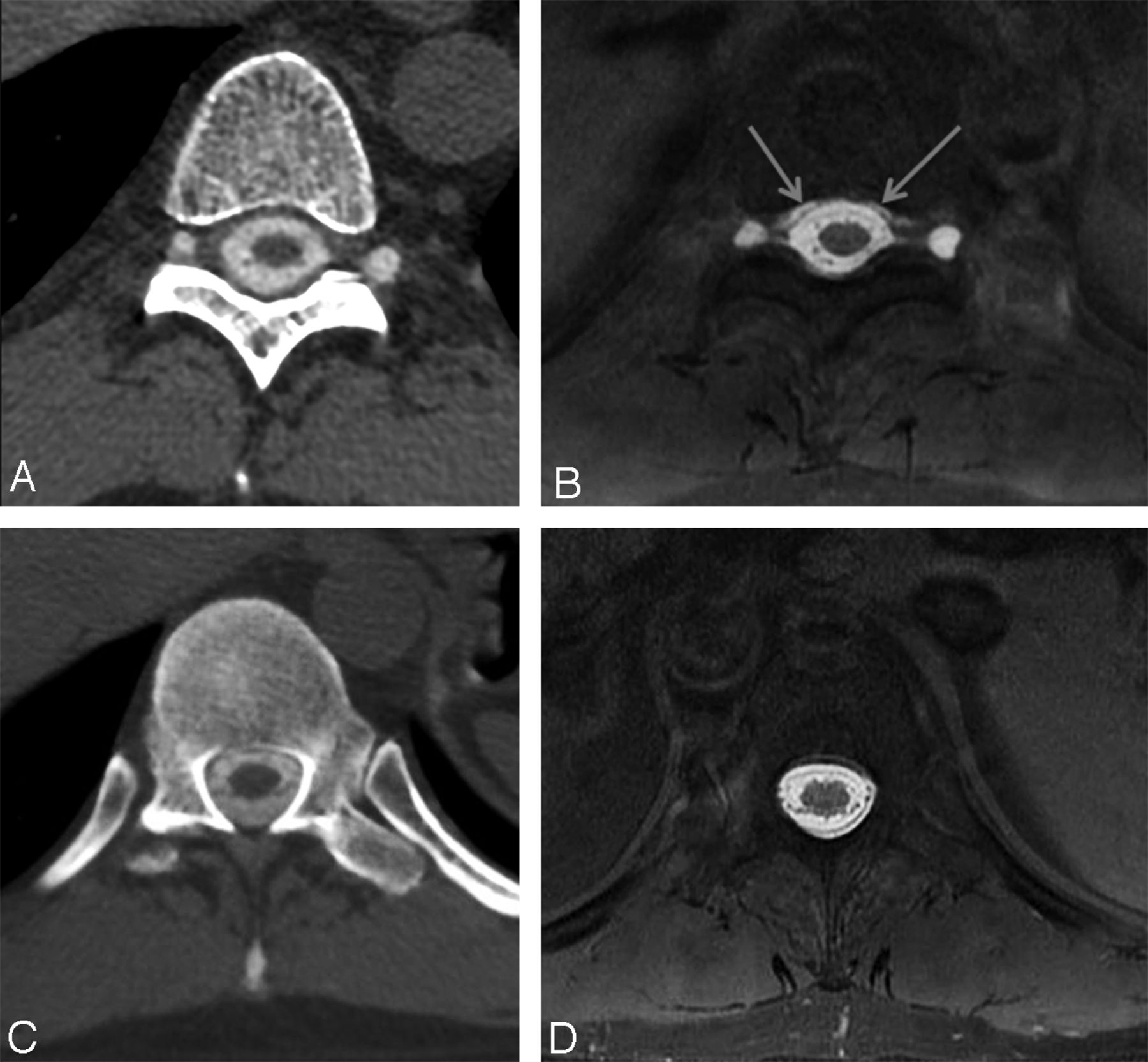
- Fig 5.
Patient 8. A 55-year-old man with postural headache and low CSF pressure on lumbar puncture. Axial CTM (A), axial MRM (B), and coronal MRM (C) images. Coronal MRM shows multiple spinal diverticula. However, only the right T7–T8 spinal diverticula, seen on the CTM (A), shows evidence of contrast leakage on axial MRM (B), evident by ill-defined T1 shortening surrounding the cyst. This lesion was treated with directed epidural blood patch with symptomatic improvement for 5 years following treatment.


Tables
Patients with CSF leak detected on CTM or MRMa
Patient No. Age (yr) Sex Opening Pressure (cm H2O) Leak Detected on CTM Leak Detected on MR Myelogram Time Difference (h:min) Extent of Extra-Arachnoid Contrast Same-Day Blood Patch Suspected CSF Leak Etiology 1 66 Female 5 No Yes 0:37 T6–T12 No Unclear; spinal diverticula C6–C7 through L2–L3 2 62 Male 7 No Yes 1:14 T9–L1 Yes Bilateral T9–T10 spinal diverticula 3 27 Male 10 Yes Yes 0:55 L2–L5 No Dural ectasia; marfanoid features 4 52 Male 7 No Yes 1:14 T6–T12 Yes Bilateral T9–T10 and left T11–T12 spinal diverticula 5 32 Female 0 No Yes 0:29 C3–L1 No No source of leak detected; T2–T3 leak seen on subsequent fluoroscopic subtraction myelogram 6 37 Female 6 Yes Yes 5:14 T10–L1 Yes T11–T12 disk osteophyte; blood patch performed before MRM 7 55 Male 8 No Yes 0:49 T5–T6 and T9–T10 No Right T5–T6 and left T9–T10 spinal diverticula 8 55 Male 6 No Yes 0:12 T7–T8 Yes Right T7–T8 spinal diverticula 9 58 Female 0 Yes Yes 0:46 C7–T4 No T1–T2 secondary to disk protrusion and dural tear ↵a The time difference reflects the time stamp difference between CTM acquisition and the beginning of MRM. A same-day blood patch was performed when logistically possible and a clear source of leak was identified.


