Advertisement

Main menu

  • Home
  • Content
    • Current Issue
    • Accepted Manuscripts
    • Article Preview
    • Past Issue Archive
    • Video Articles
    • AJNR Case Collection
    • Case of the Week Archive
    • Case of the Month Archive
    • Classic Case Archive
  • Special Collections
    • AJNR Awards
    • Low-Field MRI
    • Alzheimer Disease
    • ASNR Foundation Special Collection
    • Photon-Counting CT
    • View All
  • Multimedia
    • AJNR Podcasts
    • AJNR SCANtastic
    • Trainee Corner
    • MRI Safety Corner
    • Imaging Protocols
  • For Authors
    • Submit a Manuscript
    • Submit a Video Article
    • Submit an eLetter to the Editor/Response
    • Manuscript Submission Guidelines
    • Statistical Tips
    • Fast Publishing of Accepted Manuscripts
    • Graphical Abstract Preparation
    • Imaging Protocol Submission
    • Author Policies
  • About Us
    • About AJNR
    • Editorial Board
    • Editorial Board Alumni
  • More
    • Become a Reviewer/Academy of Reviewers
    • Subscribers
    • Permissions
    • Alerts
    • Feedback
    • Advertisers
    • ASNR Home

User menu

  • Alerts
  • Log in

Search

  • Advanced search
American Journal of Neuroradiology

American Journal of Neuroradiology

ASHNR American Society of Functional Neuroradiology ASHNR American Society of Pediatric Neuroradiology ASSR
  • Alerts
  • Log in

Advanced Search

  • Home
  • Content
    • Current Issue
    • Accepted Manuscripts
    • Article Preview
    • Past Issue Archive
    • Video Articles
    • AJNR Case Collection
    • Case of the Week Archive
    • Case of the Month Archive
    • Classic Case Archive
  • Special Collections
    • AJNR Awards
    • Low-Field MRI
    • Alzheimer Disease
    • ASNR Foundation Special Collection
    • Photon-Counting CT
    • View All
  • Multimedia
    • AJNR Podcasts
    • AJNR SCANtastic
    • Trainee Corner
    • MRI Safety Corner
    • Imaging Protocols
  • For Authors
    • Submit a Manuscript
    • Submit a Video Article
    • Submit an eLetter to the Editor/Response
    • Manuscript Submission Guidelines
    • Statistical Tips
    • Fast Publishing of Accepted Manuscripts
    • Graphical Abstract Preparation
    • Imaging Protocol Submission
    • Author Policies
  • About Us
    • About AJNR
    • Editorial Board
    • Editorial Board Alumni
  • More
    • Become a Reviewer/Academy of Reviewers
    • Subscribers
    • Permissions
    • Alerts
    • Feedback
    • Advertisers
    • ASNR Home
  • Follow AJNR on Twitter
  • Visit AJNR on Facebook
  • Follow AJNR on Instagram
  • Join AJNR on LinkedIn
  • RSS Feeds

AJNR Awards, New Junior Editors, and more. Read the latest AJNR updates

Research ArticleTechnical Note
Open Access

Do Early MRI Signals Predict Lesion Size in a Neonatal Stroke Rat Model?

S. Fau, C. Po, C. Goyenvalle, P. Meric and C. Charriaut-Marlangue
American Journal of Neuroradiology July 2013, 34 (7) E73-E76; DOI: https://doi.org/10.3174/ajnr.A3069
S. Fau
aFrom the CNRS 7102 (S.F., C.G., C.C.-M.), Equipe HICD, Université Pierre et Marie Curie Paris–Paris, France
  • Find this author on Google Scholar
  • Find this author on PubMed
  • Search for this author on this site
C. Po
bLaboratoire de RMN Biologique (C.P., P.M.), ICSN, CNRS UPR 2301, Gif-sur-Yvette, France
cIR4M (C.P.), CNRS UMR 8081, Université de Paris Sud, Orsay, France.
  • Find this author on Google Scholar
  • Find this author on PubMed
  • Search for this author on this site
C. Goyenvalle
aFrom the CNRS 7102 (S.F., C.G., C.C.-M.), Equipe HICD, Université Pierre et Marie Curie Paris–Paris, France
  • Find this author on Google Scholar
  • Find this author on PubMed
  • Search for this author on this site
P. Meric
bLaboratoire de RMN Biologique (C.P., P.M.), ICSN, CNRS UPR 2301, Gif-sur-Yvette, France
  • Find this author on Google Scholar
  • Find this author on PubMed
  • Search for this author on this site
C. Charriaut-Marlangue
aFrom the CNRS 7102 (S.F., C.G., C.C.-M.), Equipe HICD, Université Pierre et Marie Curie Paris–Paris, France
  • Find this author on Google Scholar
  • Find this author on PubMed
  • Search for this author on this site
  • Article
  • Figures & Data
  • Info & Metrics
  • Responses
  • References
  • PDF
Loading

Abstract

SUMMARY: In this study, we compared lesion size by using VADC and VT2 at 0, 2, 5, 24, and 48 hours and histologic lesions at 48 hours in a P7 rat stroke model. The best correlation between VHISTO and VADC was at H0, and between VHISTO and VT2, at H2-H5. Early MR imaging signals allowed excluding “no-lesion” and “no-reflow” animals to help standardize this neonatal stroke model and predict lesion size.

ABBREVIATIONS:

H0, H2, H5, H24, and H48
15 minutes and 2, 5, 24, and 48 hours after reperfusion, respectively
P7
7-day-old
RARE
rapid acquisition relaxation excitement
VADC
volume of lesion on ADC maps
VHISTO
volume of lesion on histologic sections
VT2
volume of lesion on T2 maps

Neuroimaging, particularly MR imaging, could be a tool for a rapid comprehensive assessment in acute stroke with the potential to guide treatment decisions for a better clinical outcome. One of the key issues in stroke assessment is to choose the best end points that appropriately indicate outcome. In experimental animal research, final lesion size has been used consistently as an outcome. Infarct sizes assessed by T2WI have been shown to correlate well by some and to underestimate histologic damage by others.1 In the developing brain, authors have reported that MR imaging in P7 rat pups submitted to stroke2 or hypoxia-ischemia3,4 seems correlated to final histologic lesions measured 24 hours or 3–10 days after the injury, without early MR imaging time-point evaluation.

The aim of the present study was to develop a preclinical design including routine MR imaging coupling ADC maps, T2 maps, and MRA. We suggested that this strategy could improve the statistical power of trials using our model by diminishing the variability of the outcome assessment. Early MR imaging could 1) confirm initial ischemia, 2) confirm arterial reflow, and 3) accurately predict the final histologic outcome at 48 hours.

Technique

Perinatal Ischemia

Care and use of experimental animals followed French and European Community guidelines. Ischemia was created in 24 Wistar P7 rats (Janvier, Le Genest Saint Isle, France) of both sexes.5 Briefly, anesthetized (chloral hydrate, 350 mg/kg, intraperitoneally) rats were exposed to left MCA electrocoagulation followed by 50-minute transient left common carotid artery occlusion. During ischemia and recovery the pups were placed in a humidified incubator at 37°C.

MR Imaging Acquisition

Pups were monitored at H0, H2, H5, H24, and H48 after reperfusion. For H0, H2, and H5 imaging session, pups were asleep due to the initial chloral hydrate injection. For H24 and H48 imaging sessions, pups were anesthetized by isoflurane 0.8% mixed in 30% O2, 70% N2. During imaging, animals were thermoregulated by a water blanket heated at 37°C. Images were acquired by using a 7T horizontal magnet equipped with a Bruker AVANCE console and with an actively shielded gradient 360mT device (Bruker BioSpin, Rheinstetten, Germany). DWI was performed from an EPI sequence (TR = 3 seconds, TE = 25 ms, b-values = 50, 500, 1000 s/mm2, FOV = 40 × 40 mm, matrix = 256 × 256, repetitions = 3, 25 sections, thickness = 0.5 mm, duration = 9.5 minutes). T2WI was performed with a RARE sequence (TR = 5404 ms, TE values = 25.6, 76.8, 128, 179.2 ms, RARE factor = 4, FOV = 40 × 40 mm, matrix = 128 × 128, 25 sections, thickness = 0.5 mm, duration = 3 minutes). ADC and T2 maps were computed by exponential fitting of the local signal-intensity versus b and TE, respectively, by using a homemade ImageJ plugin (http://rsbweb.nih.gov/ij/). MRA was performed with a 3D time-of-flight method based on a fast low-angle shot 3D sequence (TR = 15 ms, TE = 2.5 ms, FOV= 40 × 40 × 40 mm, matrix = 256 × 256 × 128, flip angle = 20°, duration = 3 minutes). Each animal underwent the H0 imaging and was then eventually excluded either as “no-lesion” if the DWI lesion was visible on <3 sections or as “no-reflow” if MRA showed an ipsilateral ICA signal of <40% of the contralateral ICA signal. All animals were sacrificed immediately after the H48 imaging session.

Outcome Assessments

The volume of the lesion was assessed by using histology at H48, ADC maps, and T2 maps from H0 to H48. VHISTO was delineated by a classic visual method on cresyl violet–stained coronal sections (50-μm thick, 500-μm intervals). VADC and VT2 were defined by applying a threshold (mean − 2 SDs) of the whole contralateral hemisphere ADC and T2, respectively. Volume measurements were expressed as a percentage of the ipsilateral hemisphere.

Statistical Analysis

All results are given as mean ± SD. The correlation and regression were performed with a standard statistical software package (SSYSTAT 3.1, Systat Software, San Jose, California) by using a Spearman rank-order correlation and a linear regression, respectively.

Results

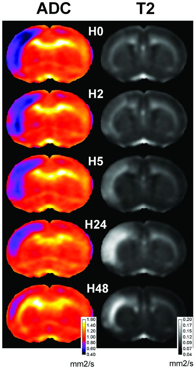
Four animals of 24 (17%) were excluded as no-lesion. All exhibited MCA interruption on H0 angiography, without a significant DWI lesion. Two animals of 24 (8%) were excluded as “no-reflow” as determined on MRA. Eighteen animals were then analyzed (Fig 1), and VADC, VT2, and VHISTO were calculated (Fig 2). An area in the ipsilateral frontoparietal cortex with low VADC was maximal at H0 in almost all the animals (Fig 1). VADC at H0 was different from 6.6%–32.1% of the ipsilateral hemisphere. A significant correlation between VADC at H0 and lesion volume (VHISTO) at 48 hours was obtained (slope: 1.09, r = 0.92, P < .001; Fig 2). In some animals, VADC slightly decreased between H2 and H5, whereas it remained similar in the others. At 24 and more markedly at 48 hours, all VADC values were highly reduced (0.2%–8.3%; Fig 1). Even if hints in the deeper cortical and subcortical layers (Fig 1) were visible on T2 maps as soon as H0 (in few animals), the T2 lesion was clearly identifiable in all animals at H2 (4.4%–26.7%). A significant correlation between VT2 and VHISTO was obtained at H2 (slope: 0.97, r = 0.91, P < .001) and H5 (slope: 1.04, r = 0.91, P < .001; Fig 2). This correlation remained at H24 but with a higher slope (1.15, r = 0.89). Then VT2 slightly decreased at H48 (r = 0.888). The range of VHISTO was from 3.5% to 24% with a mean of 13.5 ± 5.7%.

Fig 1.
  • Download figure
  • Open in new tab
  • Download powerpoint
Fig 1.

Time course of ADC and T2 maps at Bregma 1.20 mm. Sum of coregistrated sections are computed at H0, H2, H5, H24, and H48. Note that low ADC was observed in the lesioned frontoparietal cortex compared with the contralateral side. Hints of T2 were observed as early as H0 in the deeper cortical layers. At H48, marked T2 signals are still detected in the deeper cortex.

Fig 2.
  • Download figure
  • Open in new tab
  • Download powerpoint
Fig 2.

Correlation between MR imaging and histology measurements. VHISTO and VADC (left) or VT2 (right) are given at different time points after reperfusion. Note that correlation was very strong at H0 on the ADC map and at H2 and H5 on the T2 maps.

Discussion

This study showed that routine MR imaging could be a toolbox able to standardize neonatal stroke models, first by excluding no-lesion and no-reflow animals in the first MR angiography, and second by predicting histologic outcome early in a neonatal stroke model.

The first set of MR images at H0 was used to exclude no-lesion animals. These animals probably developed efficient cortical arterial anastomoses during ischemia as we previously demonstrated by using sonography.6 MR imaging also allowed excluding no-reflow animals because the absence of recanalization was shown to block the development of further edema.7

All animals exhibited a quite reproducible time course of ADC or T2 volume. As previously reported, VADC reached a maximum just after reperfusion.7 The best-correlated VADC time point with VHISTO was H0. This impressive correlation lets one think that as soon as 15 minutes after reperfusion, “the die was cast,” and almost the whole interindividual variability of animals was already determined. In contrast, at H2 and H5, the slope of regression was superior to 1, suggesting that VADC overestimated histology volume. Conversely, at 24 and 48 hours, correlation between ADC and histology became less strong and the slope coefficient decreased, leading to an underestimation of the final lesion.

As previously reported, T2 lesions appeared as soon as 2 hours after ischemia.7 Most interesting, we observed that the correlation of VT2 with VHISTO was strong as early as H2 and H5, with a slope of regression close to 1, suggesting good overlap between T2 signals and histologic lesions. Thereafter, VT2 overestimated lesions at both 24 and 48 hours.

Conclusions

This whole strategy was not reported in the literature because only parts of studies reported exclusion of no-lesion animals with intraischemic MR imaging.8 In addition, early MR imaging signals appear to be good surrogates for final stroke lesions in the developing brain.

Footnotes

  • Disclosures: Christiane Charriaut-Marlangue—RELATED: Support for Travel to Meetings for the Study or Other Purposes: Société Française sur la Recherche du Handicap de l'Enfant.* Sébastien Fau—RELATED: Grant: CNRS, Comments: French National Scientific Research Center. *Money paid to the institution.

  • This study was supported by the Institut pour la Recherche sur la Moelle Epinière, Fondation NRJ and Société Française sur la Recherche du Handicap de l'Enfant. S. Fau was the recipient of a “poste d'accueil“ from AP-HP and CNRS.

  • C.C.M., S.F., and P.M. designed and organized this study; S.F., C.P., and C.G. performed the experiments and analyzed data. C.C.M., S.F., and C.P. wrote the manuscript.

  • Paper previously presented as a poster at: Brain 09, 24th International Symposium on Cerebral Blood Flow, Metabolism, and Function, Chicago, Illinois, June 29–July 3, 2009.

Indicates open access to non-subscribers at www.ajnr.org

REFERENCES

  1. 1.↵
    1. Farr TD,
    2. Wegener S
    . Use of magnetic resonance imaging to predict outcome after stroke: a review of experimental and clinical evidence. J Cereb Blood Flow Metab 2010;30:703–17
    CrossRefPubMed
  2. 2.↵
    1. Derugin N,
    2. Wendland M,
    3. Muramatsu K,
    4. et al
    . Evolution of brain injury after transient middle cerebral artery occlusion in neonatal rats. Stroke 2000;31:1752–61
    Abstract/FREE Full Text
  3. 3.↵
    1. Wang Y,
    2. Cheung PT,
    3. Shen GX,
    4. et al
    . Hypoxic-ischemic brain injury in the neonatal rat model: relationship between lesion size at early MR imaging and irreversible infarction. AJNR Am J Neuroradiol 2006;27:51–54
    Abstract/FREE Full Text
  4. 4.↵
    1. Lodygensky GA,
    2. Inder TE,
    3. Neil JJ
    . Application of magnetic resonance imaging in animal models of perinatal hypoxic-ischemic cerebral injury. Int J Dev Neurosci 2008;26:13–25
    CrossRefPubMed
  5. 5.↵
    1. Renolleau S,
    2. Fau S,
    3. Goyenvalle C,
    4. et al
    . Specific caspase inhibitor Q-VD-OPh prevents neonatal stroke in P7 rat: a role for gender. J Neurochem 2007;100:1062–71
    CrossRefPubMed
  6. 6.↵
    1. Bonnin P,
    2. Leger PL,
    3. Deroide N,
    4. et al
    . Impact of intracranial blood-flow redistribution on stroke size during ischemia-reperfusion in 7-day-old rats. J Neurosci Methods 2011;198:103–09
    CrossRefPubMed
  7. 7.↵
    1. Fau S,
    2. Po C,
    3. Gillet B,
    4. et al
    . Effect of the reperfusion after cerebral ischemia in neonatal rats using MRI monitoring. Exp Neurol 2007;208:297–304
    CrossRefPubMed
  8. 8.↵
    1. Derugin N,
    2. Dingman A,
    3. Wendland MF,
    4. et al
    . Magnetic resonance imaging as a surrogate measure for histological sub-chronic endpoint in a neonatal rat stroke model. Brain Res 2005;1066:49–56
    CrossRefPubMed
  • Received December 6, 2011.
  • Accepted after revision January 6, 2012.
  • © 2013 by American Journal of Neuroradiology
PreviousNext
Back to top

In this issue

American Journal of Neuroradiology: 34 (7)
American Journal of Neuroradiology
Vol. 34, Issue 7
1 Jul 2013
  • Table of Contents
  • Index by author
Advertisement
Print
Download PDF
Email Article

Thank you for your interest in spreading the word on American Journal of Neuroradiology.

NOTE: We only request your email address so that the person you are recommending the page to knows that you wanted them to see it, and that it is not junk mail. We do not capture any email address.

Enter multiple addresses on separate lines or separate them with commas.
Do Early MRI Signals Predict Lesion Size in a Neonatal Stroke Rat Model?
(Your Name) has sent you a message from American Journal of Neuroradiology
(Your Name) thought you would like to see the American Journal of Neuroradiology web site.
CAPTCHA
This question is for testing whether or not you are a human visitor and to prevent automated spam submissions.
Cite this article
S. Fau, C. Po, C. Goyenvalle, P. Meric, C. Charriaut-Marlangue
Do Early MRI Signals Predict Lesion Size in a Neonatal Stroke Rat Model?
American Journal of Neuroradiology Jul 2013, 34 (7) E73-E76; DOI: 10.3174/ajnr.A3069

Citation Manager Formats

  • BibTeX
  • Bookends
  • EasyBib
  • EndNote (tagged)
  • EndNote 8 (xml)
  • Medlars
  • Mendeley
  • Papers
  • RefWorks Tagged
  • Ref Manager
  • RIS
  • Zotero
0 Responses
Respond to this article
Share
Bookmark this article
Do Early MRI Signals Predict Lesion Size in a Neonatal Stroke Rat Model?
S. Fau, C. Po, C. Goyenvalle, P. Meric, C. Charriaut-Marlangue
American Journal of Neuroradiology Jul 2013, 34 (7) E73-E76; DOI: 10.3174/ajnr.A3069
del.icio.us logo Twitter logo Facebook logo Mendeley logo
  • Tweet Widget
  • Facebook Like
  • Google Plus One
Purchase

Jump to section

  • Article
    • Abstract
    • ABBREVIATIONS:
    • Technique
    • Results
    • Discussion
    • Conclusions
    • Footnotes
    • REFERENCES
  • Figures & Data
  • Info & Metrics
  • Responses
  • References
  • PDF

Related Articles

  • No related articles found.
  • PubMed
  • Google Scholar

Cited By...

  • No citing articles found.
  • Crossref (9)
  • Google Scholar

This article has been cited by the following articles in journals that are participating in Crossref Cited-by Linking.

  • Sex differences in brain MRI abnormalities and neurodevelopmental outcomes in a rat model of neonatal hypoxia-ischemia
    Hui-Zhi Huang, Xiao-Hong Wen, Hui Liu
    International Journal of Neuroscience 2016 126 7
  • Dynamic Spatio-Temporal Imaging of Early Reflow in a Neonatal Rat Stroke Model
    Pierre-Louis Leger, Philippe Bonnin, Pierre Lacombe, Elisabeth Couture-Lepetit, Sebastien Fau, Sylvain Renolleau, Abdallah Gharib, Olivier Baud, Christiane Charriaut-Marlangue
    Journal of Cerebral Blood Flow & Metabolism 2013 33 1
  • Early Detection of Hypothermic Neuroprotection Using T2-Weighted Magnetic Resonance Imaging in a Mouse Model of Hypoxic Ischemic Encephalopathy
    Sydney E. Doman, Akanksha Girish, Christina L. Nemeth, Gabrielle T. Drummond, Patrice Carr, Maxine S. Garcia, Michael V. Johnston, Sujatha Kannan, Ali Fatemi, Jiangyang Zhang, Mary Ann Wilson
    Frontiers in Neurology 2018 9
  • A Model of Perinatal Ischemic Stroke in the Rat: 20 Years Already and What Lessons?
    Christiane Charriaut-Marlangue, Olivier Baud
    Frontiers in Neurology 2018 9
  • Prediction of hemorrhagic transformation after acute thrombolysis following major artery occlusion using relative ADC ratio: A retrospective study
    N. Shinoda, S. Hori, K. Mikami, T. Bando, D. Shimo, T. Kuroyama, Y. Kuramoto, M. Matsumoto, O. Hirai, Y. Ueno
    Journal of Neuroradiology 2017 44 6
  • Utility of relative ADC ratio in patient selection for endovascular revascularization of large vessel occlusion
    Narihide Shinoda, Shinya Hori, Kazuyuki Mikami, Toshiaki Bando, Daisuke Shimo, Takahiro Kuroyama, Yoji Kuramoto, Masato Matsumoto, Osamu Hirai, Yasushi Ueno
    Journal of Neuroradiology 2017 44 3
  • Inter-alpha Inhibitor Proteins Ameliorate Brain Injury and Improve Behavioral Outcomes in a Sex-Dependent Manner After Exposure to Neonatal Hypoxia Ischemia in Newborn and Young Adult Rats
    Xiaodi Chen, Jiyong Zhang, Yuqi Wu, Richard Tucker, Grayson L. Baird, Rose Domonoske, Adriel Barrios-Anderson, Yow-Pin Lim, Kevin Bath, Edward G. Walsh, Barbara S. Stonestreet
    Neurotherapeutics 2022 19 2
  • Spatiotemporal characterization of brain infarction by sequential multimodal MR imaging following transient focal ischemia in a Rat model of intra-arterial middle cerebral artery occlusion
    Benjamin Gory, Fabien Chauveau, Radu Bolbos, Jean-Baptiste Langlois, Paul-Emile Labeyrie, Francesco Signorelli, Alexis Turjman, Francis Turjman
    European Radiology 2016 26 12
  • Using MRI to predict the fate of excitotoxic lesions in rats
    Thibault Cholvin, Lisa Giorgi, Nathalie Baril, Jean-Michel Brezun, Bruno Poucet, Franck A. Chaillan, Giuseppe Biagini
    PLOS ONE 2018 13 7

More in this TOC Section

  • 3D Fast Spin-Echo T1 Black-Blood Imaging for the Diagnosis of Cervical Artery Dissection
  • The Kety-Schmidt Technique for Quantitative Perfusion and Oxygen Metabolism Measurements in the MR Imaging Environment
  • A Swine Model to Analyze Arterial Structural Changes Induced by Mechanical Thrombectomy
Show more Technical Notes

Similar Articles

Advertisement

Indexed Content

  • Current Issue
  • Accepted Manuscripts
  • Article Preview
  • Past Issues
  • Editorials
  • Editor's Choice
  • Fellows' Journal Club
  • Letters to the Editor
  • Video Articles

Cases

  • Case Collection
  • Archive - Case of the Week
  • Archive - Case of the Month
  • Archive - Classic Case

More from AJNR

  • Trainee Corner
  • Imaging Protocols
  • MRI Safety Corner
  • Book Reviews

Multimedia

  • AJNR Podcasts
  • AJNR Scantastics

Resources

  • Turnaround Time
  • Submit a Manuscript
  • Submit a Video Article
  • Submit an eLetter to the Editor/Response
  • Manuscript Submission Guidelines
  • Statistical Tips
  • Fast Publishing of Accepted Manuscripts
  • Graphical Abstract Preparation
  • Imaging Protocol Submission
  • Evidence-Based Medicine Level Guide
  • Publishing Checklists
  • Author Policies
  • Become a Reviewer/Academy of Reviewers
  • News and Updates

About Us

  • About AJNR
  • Editorial Board
  • Editorial Board Alumni
  • Alerts
  • Permissions
  • Not an AJNR Subscriber? Join Now
  • Advertise with Us
  • Librarian Resources
  • Feedback
  • Terms and Conditions
  • AJNR Editorial Board Alumni

American Society of Neuroradiology

  • Not an ASNR Member? Join Now

© 2025 by the American Society of Neuroradiology All rights, including for text and data mining, AI training, and similar technologies, are reserved.
Print ISSN: 0195-6108 Online ISSN: 1936-959X

Powered by HighWire