Advertisement

Main menu

  • Home
  • Content
    • Current Issue
    • Accepted Manuscripts
    • Article Preview
    • Past Issue Archive
    • Video Articles
    • AJNR Case Collection
    • Case of the Week Archive
    • Case of the Month Archive
    • Classic Case Archive
  • Special Collections
    • AJNR Awards
    • Low-Field MRI
    • Alzheimer Disease
    • ASNR Foundation Special Collection
    • Photon-Counting CT
    • View All
  • Multimedia
    • AJNR Podcasts
    • AJNR SCANtastic
    • Trainee Corner
    • MRI Safety Corner
    • Imaging Protocols
  • For Authors
    • Submit a Manuscript
    • Submit a Video Article
    • Submit an eLetter to the Editor/Response
    • Manuscript Submission Guidelines
    • Statistical Tips
    • Fast Publishing of Accepted Manuscripts
    • Graphical Abstract Preparation
    • Imaging Protocol Submission
    • Author Policies
  • About Us
    • About AJNR
    • Editorial Board
    • Editorial Board Alumni
  • More
    • Become a Reviewer/Academy of Reviewers
    • Subscribers
    • Permissions
    • Alerts
    • Feedback
    • Advertisers
    • ASNR Home

User menu

  • Alerts
  • Log in

Search

  • Advanced search
American Journal of Neuroradiology

American Journal of Neuroradiology

ASHNR American Society of Functional Neuroradiology ASHNR American Society of Pediatric Neuroradiology ASSR
  • Alerts
  • Log in

Advanced Search

  • Home
  • Content
    • Current Issue
    • Accepted Manuscripts
    • Article Preview
    • Past Issue Archive
    • Video Articles
    • AJNR Case Collection
    • Case of the Week Archive
    • Case of the Month Archive
    • Classic Case Archive
  • Special Collections
    • AJNR Awards
    • Low-Field MRI
    • Alzheimer Disease
    • ASNR Foundation Special Collection
    • Photon-Counting CT
    • View All
  • Multimedia
    • AJNR Podcasts
    • AJNR SCANtastic
    • Trainee Corner
    • MRI Safety Corner
    • Imaging Protocols
  • For Authors
    • Submit a Manuscript
    • Submit a Video Article
    • Submit an eLetter to the Editor/Response
    • Manuscript Submission Guidelines
    • Statistical Tips
    • Fast Publishing of Accepted Manuscripts
    • Graphical Abstract Preparation
    • Imaging Protocol Submission
    • Author Policies
  • About Us
    • About AJNR
    • Editorial Board
    • Editorial Board Alumni
  • More
    • Become a Reviewer/Academy of Reviewers
    • Subscribers
    • Permissions
    • Alerts
    • Feedback
    • Advertisers
    • ASNR Home
  • Follow AJNR on Twitter
  • Visit AJNR on Facebook
  • Follow AJNR on Instagram
  • Join AJNR on LinkedIn
  • RSS Feeds

AJNR Awards, New Junior Editors, and more. Read the latest AJNR updates

Research ArticleTechnical Note

Angiographic CT for Intraprocedural Monitoring of Complex Neuroendovascular Procedures

J. Pfaff, T. Struffert, P. Gölitz and A. Doerfler
American Journal of Neuroradiology July 2013, 34 (7) E77-E80; DOI: https://doi.org/10.3174/ajnr.A3014
J. Pfaff
aFrom the Department of Neuroradiology, Friedrich-Alexander University Erlangen-Nuremberg, Erlangen, Germany.
  • Find this author on Google Scholar
  • Find this author on PubMed
  • Search for this author on this site
T. Struffert
aFrom the Department of Neuroradiology, Friedrich-Alexander University Erlangen-Nuremberg, Erlangen, Germany.
  • Find this author on Google Scholar
  • Find this author on PubMed
  • Search for this author on this site
P. Gölitz
aFrom the Department of Neuroradiology, Friedrich-Alexander University Erlangen-Nuremberg, Erlangen, Germany.
  • Find this author on Google Scholar
  • Find this author on PubMed
  • Search for this author on this site
A. Doerfler
aFrom the Department of Neuroradiology, Friedrich-Alexander University Erlangen-Nuremberg, Erlangen, Germany.
  • Find this author on Google Scholar
  • Find this author on PubMed
  • Search for this author on this site
  • Article
  • Figures & Data
  • Info & Metrics
  • Responses
  • References
  • PDF
Loading

Abstract

SUMMARY Evolving techniques in interventional neuroradiology have widened therapeutic options, allowing treatment even in complex cases. Complex neuroendovascular procedures (eg, stent-assisted coiling, stent placement in X- or Y-techniques) require precise delineation of cerebral vasculature and devices. However, because of the complex anatomy or if an ideal projection is not possible, visualization of the parent artery might be difficult. We present 2 complex cases of basilar tip aneurysm in which ACT proved to be beneficial in the intraprocedural monitoring of stent-assisted coil embolization.

ABBREVIATIONS:

ACT
angiographic CT
GDC
Guglielmi detachable coil
PCA
posterior cerebral artery

Evolving techniques in interventional neuroradiology have widened therapeutic options. Therefore, interventional neuroradiologists also treat cases that, to some extent, necessitate a combination of methods (eg, stent-assisted coiling, stent placement in X- or Y-techniques1⇓–3). These complex neuroendovascular procedures require precise delineation of cerebral vasculature and devices. However, because of complex anatomy or if an ideal projection is not possible, visualization of the parent artery might be difficult. ACT uses rotational C-arm flat panel detector technology capable of high-spatial-resolution volumetric imaging. Within the angiosuite, ACT allows immediate detection or exclusion of peri- or postprocedural complications such as intracranial hemorrhage, for example.4,5 In addition, ACT has proved to be a reliable technique in neuroendovascular procedures such as stent-assisted coiling.6 Here we present 2 cases of basilar tip aneurysms in which ACT proved to be beneficial in the intraprocedural monitoring of stent-assisted coil embolization.

Technique

ACT imaging was performed on a biplane flat panel detector angiographic system (Axiom Artis dBA; Siemens Healthcare, Erlangen, Germany). ACT acquisition was obtained by using standard parameters provided by commercially available software (syngo XWP, DynaCT, InSpace 3D software; Siemens), which have been described in a previous article7: briefly, 20-second rotation; 0.4° increment; 1240 × 960 matrix in projections at zoom 0 after resampling; 200° total angle; system dose, 1.2 μGy/frame. Images were automatically corrected for gain of the image intensifier during the acquisition. There is no contrast medium required for this technique.

Postprocessing consisted of computed correction of beam-hardening, ring artifacts, and scattered radiation (ACT-preset). The selectable postprocessing algorithm included setting “bone-kernel” and “hard” image characteristics. Secondary calculations of image data were performed with a volume of approximately 7.5 × 7.5 × 7.5 cm and a 512 × 512 matrix, with a resulting isotropic voxel size of approximately 0.15 mm in each plane. Multiplanar reconstructions and maximum intensity projections in axial and sagittal orientations to the stent and coil package were obtained for better visualization of stent or arterial patency or potential migration of coil material. Overall, ACT acquisition requires only 2–3 minutes (40 seconds for positioning and rotation, 1–2 minutes for postprocessing) and could be displayed simultaneously within the angiosuite, rendering patient transfer to CT unnecessary.

Case 1

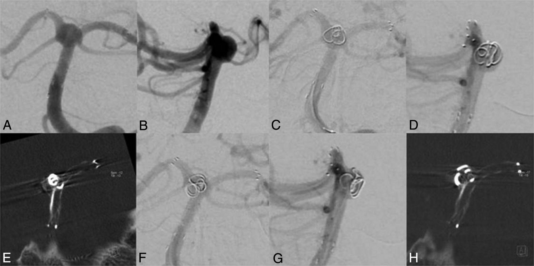
A 56-year-old women was scheduled for coil embolization of an incidental wide-neck basilar tip aneurysm (6 × 10 mm), incorporating both P1 segments into the aneurysm (Fig 1A, -B). Before the coiling, a stent (Neuroform3 Microdelivery Stent, 4.0 × 20 mm; Boston Scientific, Natick, Massachusetts) was deployed in the right PCA and the basilar artery. Subsequently, a second stent (Solitaire AB, 4.0 × 20 mm; ev3, Irvine, California) was passed through the stent struts of the Neuroform stent. The distal end of the stent was positioned in the left PCA, causing a Y-configuration and therefore recreating an aneurysm neck. After we positioned a microcatheter through the stent struts into the aneurysm, a 3D coil was inserted (Cashmere Microcoil, 4 × 45; Micrus Endovascular, San Jose, California). Because stents are, with the exception of proximal and distal radiopaque markers, hardly visible in standard techniques such as DSA, even with multiple oblique projections, it was not possible to ascertain coil deployment exclusively within the aneurysm (Fig 1C, -D). Thus, ACT was performed to accurately evaluate coil position before detachment, revealing a displaced coil loop in the P1 segment of the left PCA (Fig 1E). Repeat ACT after repositioning confirmed the correct coil position in the basilar tip aneurysm without compromising a parent artery (Fig 1F - H). Detachment of subsequent coils resulted in complete occlusion of the aneurysm (not shown).

Fig 1.
  • Download figure
  • Open in new tab
  • Download powerpoint
Fig 1.

A and B, Basilar tip aneurysm before coiling. C and D, The first coil in a basilar tip aneurysm before detachment. E, ACT scan shows coil protrusion into the left P1 segment. F and G, Delineation of the coil after repositioning without evidence suggesting coil protrusion in ACT (H).

Case 2

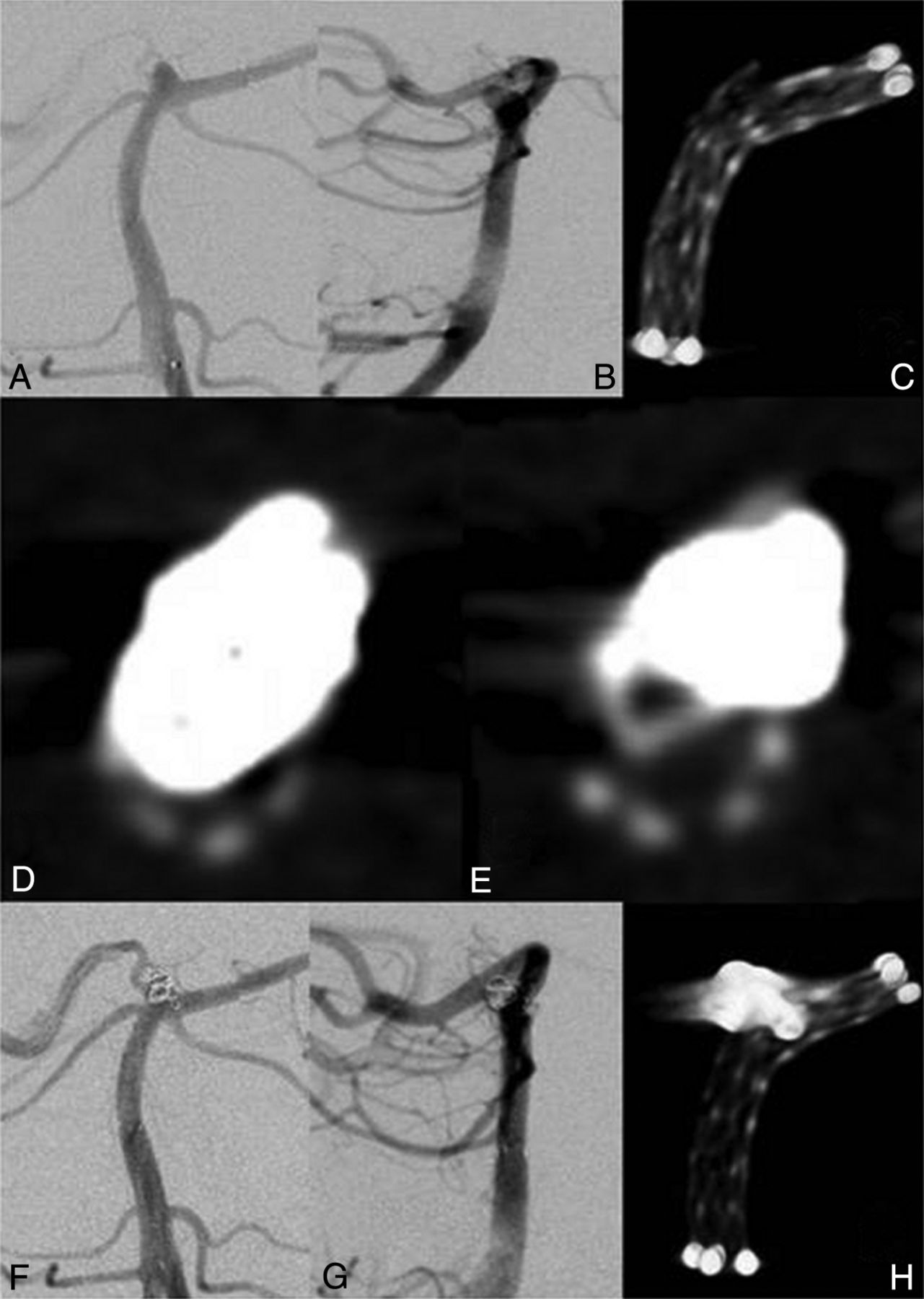
A 66-year-old man presented with an incidental wide-neck basilar tip aneurysm (2 × 3 mm) incorporating the left P1 segment (Fig 2A, -B). A stent (Neuroform3, 3.5 × 20 mm; Boston Scientific) was placed in the left PCA and the basilar artery. Because of the curved position, there was an opening of the stent struts at the sharpest angle (Fig 2C). Then, a microcatheter was passed through the stent struts into the aneurysm, and 2 coils (GDC-US, 2.5 × 30 mm, GDC-US, 2 × 20 mm; Boston Scientific) were inserted. Because there was still perfusion of the aneurysm, a third coil (GDC-US 2 × 10 mm; Boston Scientific) was inserted. Before detachment, DSA was suggestive of coil protrusion into the left P1 segment. ACT confirmed coil protrusion, most likely through the opened stent struts, resulting in an approximately 80% stenosis of the stent (Fig 2D). After multiple attempts at coil repositioning, there was still a small protrusion through the stent struts into the left P1 segment. Compared to previous coil positions, however, there was only a 25%–30% narrowing of the parent artery detectable (Fig 2E). Because additional attempts at coil repositioning or removal of the third coil were considered a disproportionate risk for the patient, detaching the coil in the current position (Fig 2F, -H) was preferred. To our knowledge, there were no neurologic symptoms present after the intervention or at regular follow-up visits within 2 years.

Fig 2.
  • Download figure
  • Open in new tab
  • Download powerpoint
Fig 2.

A and B, A 2 × 3 mm basilar tip aneurysm. C, ACT of the stent showing the opening of the stent struts. D, ACT image axial to the stent showing a profound narrowing of the stent. E, After repositioning, there is less protrusion of the coil loop, resulting in a decreased stenosis of the stent. F–H, Final imaging after coiling.

Discussion

Here we presented 2 cases of wide-neck basilar tip aneurysms incorporating 1 or both P1 segments, thus needing stent-assisted coiling. Because of the complex anatomy, especially for the first case, visualization of the parent artery or the stent was not feasible even with multiple oblique projections and DSA could not sufficiently exclude coil protrusion. Intraprocedural ACT clearly showed coil protrusion compromising arterial patency before detachment, which could be corrected, and it ascertained complete or almost complete arterial patency after coil repositioning.

Although ACT is also an established technique to identify pre-, peri-, or postprocedural complications like hemorrhage or hydrocephalus,5,8 in our cases, ACT was primarily performed for multiplanar visualization of endovascular stents and coil material.6 Beam-hardening artifacts caused by the coil package impair visibility of the stent. As demonstrated in Figs 1E and 2D, -E, beam-hardening artifacts impair visibility right after the first coil is inserted and increase with consecutively inserted coils. This problem limits the usefulness of ACT in cases with aneurysm (coil package) size mainly >10 mm in diameter but allows an excellent or at least favorable overview of the stent with adjacent small coil packages of ≤10 mm in diameter.6 When DSA cannot reliably delineate the coil position, intraprocedural ACT could help to identify even small or “silent” protruded coil loops in small coil packages.

Regarding radiation dose, preliminary results from internal investigations (T.S., 2011, unpublished data) with a calibrated ionization chamber and thermoluminescence dosimetry as well as Monte Carlo simulations and virtual phantom models show the effective dose of an ACT at a level of approximately 1.5 mSv. With collimation, this dose could be further reduced. Overall, we consider this as an acceptable dose to achieve a significant diagnostic benefit in the setting of this disease. Thus, ACT may provide the best positioning of coil material in a timely suitable manner and, compared with multiple oblique projections by using DSA, without the necessity of contrast medium.

Conclusions

Intraprocedural ACT might be helpful to accurately determine coil position during complex neuroendovascular procedures when it is difficult to visualize vasculature in DSA. Thus, intraprocedural visualization of vessel patency as well as coils and stents by using ACT may reduce procedural complications and increase safety.

REFERENCES

  1. 1.↵
    1. Saatci I,
    2. Geyik S,
    3. Yavuz K,
    4. et al
    . X-configured stent-assisted coiling in the endovascular treatment of complex anterior communicating artery aneurysms: a novel reconstructive technique. AJNR Am J Neuroradiol 2011;32:E113–17
    Abstract/FREE Full Text
  2. 2.↵
    1. Akgul E,
    2. Aksungur E,
    3. Balli T,
    4. et al
    . Y-stent-assisted coil embolization of wide-neck intracranial aneurysms: a single center experience. Interv Neuroradiol 2011;17:36–48
    Abstract/FREE Full Text
  3. 3.↵
    1. Cekirge HS,
    2. Yavuz K,
    3. Geyik S,
    4. et al
    . A Novel “Y” stent flow diversion technique for the endovascular treatment of bifurcation aneurysms without endosaccular coiling. AJNR Am J Neuroradiol 2011;32:1262–68
    Abstract/FREE Full Text
  4. 4.↵
    1. Psychogios MN,
    2. Buhk JH,
    3. Schramm P,
    4. et al
    . Feasibility of angiographic CT in peri-interventional diagnostic imaging: a comparative study with multidetector CT. AJNR Am J Neuroradiol 2010;31:1226–31. Epub 2010 Apr 1
    Abstract/FREE Full Text
  5. 5.↵
    1. Struffert T,
    2. Richter G,
    3. Engelhorn T,
    4. et al
    . Visualisation of intracerebral haemorrhage with flat-detector CT compared to multislice CT: results in 44 cases. Eur Radiol 2009;19:619–25
    PubMed
  6. 6.↵
    1. Richter G,
    2. Engelhorn T,
    3. Struffert T,
    4. et al
    . Flat panel detector angiographic CT for stent-assisted coil embolization of broad-based cerebral aneurysms. AJNR Am J Neuroradiol 2007;28:1902–08
    Abstract/FREE Full Text
  7. 7.↵
    1. Benndorf G,
    2. Strother CM,
    3. Claus B,
    4. et al
    . Angiographic CT in cerebrovascular stenting. AJNR Am J Neuroradiol 2005;26:1813–18
    Abstract/FREE Full Text
  8. 8.↵
    1. Engelhorn T,
    2. Struffert T,
    3. Richter G,
    4. et al
    . Flat panel detector angiographic CT in the management of aneurysmal rupture during coil embolization. AJNR Am J Neuroradiol 2008;29:1581–84
    Abstract/FREE Full Text
  • Received October 31, 2011.
  • Accepted after revision December 21, 2011.
  • © 2013 by American Journal of Neuroradiology
View Abstract
PreviousNext
Back to top

In this issue

American Journal of Neuroradiology: 34 (7)
American Journal of Neuroradiology
Vol. 34, Issue 7
1 Jul 2013
  • Table of Contents
  • Index by author
Advertisement
Print
Download PDF
Email Article

Thank you for your interest in spreading the word on American Journal of Neuroradiology.

NOTE: We only request your email address so that the person you are recommending the page to knows that you wanted them to see it, and that it is not junk mail. We do not capture any email address.

Enter multiple addresses on separate lines or separate them with commas.
Angiographic CT for Intraprocedural Monitoring of Complex Neuroendovascular Procedures
(Your Name) has sent you a message from American Journal of Neuroradiology
(Your Name) thought you would like to see the American Journal of Neuroradiology web site.
CAPTCHA
This question is for testing whether or not you are a human visitor and to prevent automated spam submissions.
Cite this article
J. Pfaff, T. Struffert, P. Gölitz, A. Doerfler
Angiographic CT for Intraprocedural Monitoring of Complex Neuroendovascular Procedures
American Journal of Neuroradiology Jul 2013, 34 (7) E77-E80; DOI: 10.3174/ajnr.A3014

Citation Manager Formats

  • BibTeX
  • Bookends
  • EasyBib
  • EndNote (tagged)
  • EndNote 8 (xml)
  • Medlars
  • Mendeley
  • Papers
  • RefWorks Tagged
  • Ref Manager
  • RIS
  • Zotero
0 Responses
Respond to this article
Share
Bookmark this article
Angiographic CT for Intraprocedural Monitoring of Complex Neuroendovascular Procedures
J. Pfaff, T. Struffert, P. Gölitz, A. Doerfler
American Journal of Neuroradiology Jul 2013, 34 (7) E77-E80; DOI: 10.3174/ajnr.A3014
del.icio.us logo Twitter logo Facebook logo Mendeley logo
  • Tweet Widget
  • Facebook Like
  • Google Plus One
Purchase

Jump to section

  • Article
    • Abstract
    • ABBREVIATIONS:
    • Technique
    • Discussion
    • Conclusions
    • REFERENCES
  • Figures & Data
  • Info & Metrics
  • Responses
  • References
  • PDF

Related Articles

  • No related articles found.
  • PubMed
  • Google Scholar

Cited By...

  • Application of High-Resolution C-Arm CT Combined with Streak Metal Artifact Removal Technology for the Stent-Assisted Embolization of Intracranial Aneurysms
  • Clinical evaluation of volume of interest imaging combined with metal artifact reduction reconstruction techniques in coiling and stent assisted coiling during neurointerventional procedures
  • Intravenous C-Arm Conebeam CT Angiography following Long-Term Flow-Diverter Implantation: Technologic Evaluation and Preliminary Results
  • Crossref (9)
  • Google Scholar

This article has been cited by the following articles in journals that are participating in Crossref Cited-by Linking.

  • Effective dose to patient measurements in flat-detector and multislice computed tomography: a comparison of applications in neuroradiology
    Tobias Struffert, Michael Hauer, Rosemarie Banckwitz, Christoph Köhler, Kevin Royalty, Arnd Doerfler
    European Radiology 2014 24 6
  • Flat-Panel Computed Tomography (DYNA-CT) in Neuroradiology. From High-Resolution Imaging of Implants to One-Stop-Shopping for Acute Stroke
    A. Doerfler, P. Gölitz, T. Engelhorn, S. Kloska, T. Struffert
    Clinical Neuroradiology 2015 25 S2
  • Detection of Intraparenchymal Hemorrhage After Endovascular Therapy in Patients with Acute Ischemic Stroke Using Immediate Postprocedural Flat‐Panel Computed Tomography Scan
    Seyedmehdi Payabvash, Asif A. Khan, Mushtaq H. Qureshi, Omar Saeed, M. Fareed K. Suri, Adnan I. Qureshi
    Journal of Neuroimaging 2016 26 2
  • Intravenous C-Arm Conebeam CT Angiography following Long-Term Flow-Diverter Implantation: Technologic Evaluation and Preliminary Results
    S.C.H. Yu, K.T. Lee, T.W.W. Lau, G.K.C. Wong, V.K.Y. Pang, K.Y. Chan
    American Journal of Neuroradiology 2016 37 3
  • Clinical evaluation of volume of interest imaging combined with metal artifact reduction reconstruction techniques in coiling and stent assisted coiling during neurointerventional procedures
    Qiang Zhang, Hao Zhao, Qi Sun, Jingfeng Han, Hao Zhang, Tonghui Shan, Wenlong Pan, Chuanqi Gu, Ruxiang Xu, Gengsheng Mao
    Journal of NeuroInterventional Surgery 2019 11 2
  • Application of High-Resolution C-Arm CT Combined with Streak Metal Artifact Removal Technology for the Stent-Assisted Embolization of Intracranial Aneurysms
    T.-F. Li, J. Ma, X.-W. Han, P.-J. Fu, R.-N. Niu, W.-Z. Luo, J.-Z. Ren
    American Journal of Neuroradiology 2019
  • Clinical Study of High-Resolution C-Arm CT in Mechanical Recanalization and Stent Implantation for Chronic Cerebral Artery Occlusion
    Teng-Fei Li, Cheng-Cheng Shi, Ji Ma, Pei-Ji Fu, Ming Zhu, Jian-Zhuang Ren, Xin-Wei Han
    Neurology India 2022 70 2
  • Vessel territory mapping after cerebral revascularization surgery using selective angiographic flat detector perfusion imaging
    Thijs van der Zijden, Annelies Mondelaers, Maurits Voormolen, Laetitia Yperzeele, Tomas Menovsky
    Journal of Integrative Neuroscience 2021 20 1
  • Vascular Imaging of the Central Nervous System
    Pedro Vilela
    2014

More in this TOC Section

  • 3D Fast Spin-Echo T1 Black-Blood Imaging for the Diagnosis of Cervical Artery Dissection
  • The Kety-Schmidt Technique for Quantitative Perfusion and Oxygen Metabolism Measurements in the MR Imaging Environment
  • A Swine Model to Analyze Arterial Structural Changes Induced by Mechanical Thrombectomy
Show more Technical Notes

Similar Articles

Advertisement

Indexed Content

  • Current Issue
  • Accepted Manuscripts
  • Article Preview
  • Past Issues
  • Editorials
  • Editor's Choice
  • Fellows' Journal Club
  • Letters to the Editor
  • Video Articles

Cases

  • Case Collection
  • Archive - Case of the Week
  • Archive - Case of the Month
  • Archive - Classic Case

More from AJNR

  • Trainee Corner
  • Imaging Protocols
  • MRI Safety Corner
  • Book Reviews

Multimedia

  • AJNR Podcasts
  • AJNR Scantastics

Resources

  • Turnaround Time
  • Submit a Manuscript
  • Submit a Video Article
  • Submit an eLetter to the Editor/Response
  • Manuscript Submission Guidelines
  • Statistical Tips
  • Fast Publishing of Accepted Manuscripts
  • Graphical Abstract Preparation
  • Imaging Protocol Submission
  • Evidence-Based Medicine Level Guide
  • Publishing Checklists
  • Author Policies
  • Become a Reviewer/Academy of Reviewers
  • News and Updates

About Us

  • About AJNR
  • Editorial Board
  • Editorial Board Alumni
  • Alerts
  • Permissions
  • Not an AJNR Subscriber? Join Now
  • Advertise with Us
  • Librarian Resources
  • Feedback
  • Terms and Conditions
  • AJNR Editorial Board Alumni

American Society of Neuroradiology

  • Not an ASNR Member? Join Now

© 2025 by the American Society of Neuroradiology All rights, including for text and data mining, AI training, and similar technologies, are reserved.
Print ISSN: 0195-6108 Online ISSN: 1936-959X

Powered by HighWire