
Article Figures & Data
Figures
- Fig. 1.
Hypoplasty of the left SPS. A, Frontal view of the left vertebral angiogram shows no visualization of the left SPS in the venous phase. Note well-developed venous drainage through the anastomosis of the petrosal vein with the transverse pontine vein (black arrows) and the lateral mesencephalic vein (black arrowheads). The right SPS (white arrows) is partially demonstrated at the posterior segment (from the confluence of the petrosal vein to the transverse). Frontal (B) and lateral (C) views of left internal carotid angiogram show no demonstration of left SPS in the venous phase. The venous drainage from the cavernous sinus is through the pterygoid plexus. The superficial middle cerebral vein and deep middle cerebral vein join to the cavernous sinus (black arrows).
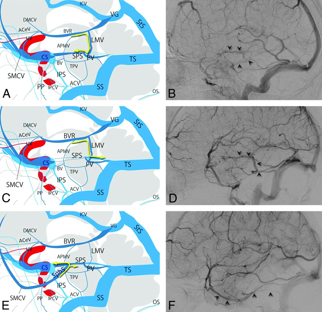
- Fig. 2.
Types of demonstration of normal SPS drainage on carotid angiography. A, C, and E, Schematic drawings of SPS demonstration on carotid angiography. B, D, and F, Lateral views of carotid angiography in the venous phase. Schematic drawing (A) and lateral view (B) of carotid angiography demonstrate the SPS drainage route (arrowheads) from the basal vein of Rosenthal (BVR) via the lateral mesencephalic vein (LMV), the petrosal vein (PV), and the anterior segment of the SPS into the cavernous sinus (CS). Schematic drawing (C) and lateral view (D) of carotid angiography demonstrate the SPS drainage route (arrowheads) from the BVR via the LMV, the PV, and the posterior segment of the SPS into the transverse sinus. Schematic drawing (E) and lateral view (F) of carotid angiography demonstrate the SPS drainage route (arrowheads) from the superficial middle cerebral vein (SMCV) via the sphenopetrosal sinus (primitive tentorial sinus) and both segments of the SPS. ACeV indicates anterior cerebral vein; ACV, anterior condylar vein; APMV, anterior pontomesencephalic vein; BV, bridging vein; DMCV, deep middle cerebral vein; ICV, internal cerebral vein; IPCV, inferior petroclival vein; IPS, inferior petrosal sinus; OS, occipital sinus; PP, pterygoid plexus; SphS, sphenopetrosal sinus; SS, sigmoid sinus; StS, straight sinus; TPV, transverse pontine vein; TS, transverse sinus; UV, uncal vein; VG, vein of Galen.
- Fig. 3.
Demonstration of the SPS on carotid angiography with the blood flow from the transverse sinus to the CS. Sequential images of frontal views of the left carotid angiogram show that the blood flow from the right transverse sinus partially fills the SPS (arrowheads) and continues to the right cavernous sinus (arrow).
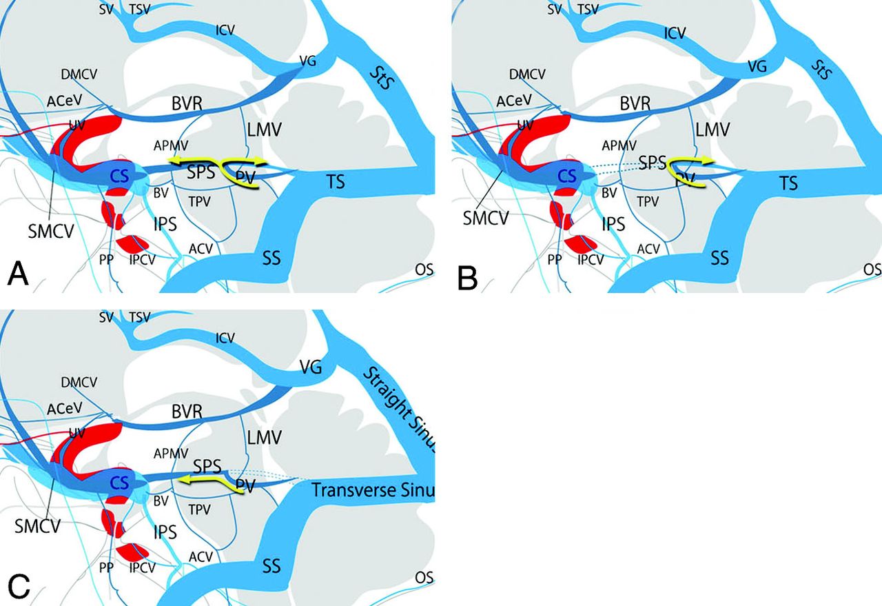
- Fig. 4.
Schematic drawing of the hemodynamic types of the SPS based on vertebral angiography. A, Drainage from the petrosal vein (PV) into both the anterior and the posterior segment of the SPS (yellow arrow). B, Drainage from the PV into the posterior segment of the SPS to the transverse sinus (TS) alone (yellow arrow). C, Drainage from the PV into the anterior segment of the SPS to the cavernous sinus (CS) alone (yellow arrow). ACeV indicates anterior cerebral vein; ACV, anterior condylar vein; APMV, anterior pontomesencephalic vein; BV, bridging vein; BVR, basal vein of Rosenthal; DMCV, deep middle cerebral vein; ICV, internal cerebral vein; IPCV, inferior petroclival vein; IPS, inferior petrosal sinus; LMV, lateral mesencephalic vein; OS, occipital sinus; PP, pterygoid plexus; SMCV, superficial middle cerebral vein; SS, sigmoid sinus; StS, straight sinus; TPV, transverse pontine vein; TS, transverse sinus; UV, uncal vein; VG, vein of Galen.
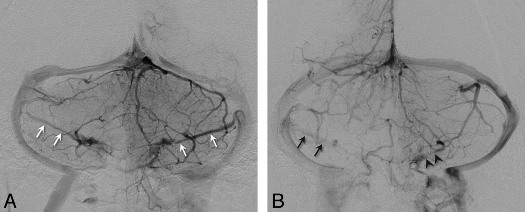
- Fig. 5.
Frontal view of the left vertebral angiography at venous phase. A, The whole SPS was demonstrated bilaterally (white arrows). B, The right SPS was demonstrated on the anterior segment alone (black arrows). The left SPS was demonstrated on the anterior segment alone (arrowheads).
- Fig. 6.
Frontal views of the vertebral angiogram show variations of the SPS. A, The right SPS reveals duplication of the superior segment (arrows) and inferior segment (arrowheads). The inferior segment is connected to the cavernous sinus. B, The right SPS reveals disconnection of the anterior and posterior segments (arrow). Both segments drain individually into the transverse sinus and the cavernous sinus.
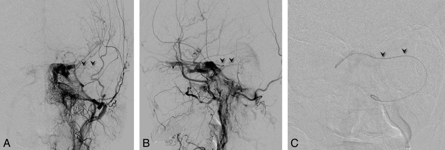
- Fig. 7.
CSDAVF with SPS drainage. Frontal (A) and lateral (B) views of the left external carotid angiogram show the AVF draining into the left SPS (arrowheads). Additional drainage routes of the superior ophthalmic vein, the inferior petrosal sinus, the intercavernous sinus, and the superficial middle cerebral vein are also noted. C, Microcatheter is inserted into the right cavernous sinus via the left SPS (arrowheads) for the transvenous embolization.
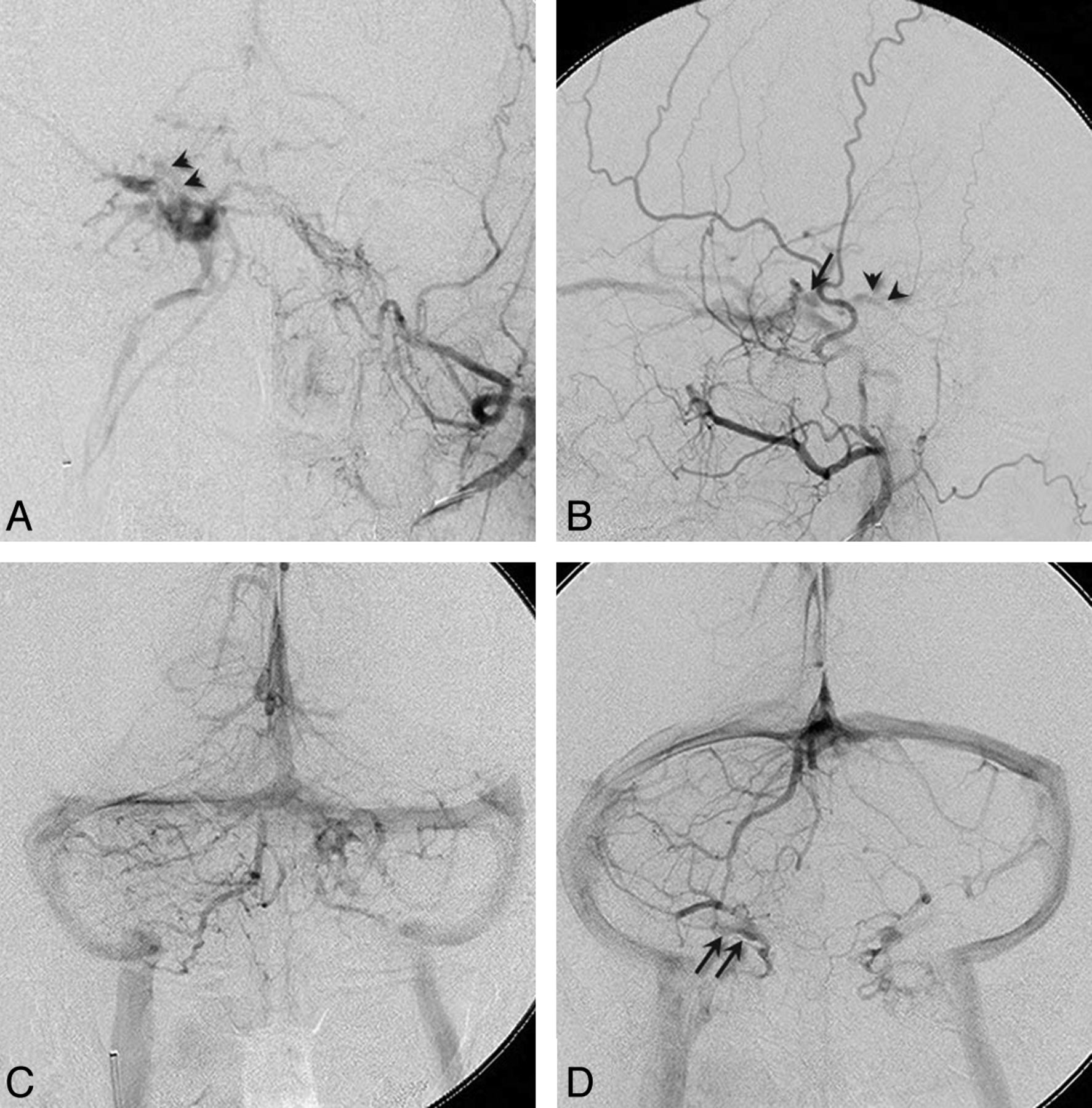
- Fig. 8.
CSDAVF with SPS drainage. Frontal (A) and lateral (B) views of the left internal maxillary angiogram show the AVF draining into the SPS (arrow) and the petrosal vein (arrowheads). Other drainage routes of the superior ophthalmic vein, the superficial middle cerebral vein, and the inferior petrosal sinus are also noted. C, Frontal view of the right vertebral angiogram shows no visualization of the right SPS and the obvious venous congestion of the right cerebellar hemisphere. D, Frontal view of the right vertebral angiogram after transvenous embolization shows normal venous drainage of the right SPS on the cavernous sinus side (arrows).
- Fig. 9.
The duplication of the SPS in CSDAVF. Frontal (A) and lateral (B) views of the left internal maxillary angiogram show the CSDAVF draining into the left duplicated SPS (arrowheads). Additional drainage routes of the superior ophthalmic vein, the inferior petrosal sinus, and the superficial middle cerebral vein are also noted. C, Selective venography by the inserted microcatheter into the SPS shows the duplication of the SPS (arrowheads).


Tables
- Table 1:
Hemodynamic status of the SPS in the cases of normal venous circulation on vertebral angiography
Demonstration/Hemodynamics Number of Sides (%) Total demonstration 116 (48.7) PV→anterior and posterior segments of SPS Partial demonstration 119 (50) PV→posterior segment of SPS 82 (34.5) PV→anterior segment of SPS 37 (15.5) No demonstration 3 (1.2) Note:—PV indicates petrosal vein.
- Table 2:
Hemodynamic status of the SPS in the cases of normal venous circulation on carotid angiography
Demonstration/Hemodynamics Number of Sides (%) Partial demonstration 10 (4.2) BVR→ LMV→ PV→ posterior segments of SPS 8 (3.4) BVR→ LMV→ PV→ anterior segment of SPS 2 (0.8) Total demonstration (SS→ SPS→ CS) 1 (0.4) Note:—BVR indicates basal vein of Rosenthal; CS, cavernous sinus; LMV, lateral mesencephalic vein; PV, petrosal vein; SS, sigmoid sinus.
Drainage Patterns Number of Cases CS→SPS→SS 3 CS→SPS→PV 3 CS→SPS→SphS 1 Note:—CS indicates cavernous sinus; PV, petrosal vein; SphS, sphenopetrosal sinus; SPS, superior petrosal sinus; SS, sigmoid sinus.


