Advertisement

Main menu

  • Home
  • Content
    • Current Issue
    • Accepted Manuscripts
    • Article Preview
    • Past Issue Archive
    • Video Articles
    • AJNR Case Collection
    • Case of the Week Archive
    • Case of the Month Archive
    • Classic Case Archive
  • Special Collections
    • AJNR Awards
    • Low-Field MRI
    • Alzheimer Disease
    • ASNR Foundation Special Collection
    • Photon-Counting CT
    • View All
  • Multimedia
    • AJNR Podcasts
    • AJNR SCANtastic
    • Trainee Corner
    • MRI Safety Corner
    • Imaging Protocols
  • For Authors
    • Submit a Manuscript
    • Submit a Video Article
    • Submit an eLetter to the Editor/Response
    • Manuscript Submission Guidelines
    • Statistical Tips
    • Fast Publishing of Accepted Manuscripts
    • Graphical Abstract Preparation
    • Imaging Protocol Submission
    • Author Policies
  • About Us
    • About AJNR
    • Editorial Board
    • Editorial Board Alumni
  • More
    • Become a Reviewer/Academy of Reviewers
    • Subscribers
    • Permissions
    • Alerts
    • Feedback
    • Advertisers
    • ASNR Home

User menu

  • Alerts
  • Log in

Search

  • Advanced search
American Journal of Neuroradiology

American Journal of Neuroradiology

ASHNR American Society of Functional Neuroradiology ASHNR American Society of Pediatric Neuroradiology ASSR
  • Alerts
  • Log in

Advanced Search

  • Home
  • Content
    • Current Issue
    • Accepted Manuscripts
    • Article Preview
    • Past Issue Archive
    • Video Articles
    • AJNR Case Collection
    • Case of the Week Archive
    • Case of the Month Archive
    • Classic Case Archive
  • Special Collections
    • AJNR Awards
    • Low-Field MRI
    • Alzheimer Disease
    • ASNR Foundation Special Collection
    • Photon-Counting CT
    • View All
  • Multimedia
    • AJNR Podcasts
    • AJNR SCANtastic
    • Trainee Corner
    • MRI Safety Corner
    • Imaging Protocols
  • For Authors
    • Submit a Manuscript
    • Submit a Video Article
    • Submit an eLetter to the Editor/Response
    • Manuscript Submission Guidelines
    • Statistical Tips
    • Fast Publishing of Accepted Manuscripts
    • Graphical Abstract Preparation
    • Imaging Protocol Submission
    • Author Policies
  • About Us
    • About AJNR
    • Editorial Board
    • Editorial Board Alumni
  • More
    • Become a Reviewer/Academy of Reviewers
    • Subscribers
    • Permissions
    • Alerts
    • Feedback
    • Advertisers
    • ASNR Home
  • Follow AJNR on Twitter
  • Visit AJNR on Facebook
  • Follow AJNR on Instagram
  • Join AJNR on LinkedIn
  • RSS Feeds

AJNR Awards, New Junior Editors, and more. Read the latest AJNR updates

Research ArticleBrain

Automated Determination of Brain Parenchymal Fraction in Multiple Sclerosis

M. Vågberg, T. Lindqvist, K. Ambarki, J.B.M. Warntjes, P. Sundström, R. Birgander and A. Svenningsson
American Journal of Neuroradiology March 2013, 34 (3) 498-504; DOI: https://doi.org/10.3174/ajnr.A3262
M. Vågberg
aFrom the Department of Pharmacology and Clinical Neuroscience, Section of Neuroscience (M.V., P.S., A.S.)
  • Find this author on Google Scholar
  • Find this author on PubMed
  • Search for this author on this site
T. Lindqvist
bDepartment of Radiation Sciences, Diagnostic Radiology (T.L., K.A., R.B.)
  • Find this author on Google Scholar
  • Find this author on PubMed
  • Search for this author on this site
K. Ambarki
bDepartment of Radiation Sciences, Diagnostic Radiology (T.L., K.A., R.B.)
cCenter for Biomedical Engineering and Physics (K.A.), Umeå University, Umeå, Sweden
  • Find this author on Google Scholar
  • Find this author on PubMed
  • Search for this author on this site
J.B.M. Warntjes
dCenter for Medical Imaging Science and Visualization and Division of Clinical Physiology, Department of Medicine and Health (J.B.M.W.), Linköping University, Linköping, Sweden.
  • Find this author on Google Scholar
  • Find this author on PubMed
  • Search for this author on this site
P. Sundström
aFrom the Department of Pharmacology and Clinical Neuroscience, Section of Neuroscience (M.V., P.S., A.S.)
  • Find this author on Google Scholar
  • Find this author on PubMed
  • Search for this author on this site
R. Birgander
bDepartment of Radiation Sciences, Diagnostic Radiology (T.L., K.A., R.B.)
  • Find this author on Google Scholar
  • Find this author on PubMed
  • Search for this author on this site
A. Svenningsson
aFrom the Department of Pharmacology and Clinical Neuroscience, Section of Neuroscience (M.V., P.S., A.S.)
  • Find this author on Google Scholar
  • Find this author on PubMed
  • Search for this author on this site
  • Article
  • Figures & Data
  • Info & Metrics
  • Responses
  • References
  • PDF
Loading

Article Figures & Data

Figures

  • Fig. 1.
    • Download figure
    • Open in new tab
    • Download powerpoint
    Fig. 1.

    Overview of synthetic tissue mapping. A, MR quantification scan of an axial section of the brain results in a pixel-wise measurement of the R1 and R2 relaxation rates and PD. Data of the ROI (gray box) is plotted on the R1-R2 projection of this space in B. CSF, gray matter, and WM have unique combinations of R1, R2, and PD and thus a fixed position in R1-R2-PD space (circles), containing 100% of the indicated tissue types. Partial volume data are located between these identified positions. For CSF, the partial volume values are shown as the white gradient. All voxels containing CSF, gray matter, or WM are included in the ICV mask, followed by a region growing algorithm to ensure that the ICV is represented as a continuous volume (C). D, Within the ICV mask, the partial volume CSF is calculated. The brain parenchymal volume corresponds to the ICV minus the total CSF volume. Finally, the BPF is found as the ratio between brain parenchymal volume and ICV.
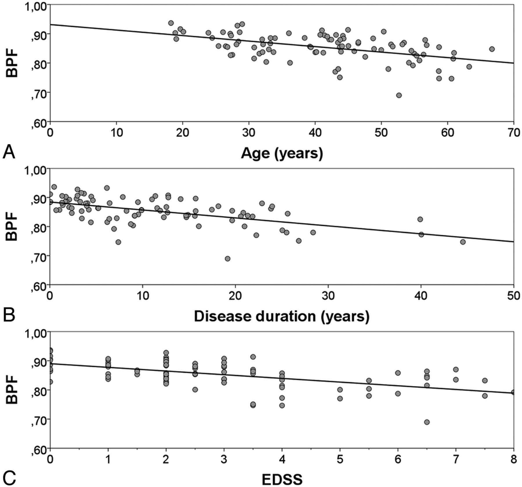
  • Fig. 2.
    • Download figure
    • Open in new tab
    • Download powerpoint
    Fig. 2.

    BPF in relation to age, disease duration, and functional impairment. BPF of the patients with relapsing onset MS (relapsing-remitting MS and secondary-progressive MS) plotted as a function of (A) age (r2 = 0.24; P < .001), (B) disease duration (r2 = 0.33; P < .001), and (C) Expanded Disability Status Scale (r2 = 0.32; P < .001).
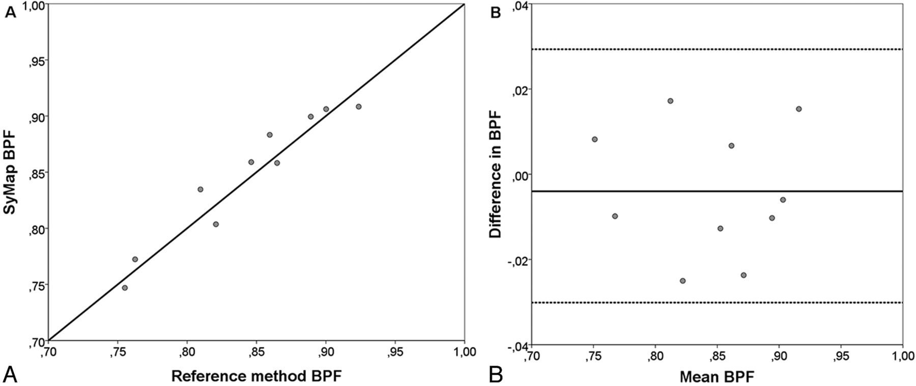
  • Fig. 3.
    • Download figure
    • Open in new tab
    • Download powerpoint
    Fig. 3.

    Comparison between SyMap and QBrain. A, BPF values calculated with the SyMap method plotted against the value calculated with the manual reference method for the same subject. B, Difference in BPF between the methods plotted for each subject against the mean BPF for each subject as a Bland-Altman plot. Mean difference (solid line), limits of agreement (dotted lines, mean difference ± 1.96 SD).
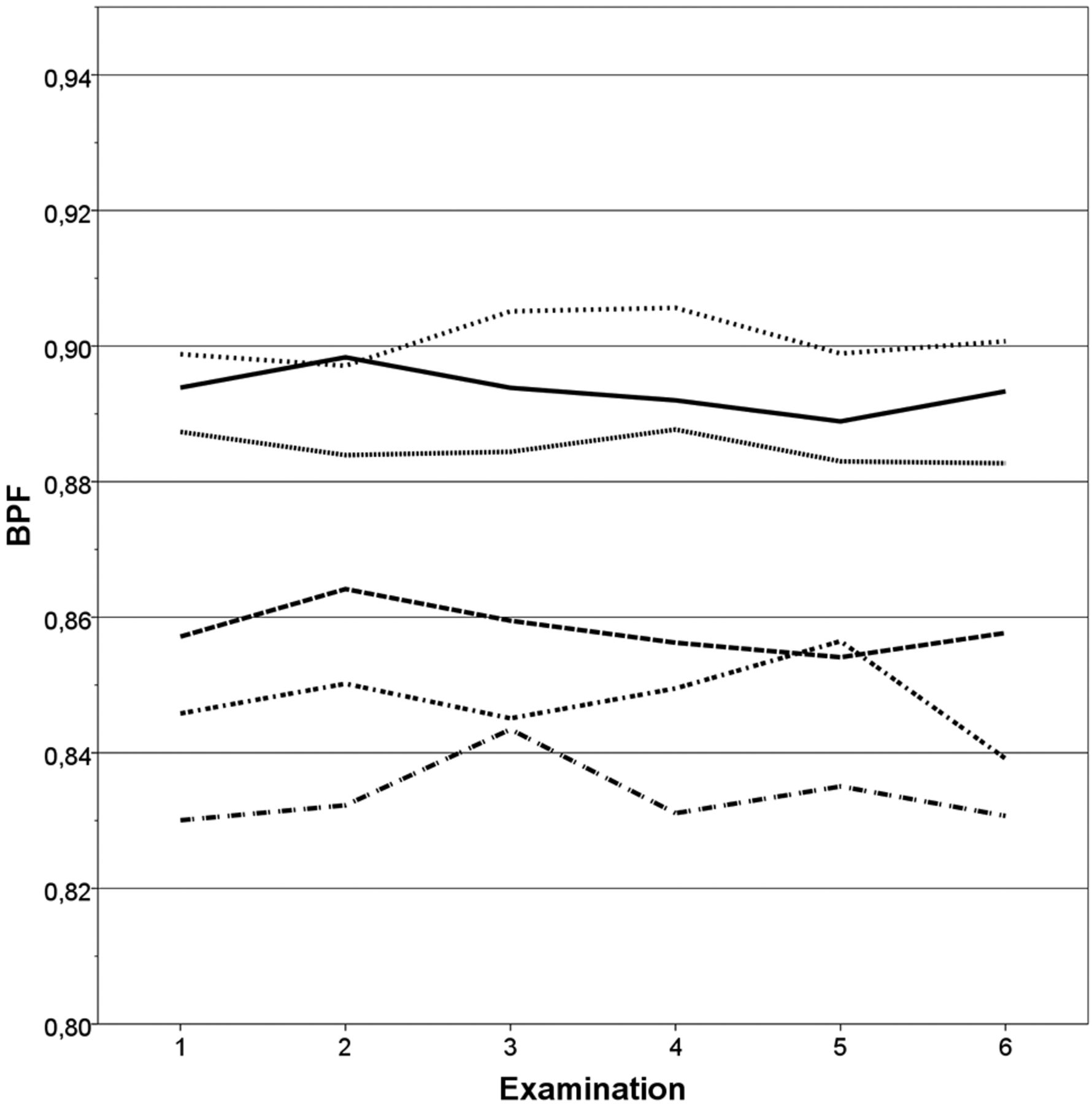
  • Fig. 4.
    • Download figure
    • Open in new tab
    • Download powerpoint
    Fig. 4.

    Reproducibility of SyMap. BPF of each of the repeated scans plotted for each of the 6 subjects. Coefficient of variation = 0.45% (SE = 0.068%).

PreviousNext
Back to top

In this issue

American Journal of Neuroradiology: 34 (3)
American Journal of Neuroradiology
Vol. 34, Issue 3
1 Mar 2013
  • Table of Contents
  • Index by author
Advertisement
Print
Download PDF
Email Article

Thank you for your interest in spreading the word on American Journal of Neuroradiology.

NOTE: We only request your email address so that the person you are recommending the page to knows that you wanted them to see it, and that it is not junk mail. We do not capture any email address.

Enter multiple addresses on separate lines or separate them with commas.
Automated Determination of Brain Parenchymal Fraction in Multiple Sclerosis
(Your Name) has sent you a message from American Journal of Neuroradiology
(Your Name) thought you would like to see the American Journal of Neuroradiology web site.
CAPTCHA
This question is for testing whether or not you are a human visitor and to prevent automated spam submissions.
Cite this article
M. Vågberg, T. Lindqvist, K. Ambarki, J.B.M. Warntjes, P. Sundström, R. Birgander, A. Svenningsson
Automated Determination of Brain Parenchymal Fraction in Multiple Sclerosis
American Journal of Neuroradiology Mar 2013, 34 (3) 498-504; DOI: 10.3174/ajnr.A3262

Citation Manager Formats

  • BibTeX
  • Bookends
  • EasyBib
  • EndNote (tagged)
  • EndNote 8 (xml)
  • Medlars
  • Mendeley
  • Papers
  • RefWorks Tagged
  • Ref Manager
  • RIS
  • Zotero
0 Responses
Respond to this article
Share
Bookmark this article
Automated Determination of Brain Parenchymal Fraction in Multiple Sclerosis
M. Vågberg, T. Lindqvist, K. Ambarki, J.B.M. Warntjes, P. Sundström, R. Birgander, A. Svenningsson
American Journal of Neuroradiology Mar 2013, 34 (3) 498-504; DOI: 10.3174/ajnr.A3262
del.icio.us logo Twitter logo Facebook logo Mendeley logo
  • Tweet Widget
  • Facebook Like
  • Google Plus One
Purchase

Jump to section

  • Article
    • Abstract
    • ABBREVIATIONS:
    • Materials and Methods
    • Results
    • Discussion
    • Conclusions
    • Acknowledgments
    • Footnotes
    • References
  • Figures & Data
  • Info & Metrics
  • Responses
  • References
  • PDF

Related Articles

  • No related articles found.
  • PubMed
  • Google Scholar

Cited By...

  • Synthetic MRI in Progressive MS: Associations with Disability
  • Aging and the Brain: A Quantitative Study of Clinical CT Images
  • Gadolinium Retention in the Brain: An MRI Relaxometry Study of Linear and Macrocyclic Gadolinium-Based Contrast Agents in Multiple Sclerosis
  • Normal Values of Magnetic Relaxation Parameters of Spine Components with the Synthetic MRI Sequence
  • Improved Precision of Automatic Brain Volume Measurements in Patients with Clinically Isolated Syndrome and Multiple Sclerosis Using Edema Correction
  • Loss of corticospinal tract integrity in early MS disease stages
  • Rituximab in multiple sclerosis: A retrospective observational study on safety and efficacy
  • Clinical Feasibility of Synthetic MRI in Multiple Sclerosis: A Diagnostic and Volumetric Validation Study
  • Quantitative MRI for Rapid and User-Independent Monitoring of Intracranial CSF Volume in Hydrocephalus
  • Quantitative MRI for Analysis of Active Multiple Sclerosis Lesions without Gadolinium-Based Contrast Agent
  • Effects of Gadolinium Contrast Agent Administration on Automatic Brain Tissue Classification of Patients with Multiple Sclerosis
  • Crossref (49)
  • Google Scholar

This article has been cited by the following articles in journals that are participating in Crossref Cited-by Linking.

  • Rituximab in multiple sclerosis
    Jonatan Salzer, Rasmus Svenningsson, Peter Alping, Lenka Novakova, Anna Björck, Katharina Fink, Protik Islam-Jakobsson, Clas Malmeström, Markus Axelsson, Mattias Vågberg, Peter Sundström, Jan Lycke, Fredrik Piehl, Anders Svenningsson
    Neurology 2016 87 20
  • SyMRI of the Brain
    Akifumi Hagiwara, Marcel Warntjes, Masaaki Hori, Christina Andica, Misaki Nakazawa, Kanako Kunishima Kumamaru, Osamu Abe, Shigeki Aoki
    Investigative Radiology 2017 52 10
  • Clinical Feasibility of Synthetic MRI in Multiple Sclerosis: A Diagnostic and Volumetric Validation Study
    T. Granberg, M. Uppman, F. Hashim, C. Cananau, L.E. Nordin, S. Shams, J. Berglund, Y. Forslin, P. Aspelin, S. Fredrikson, M. Kristoffersen-Wiberg
    American Journal of Neuroradiology 2016 37 6
  • Levels and Age Dependency of Neurofilament Light and Glial Fibrillary Acidic Protein in Healthy Individuals and Their Relation to the Brain Parenchymal Fraction
    Mattias Vågberg, Niklas Norgren, Ann Dring, Thomas Lindqvist, Richard Birgander, Henrik Zetterberg, Anders Svenningsson, Markus Reindl
    PLOS ONE 2015 10 8
  • MRI in the assessment and monitoring of multiple sclerosis: an update on best practice
    Ulrike W. Kaunzner, Susan A. Gauthier
    Therapeutic Advances in Neurological Disorders 2017 10 6
  • Neurofilament levels, disease activity and brain volume during follow-up in multiple sclerosis
    Irene Håkansson, Anders Tisell, Petra Cassel, Kaj Blennow, Henrik Zetterberg, Peter Lundberg, Charlotte Dahle, Magnus Vrethem, Jan Ernerudh
    Journal of Neuroinflammation 2018 15 1
  • Increased Concentrations of Glutamate and Glutamine in Normal-Appearing White Matter of Patients with Multiple Sclerosis and Normal MR Imaging Brain Scans
    Anders Tisell, Olof Dahlqvist Leinhard, Jan Bertus Marcel Warntjes, Anne Aalto, Örjan Smedby, Anne-Marie Landtblom, Peter Lundberg, Richard Jay Smeyne
    PLoS ONE 2013 8 4
  • Normal Appearing and Diffusely Abnormal White Matter in Patients with Multiple Sclerosis Assessed with Quantitative MR
    Janne West, Anne Aalto, Anders Tisell, Olof Dahlqvist Leinhard, Anne-Marie Landtblom, Örjan Smedby, Peter Lundberg, Pablo Villoslada
    PLoS ONE 2014 9 4
  • Quantitative MRI for Analysis of Active Multiple Sclerosis Lesions without Gadolinium-Based Contrast Agent
    I. Blystad, I. Håkansson, A. Tisell, J. Ernerudh, Ö. Smedby, P. Lundberg, E.-M. Larsson
    American Journal of Neuroradiology 2016 37 1
  • Synthetic MRI: Technologies and Applications in Neuroradiology
    Sooyeon Ji, Dongjin Yang, Jongho Lee, Seung Hong Choi, Hyeonjin Kim, Koung Mi Kang
    Journal of Magnetic Resonance Imaging 2022 55 4

More in this TOC Section

  • Enhanced Axonal Metabolism during Early Natalizumab Treatment in Relapsing-Remitting Multiple Sclerosis
  • SWI or T2*: Which MRI Sequence to Use in the Detection of Cerebral Microbleeds? The Karolinska Imaging Dementia Study
  • Progression of Microstructural Damage in Spinocerebellar Ataxia Type 2: A Longitudinal DTI Study
Show more BRAIN

Similar Articles

Advertisement

Indexed Content

  • Current Issue
  • Accepted Manuscripts
  • Article Preview
  • Past Issues
  • Editorials
  • Editor's Choice
  • Fellows' Journal Club
  • Letters to the Editor
  • Video Articles

Cases

  • Case Collection
  • Archive - Case of the Week
  • Archive - Case of the Month
  • Archive - Classic Case

More from AJNR

  • Trainee Corner
  • Imaging Protocols
  • MRI Safety Corner
  • Book Reviews

Multimedia

  • AJNR Podcasts
  • AJNR Scantastics

Resources

  • Turnaround Time
  • Submit a Manuscript
  • Submit a Video Article
  • Submit an eLetter to the Editor/Response
  • Manuscript Submission Guidelines
  • Statistical Tips
  • Fast Publishing of Accepted Manuscripts
  • Graphical Abstract Preparation
  • Imaging Protocol Submission
  • Evidence-Based Medicine Level Guide
  • Publishing Checklists
  • Author Policies
  • Become a Reviewer/Academy of Reviewers
  • News and Updates

About Us

  • About AJNR
  • Editorial Board
  • Editorial Board Alumni
  • Alerts
  • Permissions
  • Not an AJNR Subscriber? Join Now
  • Advertise with Us
  • Librarian Resources
  • Feedback
  • Terms and Conditions
  • AJNR Editorial Board Alumni

American Society of Neuroradiology

  • Not an ASNR Member? Join Now

© 2025 by the American Society of Neuroradiology All rights, including for text and data mining, AI training, and similar technologies, are reserved.
Print ISSN: 0195-6108 Online ISSN: 1936-959X

Powered by HighWire