Advertisement

Main menu

  • Home
  • Content
    • Current Issue
    • Accepted Manuscripts
    • Article Preview
    • Past Issue Archive
    • Video Articles
    • AJNR Case Collection
    • Case of the Week Archive
    • Case of the Month Archive
    • Classic Case Archive
  • Special Collections
    • AJNR Awards
    • Low-Field MRI
    • Alzheimer Disease
    • ASNR Foundation Special Collection
    • Photon-Counting CT
    • View All
  • Multimedia
    • AJNR Podcasts
    • AJNR SCANtastic
    • Trainee Corner
    • MRI Safety Corner
    • Imaging Protocols
  • For Authors
    • Submit a Manuscript
    • Submit a Video Article
    • Submit an eLetter to the Editor/Response
    • Manuscript Submission Guidelines
    • Statistical Tips
    • Fast Publishing of Accepted Manuscripts
    • Graphical Abstract Preparation
    • Imaging Protocol Submission
    • Author Policies
  • About Us
    • About AJNR
    • Editorial Board
    • Editorial Board Alumni
  • More
    • Become a Reviewer/Academy of Reviewers
    • Subscribers
    • Permissions
    • Alerts
    • Feedback
    • Advertisers
    • ASNR Home

User menu

  • Alerts
  • Log in

Search

  • Advanced search
American Journal of Neuroradiology

American Journal of Neuroradiology

ASHNR American Society of Functional Neuroradiology ASHNR American Society of Pediatric Neuroradiology ASSR
  • Alerts
  • Log in

Advanced Search

  • Home
  • Content
    • Current Issue
    • Accepted Manuscripts
    • Article Preview
    • Past Issue Archive
    • Video Articles
    • AJNR Case Collection
    • Case of the Week Archive
    • Case of the Month Archive
    • Classic Case Archive
  • Special Collections
    • AJNR Awards
    • Low-Field MRI
    • Alzheimer Disease
    • ASNR Foundation Special Collection
    • Photon-Counting CT
    • View All
  • Multimedia
    • AJNR Podcasts
    • AJNR SCANtastic
    • Trainee Corner
    • MRI Safety Corner
    • Imaging Protocols
  • For Authors
    • Submit a Manuscript
    • Submit a Video Article
    • Submit an eLetter to the Editor/Response
    • Manuscript Submission Guidelines
    • Statistical Tips
    • Fast Publishing of Accepted Manuscripts
    • Graphical Abstract Preparation
    • Imaging Protocol Submission
    • Author Policies
  • About Us
    • About AJNR
    • Editorial Board
    • Editorial Board Alumni
  • More
    • Become a Reviewer/Academy of Reviewers
    • Subscribers
    • Permissions
    • Alerts
    • Feedback
    • Advertisers
    • ASNR Home
  • Follow AJNR on Twitter
  • Visit AJNR on Facebook
  • Follow AJNR on Instagram
  • Join AJNR on LinkedIn
  • RSS Feeds

AJNR Awards, New Junior Editors, and more. Read the latest AJNR updates

Research ArticleSpine

Spatial Normalization and Regional Assessment of Cord Atrophy: Voxel-Based Analysis of Cervical Cord 3D T1-Weighted Images

P. Valsasina, M.A. Horsfield, M.A. Rocca, M. Absinta, G. Comi and M. Filippi
American Journal of Neuroradiology December 2012, 33 (11) 2195-2200; DOI: https://doi.org/10.3174/ajnr.A3139
P. Valsasina
aNeuroimaging Research Unit (P.V., M.A.R., M.A., M.F.), Institute of Experimental Neurology
  • Find this author on Google Scholar
  • Find this author on PubMed
  • Search for this author on this site
M.A. Horsfield
cMedical Physics Group (M.A.H.), Department of Cardiovascular Sciences, University of Leicester, Leicester Royal Infirmary, Leicester, United Kingdom.
  • Find this author on Google Scholar
  • Find this author on PubMed
  • Search for this author on this site
M.A. Rocca
aNeuroimaging Research Unit (P.V., M.A.R., M.A., M.F.), Institute of Experimental Neurology
bDepartment of Neurology (M.A.R., M.A., G.C., M.F.), San Raffaele Scientific Institute, “Vita-Salute” San Raffaele University, Milan, Italy
  • Find this author on Google Scholar
  • Find this author on PubMed
  • Search for this author on this site
M. Absinta
aNeuroimaging Research Unit (P.V., M.A.R., M.A., M.F.), Institute of Experimental Neurology
bDepartment of Neurology (M.A.R., M.A., G.C., M.F.), San Raffaele Scientific Institute, “Vita-Salute” San Raffaele University, Milan, Italy
  • Find this author on Google Scholar
  • Find this author on PubMed
  • Search for this author on this site
G. Comi
bDepartment of Neurology (M.A.R., M.A., G.C., M.F.), San Raffaele Scientific Institute, “Vita-Salute” San Raffaele University, Milan, Italy
  • Find this author on Google Scholar
  • Find this author on PubMed
  • Search for this author on this site
M. Filippi
aNeuroimaging Research Unit (P.V., M.A.R., M.A., M.F.), Institute of Experimental Neurology
bDepartment of Neurology (M.A.R., M.A., G.C., M.F.), San Raffaele Scientific Institute, “Vita-Salute” San Raffaele University, Milan, Italy
  • Find this author on Google Scholar
  • Find this author on PubMed
  • Search for this author on this site
  • Article
  • Figures & Data
  • Supplemental
  • Info & Metrics
  • Responses
  • References
  • PDF
Loading

Article Figures & Data

Figures

  • Tables
  • Fig 1.
    • Download figure
    • Open in new tab
    • Download powerpoint
    Fig 1.

    A, Coronal, sagittal, and axial views of the cervical cord template created as average of the 83 healthy subjects included in the study. B, Coronal, sagittal, and axial sections (at different cervical cord levels) of the corresponding color-coded normalized region-label mask reporting the 24 reference anatomic regions. Areas outside the cervical cord were masked out.
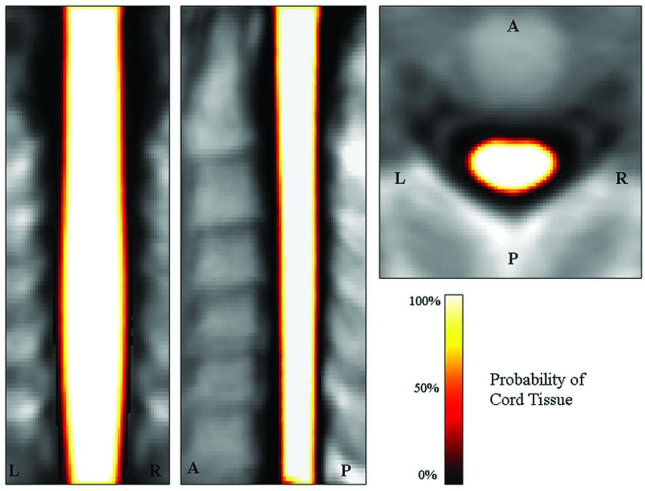
  • Fig 2.
    • Download figure
    • Open in new tab
    • Download powerpoint
    Fig 2.

    Coronal, sagittal, and axial views of the mean cord tissue probability map for the 83 study subjects.
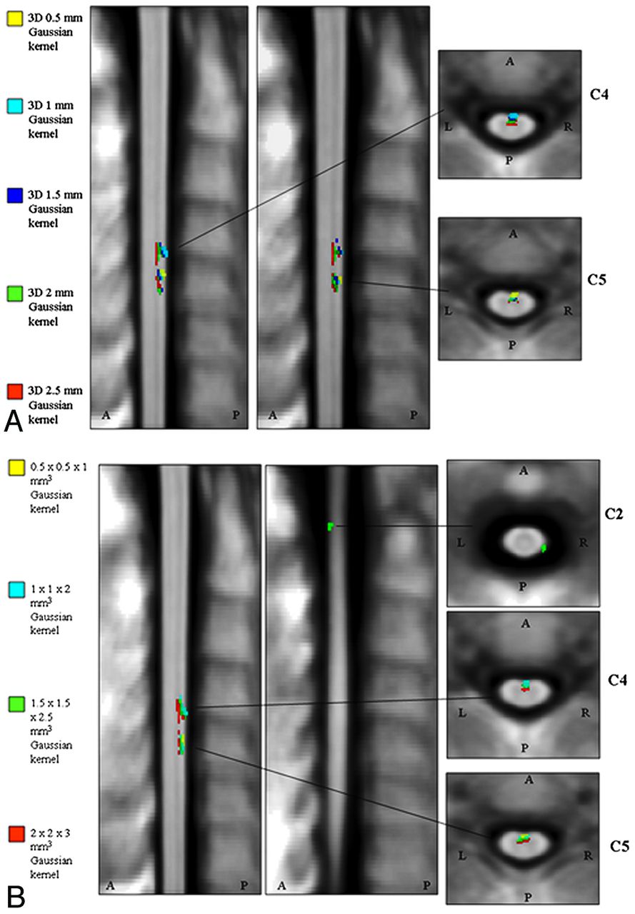
  • Fig 3.
    • Download figure
    • Open in new tab
    • Download powerpoint
    Fig 3.

    Significant clusters of correlation between cervical cord atrophy and subject age, corrected for the possible confounding effects of sex and total cord volume (P < .001, uncorrected for multiple comparisons; cluster extent, k ≥ 20) with the different smoothing options applied to our cervical cord masks. A, Isotropic Gaussian kernels with FWHM ranging from 0.5 to 2.5 mm3. B, Anisotropic Gaussian kernels with FWHM ranging from 0.5 × 0.5 × 1 mm3 to 2 × 2 × 3 mm3.

Tables

  • Figures
  • Significant clusters of correlation between cord atrophy and aging revealed by voxel-based analyses with different smoothing optionsa

    Smoothing-Kernel WidthPeak LocationsP ValueT ValueκR% Voxels% Voxels
    APR-L
    0.5 × 0.5 × 0.5 mm3AR-C5<.0013.931−0.4131/06/25
    1 × 1 × 1 mm3AR-C4<.0013.832−0.4073/08/65
    AR-C5<.0013.941−0.40
    1.5 × 1.5 × 1.5 mm3AR-C4<.0013.747−0.39103/016/87
    AR-C5<.0013.856−0.40
    2 × 2 × 2 mm3AR-C4<.0014.276−0.44140/026/115
    AL-C5<.0014.164−0.42
    2.5 × 2.5 × 2.5 mm3AL-C4<.0014.591−0.46149/033/116
    AL-C5<.0014.158−0.42
    0.5 × 0.5 × 1 mm3AR-C5<.0013.929−0.4029/05/24
    1 × 1 × 2 mm3AR-C4<.0013.842−0.3989/013/76
    AR-C5<.0013.847−0.40
    1.5 × 1.5 × 2.5 mm3PR-C1/C2<.0013.620−0.37117/2023/114
    AR-C4<.0013.752−0.39
    AR-C5<.0013.865−0.40
    2 × 2 × 3 mm3AR C4<.0014.1150−0.43150/035/115
    AL C5<.0014.3−0.45
    • ↵a Multiple regression with age corrected for sex and total cord volume, P < .001; uncorrected for multiple comparisons, cluster extent κ ≥ 20.

PreviousNext
Back to top

In this issue

American Journal of Neuroradiology: 33 (11)
American Journal of Neuroradiology
Vol. 33, Issue 11
1 Dec 2012
  • Table of Contents
  • Index by author
Advertisement
Print
Download PDF
Email Article

Thank you for your interest in spreading the word on American Journal of Neuroradiology.

NOTE: We only request your email address so that the person you are recommending the page to knows that you wanted them to see it, and that it is not junk mail. We do not capture any email address.

Enter multiple addresses on separate lines or separate them with commas.
Spatial Normalization and Regional Assessment of Cord Atrophy: Voxel-Based Analysis of Cervical Cord 3D T1-Weighted Images
(Your Name) has sent you a message from American Journal of Neuroradiology
(Your Name) thought you would like to see the American Journal of Neuroradiology web site.
CAPTCHA
This question is for testing whether or not you are a human visitor and to prevent automated spam submissions.
Cite this article
P. Valsasina, M.A. Horsfield, M.A. Rocca, M. Absinta, G. Comi, M. Filippi
Spatial Normalization and Regional Assessment of Cord Atrophy: Voxel-Based Analysis of Cervical Cord 3D T1-Weighted Images
American Journal of Neuroradiology Dec 2012, 33 (11) 2195-2200; DOI: 10.3174/ajnr.A3139

Citation Manager Formats

  • BibTeX
  • Bookends
  • EasyBib
  • EndNote (tagged)
  • EndNote 8 (xml)
  • Medlars
  • Mendeley
  • Papers
  • RefWorks Tagged
  • Ref Manager
  • RIS
  • Zotero
0 Responses
Respond to this article
Share
Bookmark this article
Spatial Normalization and Regional Assessment of Cord Atrophy: Voxel-Based Analysis of Cervical Cord 3D T1-Weighted Images
P. Valsasina, M.A. Horsfield, M.A. Rocca, M. Absinta, G. Comi, M. Filippi
American Journal of Neuroradiology Dec 2012, 33 (11) 2195-2200; DOI: 10.3174/ajnr.A3139
del.icio.us logo Twitter logo Facebook logo Mendeley logo
  • Tweet Widget
  • Facebook Like
  • Google Plus One
Purchase

Jump to section

  • Article
    • Abstract
    • ABBREVIATIONS:
    • Materials and Methods
    • Results
    • Discussion
    • Conclusions
    • Footnotes
    • References
  • Figures & Data
  • Supplemental
  • Info & Metrics
  • Responses
  • References
  • PDF

Related Articles

  • PubMed
  • Google Scholar

Cited By...

  • Connectome Spatial Smoothing (CSS): concepts, methods, and evaluation
  • Heritability of cervical spinal cord structure
  • Considerations for Mean Upper Cervical Cord Area Implementation in a Longitudinal MRI Setting: Methods, Interrater Reliability, and MRI Quality Control
  • Anatomical Changes at the Level of the Primary Synapse in Neuropathic Pain: Evidence from the Spinal Trigeminal Nucleus
  • Spinal cord imaging in multiple sclerosis: Filling the gap with the brain
  • Crossref (35)
  • Google Scholar

This article has been cited by the following articles in journals that are participating in Crossref Cited-by Linking.

  • PAM50: Unbiased multimodal template of the brainstem and spinal cord aligned with the ICBM152 space
    Benjamin De Leener, Vladimir S. Fonov, D. Louis Collins, Virginie Callot, Nikola Stikov, Julien Cohen-Adad
    NeuroImage 2018 165
  • MRI monitoring of pathological changes in the spinal cord in patients with multiple sclerosis
    Achim Gass, Maria A Rocca, Federica Agosta, Olga Ciccarelli, Declan Chard, Paola Valsasina, Jonathan C W Brooks, Antje Bischof, Philipp Eisele, Ludwig Kappos, Frederik Barkhof, Massimo Filippi
    The Lancet Neurology 2015 14 4
  • Framework for integrated MRI average of the spinal cord white and gray matter: The MNI–Poly–AMU template
    V.S. Fonov, A. Le Troter, M. Taso, B. De Leener, G. Lévêque, M. Benhamou, M. Sdika, H. Benali, P.-F. Pradat, D.L. Collins, V. Callot, J. Cohen-Adad
    NeuroImage 2014 102
  • Spatial distribution of multiple sclerosis lesions in the cervical spinal cord
    Dominique Eden, Charley Gros, Atef Badji, Sara M Dupont, Benjamin De Leener, Josefina Maranzano, Ren Zhuoquiong, Yaou Liu, Tobias Granberg, Russell Ouellette, Leszek Stawiarz, Jan Hillert, Jason Talbott, Elise Bannier, Anne Kerbrat, Gilles Edan, Pierre Labauge, Virginie Callot, Jean Pelletier, Bertrand Audoin, Henitsoa Rasoanandrianina, Jean-Christophe Brisset, Paola Valsasina, Maria A Rocca, Massimo Filippi, Rohit Bakshi, Shahamat Tauhid, Ferran Prados, Marios Yiannakas, Hugh Kearney, Olga Ciccarelli, Seth A Smith, Constantina Andrada Treaba, Caterina Mainero, Jennifer Lefeuvre, Daniel S Reich, Govind Nair, Timothy M Shepherd, Erik Charlson, Yasuhiko Tachibana, Masaaki Hori, Kouhei Kamiya, Lydia Chougar, Sridar Narayanan, Julien Cohen-Adad
    Brain 2019 142 3
  • Automatic Segmentation of the Spinal Cord and Spinal Canal Coupled With Vertebral Labeling
    Benjamin De Leener, Julien Cohen-Adad, Samuel Kadoury
    IEEE Transactions on Medical Imaging 2015 34 8
  • Tract-specific and age-related variations of the spinal cord microstructure: a multi-parametric MRI study using diffusion tensor imaging (DTI) and inhomogeneous magnetization transfer (ihMT)
    Manuel Taso, Olivier M. Girard, Guillaume Duhamel, Arnaud Le Troter, Thorsten Feiweier, Maxime Guye, Jean-Philippe Ranjeva, Virginie Callot
    NMR in Biomedicine 2016 29 6
  • Segmentation of the human spinal cord
    Benjamin De Leener, Manuel Taso, Julien Cohen-Adad, Virginie Callot
    Magnetic Resonance Materials in Physics, Biology and Medicine 2016 29 2
  • Microstructural imaging in the spinal cord and validation strategies
    J. Cohen-Adad
    NeuroImage 2018 182
  • Cervical Cord T1-weighted Hypointense Lesions at MR Imaging in Multiple Sclerosis: Relationship to Cord Atrophy and Disability
    Paola Valsasina, Mohammed Aboulwafa, Paolo Preziosa, Roberta Messina, Andrea Falini, Giancarlo Comi, Massimo Filippi, Maria A. Rocca
    Radiology 2018 288 1
  • A review on Alzheimer’s disease classification from normal controls and mild cognitive impairment using structural MR images
    Neha Garg, Mahipal Singh Choudhry, Rajesh M Bodade
    Journal of Neuroscience Methods 2023 384

More in this TOC Section

  • Bern Score Validity for SIH
  • MP2RAGE 7T in MS Lesions of the Cervical Spine
  • Resisted Inspiration for CSF-Venous Fistula
Show more Spine

Similar Articles

Advertisement

Indexed Content

  • Current Issue
  • Accepted Manuscripts
  • Article Preview
  • Past Issues
  • Editorials
  • Editor's Choice
  • Fellows' Journal Club
  • Letters to the Editor
  • Video Articles

Cases

  • Case Collection
  • Archive - Case of the Week
  • Archive - Case of the Month
  • Archive - Classic Case

More from AJNR

  • Trainee Corner
  • Imaging Protocols
  • MRI Safety Corner
  • Book Reviews

Multimedia

  • AJNR Podcasts
  • AJNR Scantastics

Resources

  • Turnaround Time
  • Submit a Manuscript
  • Submit a Video Article
  • Submit an eLetter to the Editor/Response
  • Manuscript Submission Guidelines
  • Statistical Tips
  • Fast Publishing of Accepted Manuscripts
  • Graphical Abstract Preparation
  • Imaging Protocol Submission
  • Evidence-Based Medicine Level Guide
  • Publishing Checklists
  • Author Policies
  • Become a Reviewer/Academy of Reviewers
  • News and Updates

About Us

  • About AJNR
  • Editorial Board
  • Editorial Board Alumni
  • Alerts
  • Permissions
  • Not an AJNR Subscriber? Join Now
  • Advertise with Us
  • Librarian Resources
  • Feedback
  • Terms and Conditions
  • AJNR Editorial Board Alumni

American Society of Neuroradiology

  • Not an ASNR Member? Join Now

© 2025 by the American Society of Neuroradiology All rights, including for text and data mining, AI training, and similar technologies, are reserved.
Print ISSN: 0195-6108 Online ISSN: 1936-959X

Powered by HighWire