Advertisement

Main menu

  • Home
  • Content
    • Current Issue
    • Accepted Manuscripts
    • Article Preview
    • Past Issue Archive
    • Video Articles
    • AJNR Case Collection
    • Case of the Week Archive
    • Case of the Month Archive
    • Classic Case Archive
  • Special Collections
    • AJNR Awards
    • Low-Field MRI
    • Alzheimer Disease
    • ASNR Foundation Special Collection
    • Photon-Counting CT
    • View All
  • Multimedia
    • AJNR Podcasts
    • AJNR SCANtastic
    • Trainee Corner
    • MRI Safety Corner
    • Imaging Protocols
  • For Authors
    • Submit a Manuscript
    • Submit a Video Article
    • Submit an eLetter to the Editor/Response
    • Manuscript Submission Guidelines
    • Statistical Tips
    • Fast Publishing of Accepted Manuscripts
    • Graphical Abstract Preparation
    • Imaging Protocol Submission
    • Author Policies
  • About Us
    • About AJNR
    • Editorial Board
    • Editorial Board Alumni
  • More
    • Become a Reviewer/Academy of Reviewers
    • Subscribers
    • Permissions
    • Alerts
    • Feedback
    • Advertisers
    • ASNR Home

User menu

  • Alerts
  • Log in

Search

  • Advanced search
American Journal of Neuroradiology

American Journal of Neuroradiology

ASHNR American Society of Functional Neuroradiology ASHNR American Society of Pediatric Neuroradiology ASSR
  • Alerts
  • Log in

Advanced Search

  • Home
  • Content
    • Current Issue
    • Accepted Manuscripts
    • Article Preview
    • Past Issue Archive
    • Video Articles
    • AJNR Case Collection
    • Case of the Week Archive
    • Case of the Month Archive
    • Classic Case Archive
  • Special Collections
    • AJNR Awards
    • Low-Field MRI
    • Alzheimer Disease
    • ASNR Foundation Special Collection
    • Photon-Counting CT
    • View All
  • Multimedia
    • AJNR Podcasts
    • AJNR SCANtastic
    • Trainee Corner
    • MRI Safety Corner
    • Imaging Protocols
  • For Authors
    • Submit a Manuscript
    • Submit a Video Article
    • Submit an eLetter to the Editor/Response
    • Manuscript Submission Guidelines
    • Statistical Tips
    • Fast Publishing of Accepted Manuscripts
    • Graphical Abstract Preparation
    • Imaging Protocol Submission
    • Author Policies
  • About Us
    • About AJNR
    • Editorial Board
    • Editorial Board Alumni
  • More
    • Become a Reviewer/Academy of Reviewers
    • Subscribers
    • Permissions
    • Alerts
    • Feedback
    • Advertisers
    • ASNR Home
  • Follow AJNR on Twitter
  • Visit AJNR on Facebook
  • Follow AJNR on Instagram
  • Join AJNR on LinkedIn
  • RSS Feeds

AJNR Awards, New Junior Editors, and more. Read the latest AJNR updates

Research ArticleSpine Imaging and Spine Image-Guided Interventions
Open Access

Normal Ossification Patterns of Atlas and Axis: A CT Study

G.M. Karwacki and J.F. Schneider
American Journal of Neuroradiology November 2012, 33 (10) 1882-1887; DOI: https://doi.org/10.3174/ajnr.A3105
G.M. Karwacki
aFrom the Departments of Radiology (G.M.K.)
bPediatric Radiology (G.M.K., J.F.S.), University Hospital Basel, Basel, Switzerland.
  • Find this author on Google Scholar
  • Find this author on PubMed
  • Search for this author on this site
J.F. Schneider
bPediatric Radiology (G.M.K., J.F.S.), University Hospital Basel, Basel, Switzerland.
  • Find this author on Google Scholar
  • Find this author on PubMed
  • Search for this author on this site
  • Article
  • Figures & Data
  • Info & Metrics
  • Responses
  • References
  • PDF
Loading

Article Figures & Data

Figures

  • Tables
  • Fig 1.
    • Download figure
    • Open in new tab
    • Download powerpoint
    Fig 1.

    Two examples of absence of ventral ossification center of the AAA. A, Large nonossified AAA in a 2-year-old child. B, Midline cartilaginous gap at the AAA in a 12-year-old child.
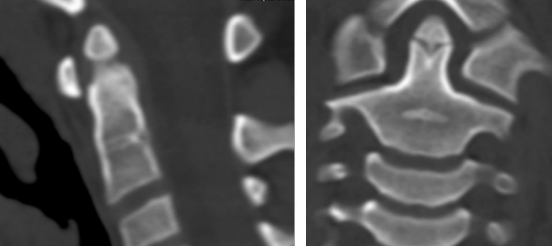
  • Fig 2.
    • Download figure
    • Open in new tab
    • Download powerpoint
    Fig 2.

    Singular midline OC pattern at the AAA. A, 5-month-old child with already large midline OC and large ventrolateral synchondroses. B, A 2.5-year-old child with progressive ossification of the chondral AAA anlage from both the central OC and the lateral masses of the atlas. C, 7-year-old child with complete ossification of the AAA but still-visible ventrolateral synchondroses.
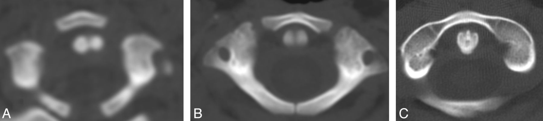
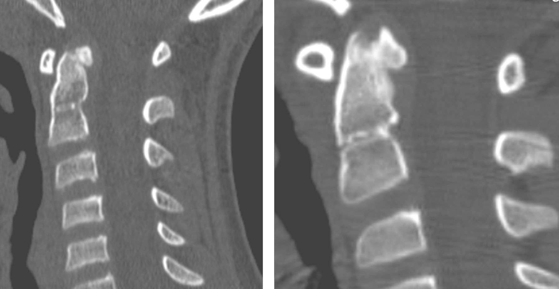
  • Fig 3.
    • Download figure
    • Open in new tab
    • Download powerpoint
    Fig 3.

    Multiple OC patterns at the AAA. A, 3-year-old child with 2 OCs paramedially located. B, A 2.5-year-old child with 3 irregular OCs within the AAA.
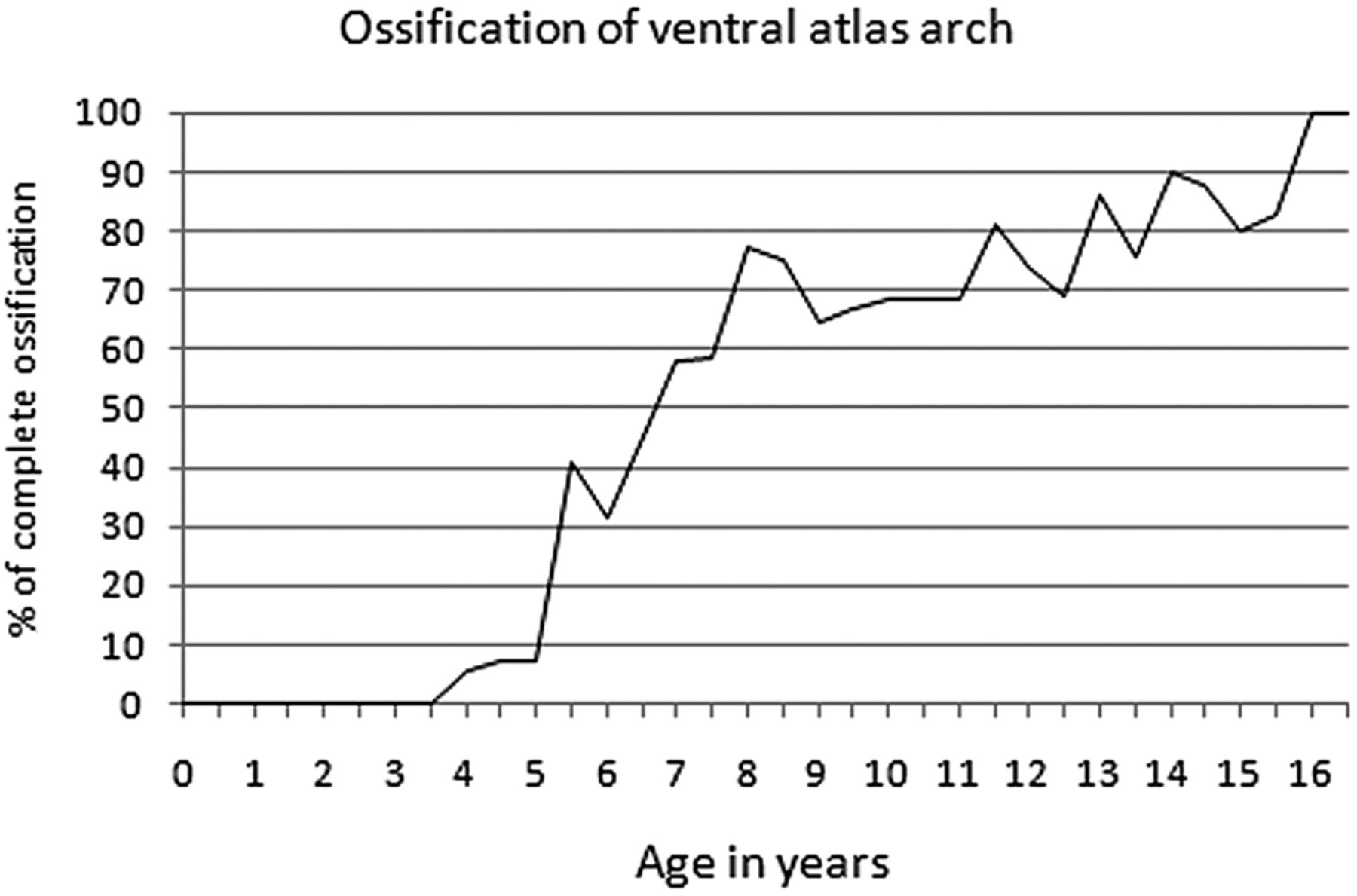
  • Fig 4.
    • Download figure
    • Open in new tab
    • Download powerpoint
    Fig 4.

    Ossification timetable of the anterior atlas arch.
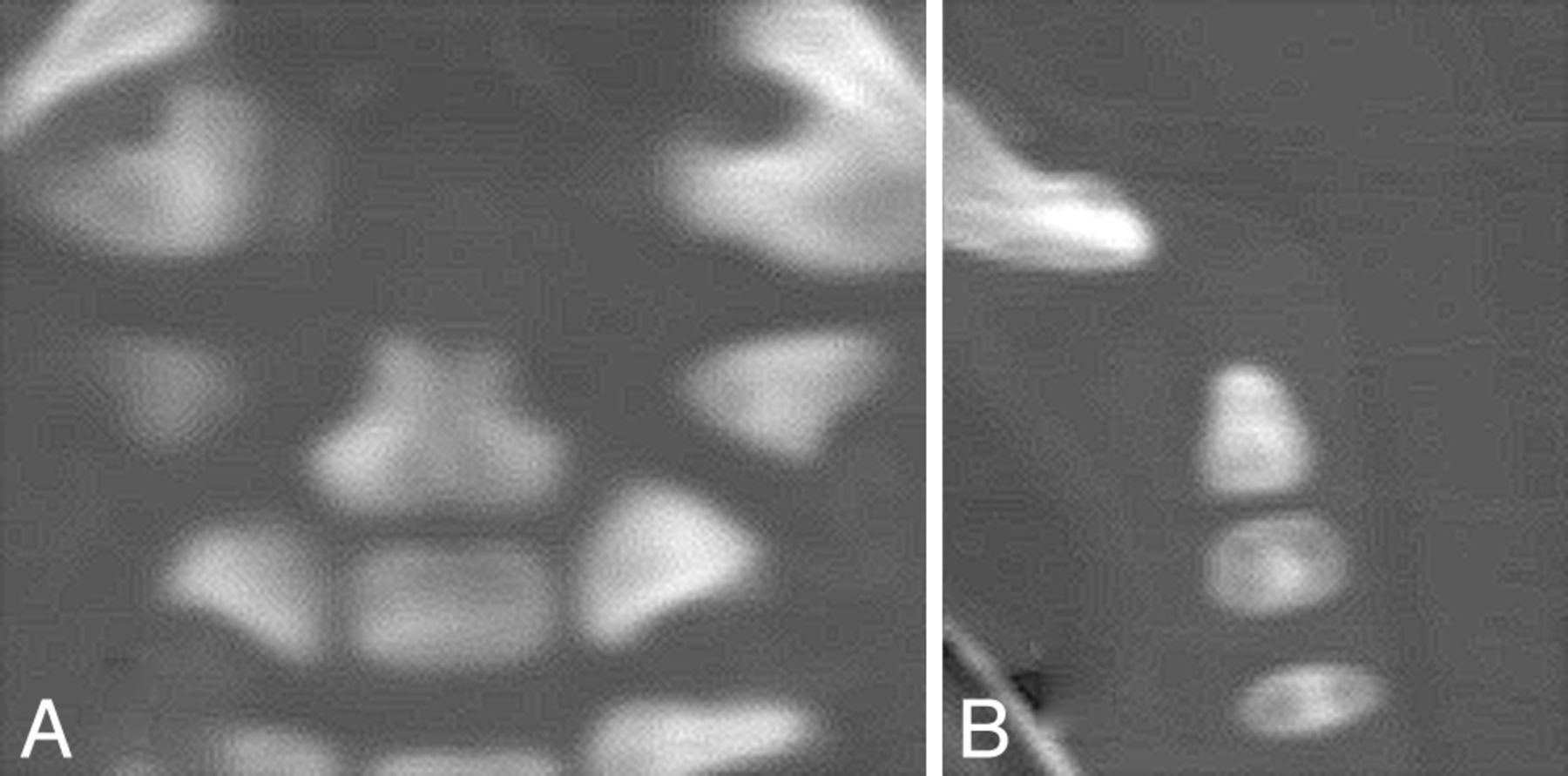
  • Fig 5.
    • Download figure
    • Open in new tab
    • Download powerpoint
    Fig 5.

    Different aspects of the PAA. A, Normal midline synchondrosis. B, Rare paramedial posterior synchondrosis. C, Rare midline OC within the PAA.

  • Fig 6.
    • Download figure
    • Open in new tab
    • Download powerpoint
    Fig 6.

    Ossification timetable of the posterior atlas arch.

  • Fig 7.
    • Download figure
    • Open in new tab
    • Download powerpoint
    Fig 7.

    Coronal (A) and sagittal (B) view of the neonatal axis with 5 OCs. A, While both club-shaped centers of the dens are already fused at birth, a midline vertical fusion line can still be perceived. B, Smooth cartilaginous rounded CHT at the top of the dens is best appreciated on the sagittal view.
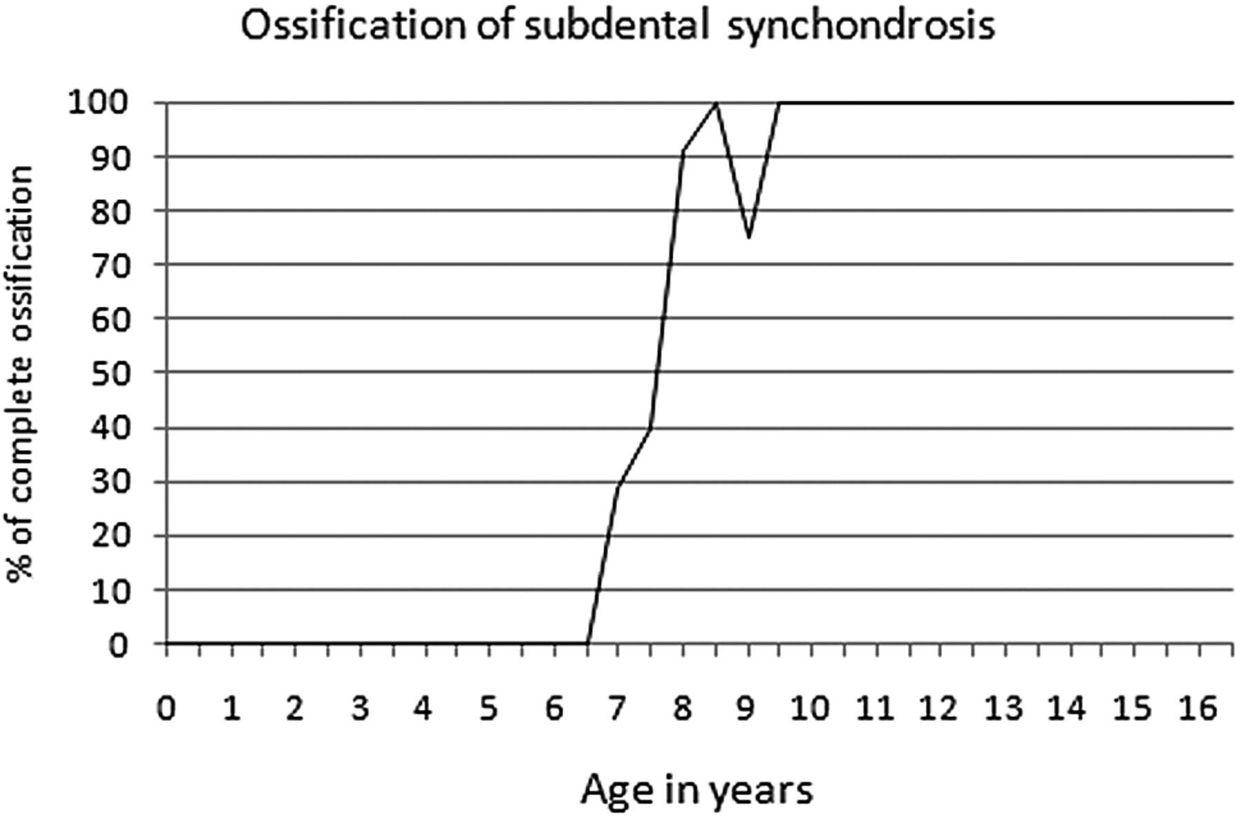
  • Fig 8.
    • Download figure
    • Open in new tab
    • Download powerpoint
    Fig 8.

    Ossification timetable of the subdental synchondrosis.
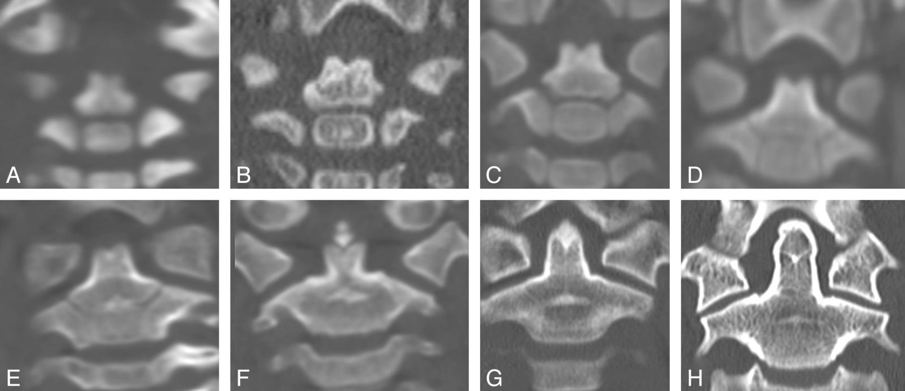
  • Fig 9.
    • Download figure
    • Open in new tab
    • Download powerpoint
    Fig 9.

    Coronal view of ossification development of the axis. Children are of the following ages (A) 30 days, (B) 5 months, (C) 1.5 years, (D) 2 years, (E) 3 years, (F) 5 years, (G) 8.5 years, and (H) 14 years.
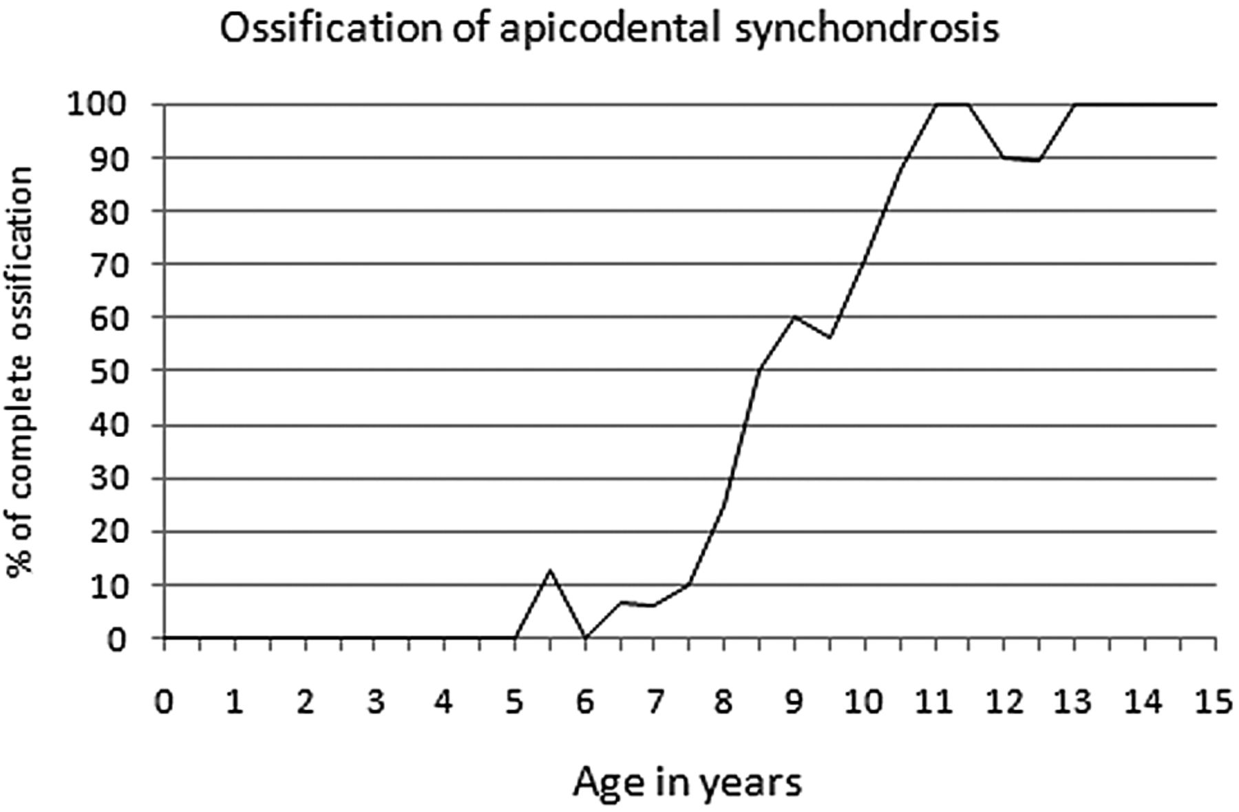
  • Fig 10.
    • Download figure
    • Open in new tab
    • Download powerpoint
    Fig 10.

    Ossification timetable of the apicodental synchondrosis.
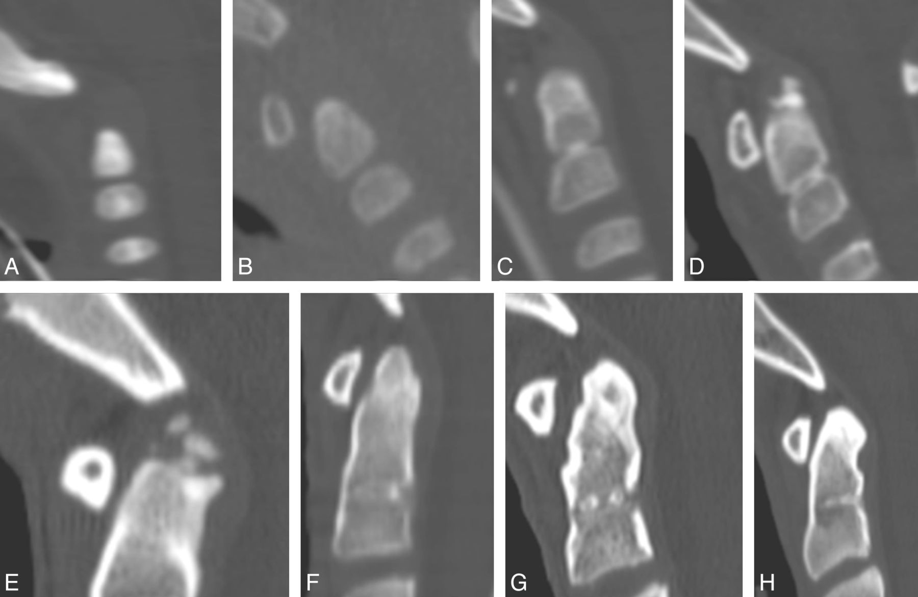
  • Fig 11.
    • Download figure
    • Open in new tab
    • Download powerpoint
    Fig 11.

    Sagittal view of ossification development of the axis. Children are of the following ages: (A) 30 days, (B) 3 months, (C) 3 years, (D) 5 years, (E) 6.5 years, (F) 8 years, (G) 13 years, and (H) 16 years.

  • Fig 12.
    • Download figure
    • Open in new tab
    • Download powerpoint
    Fig 12.

    Rare posterior ossification of the CHT resembled a dislocated fragment.

  • Fig 13.
    • Download figure
    • Open in new tab
    • Download powerpoint
    Fig 13.

    Large ossiculum terminale in an 8-year-old child and incomplete ossification of the ADS.

Tables

  • Figures
  • Number of CT examinations per age category

    Age (yrs)Patients
    046
    138
    235
    336
    431
    531
    625
    725
    833
    928
    1030
    1126
    1239
    1341
    1441
    1535
    166
    174
    Total550
PreviousNext
Back to top

In this issue

American Journal of Neuroradiology: 33 (10)
American Journal of Neuroradiology
Vol. 33, Issue 10
1 Nov 2012
  • Table of Contents
  • Index by author
Advertisement
Print
Download PDF
Email Article

Thank you for your interest in spreading the word on American Journal of Neuroradiology.

NOTE: We only request your email address so that the person you are recommending the page to knows that you wanted them to see it, and that it is not junk mail. We do not capture any email address.

Enter multiple addresses on separate lines or separate them with commas.
Normal Ossification Patterns of Atlas and Axis: A CT Study
(Your Name) has sent you a message from American Journal of Neuroradiology
(Your Name) thought you would like to see the American Journal of Neuroradiology web site.
CAPTCHA
This question is for testing whether or not you are a human visitor and to prevent automated spam submissions.
Cite this article
G.M. Karwacki, J.F. Schneider
Normal Ossification Patterns of Atlas and Axis: A CT Study
American Journal of Neuroradiology Nov 2012, 33 (10) 1882-1887; DOI: 10.3174/ajnr.A3105

Citation Manager Formats

  • BibTeX
  • Bookends
  • EasyBib
  • EndNote (tagged)
  • EndNote 8 (xml)
  • Medlars
  • Mendeley
  • Papers
  • RefWorks Tagged
  • Ref Manager
  • RIS
  • Zotero
0 Responses
Respond to this article
Share
Bookmark this article
Normal Ossification Patterns of Atlas and Axis: A CT Study
G.M. Karwacki, J.F. Schneider
American Journal of Neuroradiology Nov 2012, 33 (10) 1882-1887; DOI: 10.3174/ajnr.A3105
del.icio.us logo Twitter logo Facebook logo Mendeley logo
  • Tweet Widget
  • Facebook Like
  • Google Plus One
Purchase

Jump to section

  • Article
    • Abstract
    • ABBREVIATIONS:
    • Materials and Methods
    • Results
    • Discussion
    • Conclusions
    • References
  • Figures & Data
  • Info & Metrics
  • Responses
  • References
  • PDF

Related Articles

  • No related articles found.
  • PubMed
  • Google Scholar

Cited By...

  • The growth pattern of Neandertals, reconstructed from a juvenile skeleton from El Sidron (Spain)
  • Crossref
  • Google Scholar

This article has not yet been cited by articles in journals that are participating in Crossref Cited-by Linking.

More in this TOC Section

  • Management Outcomes For VO Spine Biopsy
  • Characteristics of SIH Type I Culprit Lesions
  • Advanced Imaging of Type 2 Spinal CSF Leaks
Show more Spine Imaging and Spine Image-Guided Interventions

Similar Articles

Advertisement

Indexed Content

  • Current Issue
  • Accepted Manuscripts
  • Article Preview
  • Past Issues
  • Editorials
  • Editor's Choice
  • Fellows' Journal Club
  • Letters to the Editor
  • Video Articles

Cases

  • Case Collection
  • Archive - Case of the Week
  • Archive - Case of the Month
  • Archive - Classic Case

More from AJNR

  • Trainee Corner
  • Imaging Protocols
  • MRI Safety Corner
  • Book Reviews

Multimedia

  • AJNR Podcasts
  • AJNR Scantastics

Resources

  • Turnaround Time
  • Submit a Manuscript
  • Submit a Video Article
  • Submit an eLetter to the Editor/Response
  • Manuscript Submission Guidelines
  • Statistical Tips
  • Fast Publishing of Accepted Manuscripts
  • Graphical Abstract Preparation
  • Imaging Protocol Submission
  • Evidence-Based Medicine Level Guide
  • Publishing Checklists
  • Author Policies
  • Become a Reviewer/Academy of Reviewers
  • News and Updates

About Us

  • About AJNR
  • Editorial Board
  • Editorial Board Alumni
  • Alerts
  • Permissions
  • Not an AJNR Subscriber? Join Now
  • Advertise with Us
  • Librarian Resources
  • Feedback
  • Terms and Conditions
  • AJNR Editorial Board Alumni

American Society of Neuroradiology

  • Not an ASNR Member? Join Now

© 2025 by the American Society of Neuroradiology All rights, including for text and data mining, AI training, and similar technologies, are reserved.
Print ISSN: 0195-6108 Online ISSN: 1936-959X

Powered by HighWire