
Article Figures & Data
Figures
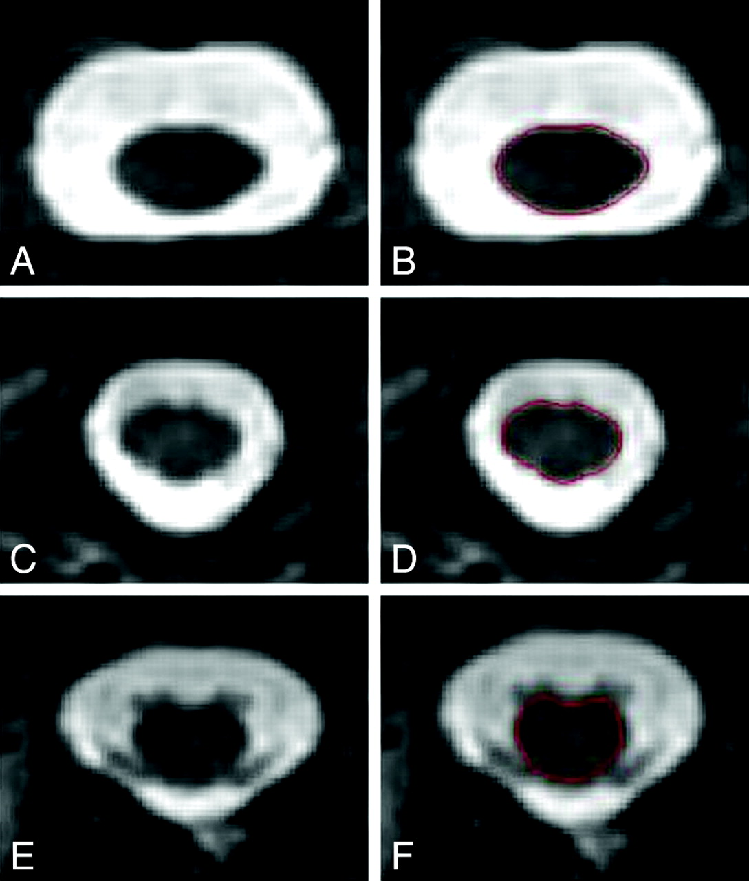
- Fig 1.
Representative spinal MR imaging scans and segmentation. T2-weighted fast spin-echo axial images obtained at 3T of the upper cervical (A), lower cervical (C), and thoracic (E) spinal cord are shown. High contrast between the spinal cord (hypointense) and the surrounding CSF (hyperintense) is noted. Semiautomated spinal cord contour maps are shown for the identical sections in B, D, and F. The patient is a 29-year-old woman with RRMS of 7 years' disease duration on treatment with interferon β-1a at the time of imaging, with no physical disability (EDSS score of 0).
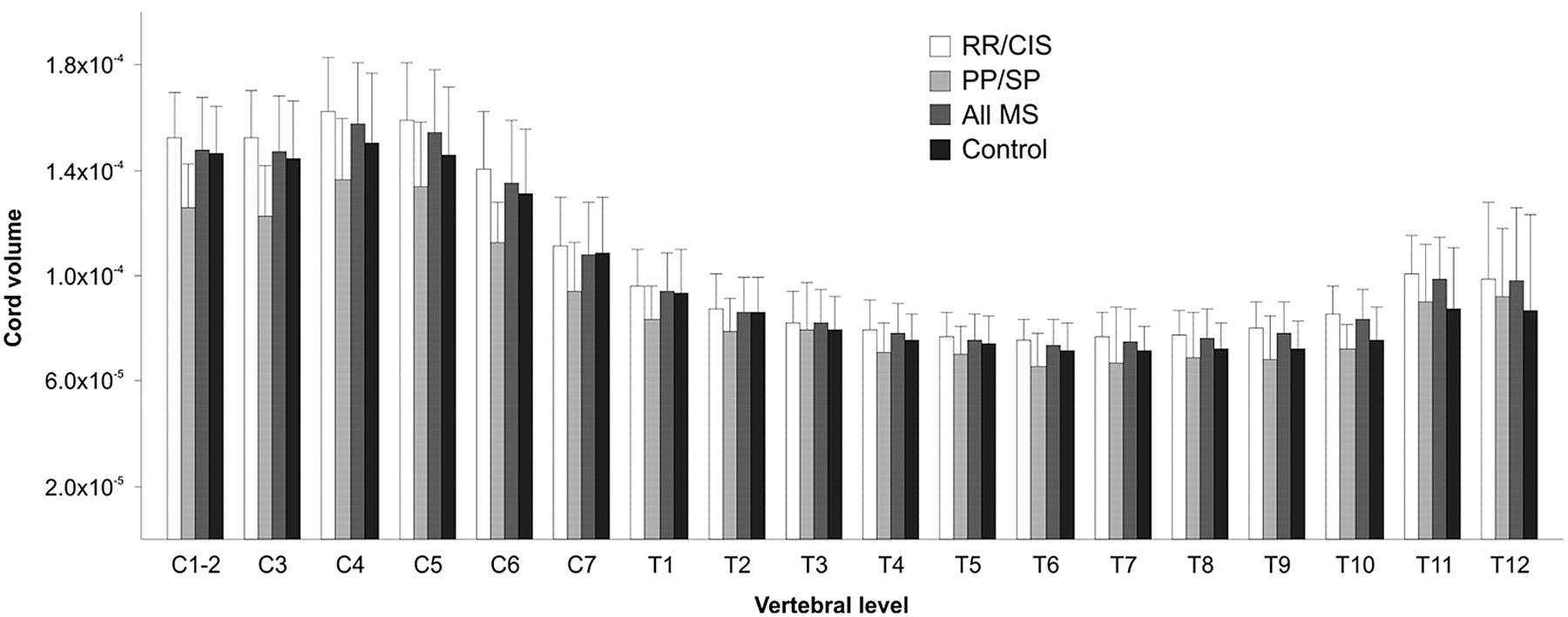
- Fig 2.
Spinal cord volume (plotted as volume/section/ICV) segmented by vertebral level. Group comparison P values with and without correction for multiple statistical tests are presented in On-line Tables 2 and 3. Bar heights represent means; error bars represent SDs.
- Fig 3.
Whole spinal cord volume (plotted as volume/section/ICV). Whole spinal cord volume is 12.6% smaller in PPMS/SPMS compared with RRMS/CIS. No significant difference in whole spinal cord volume is detected between RRMS/CIS and controls, PPMS/SPMS and controls, or all MS and controls. Group comparison P values with and without correction for multiple statistical tests are presented in On-line Tables 2 and 3. Bar heights represent means; error bars represent SDs.


In this issue


Jump to section
Related Articles
Cited By...
- What are the gray and white matter volumes of the human spinal cord?
- Clinically relevant cranio-caudal patterns of cervical cord atrophy evolution in MS
- Cervical spinal cord atrophy: An early marker of progressive MS onset
- Multicenter Validation of Mean Upper Cervical Cord Area Measurements from Head 3D T1-Weighted MR Imaging in Patients with Multiple Sclerosis
- Voxel-wise mapping of cervical cord damage in multiple sclerosis patients with different clinical phenotypes