
Article Figures & Data
Figures
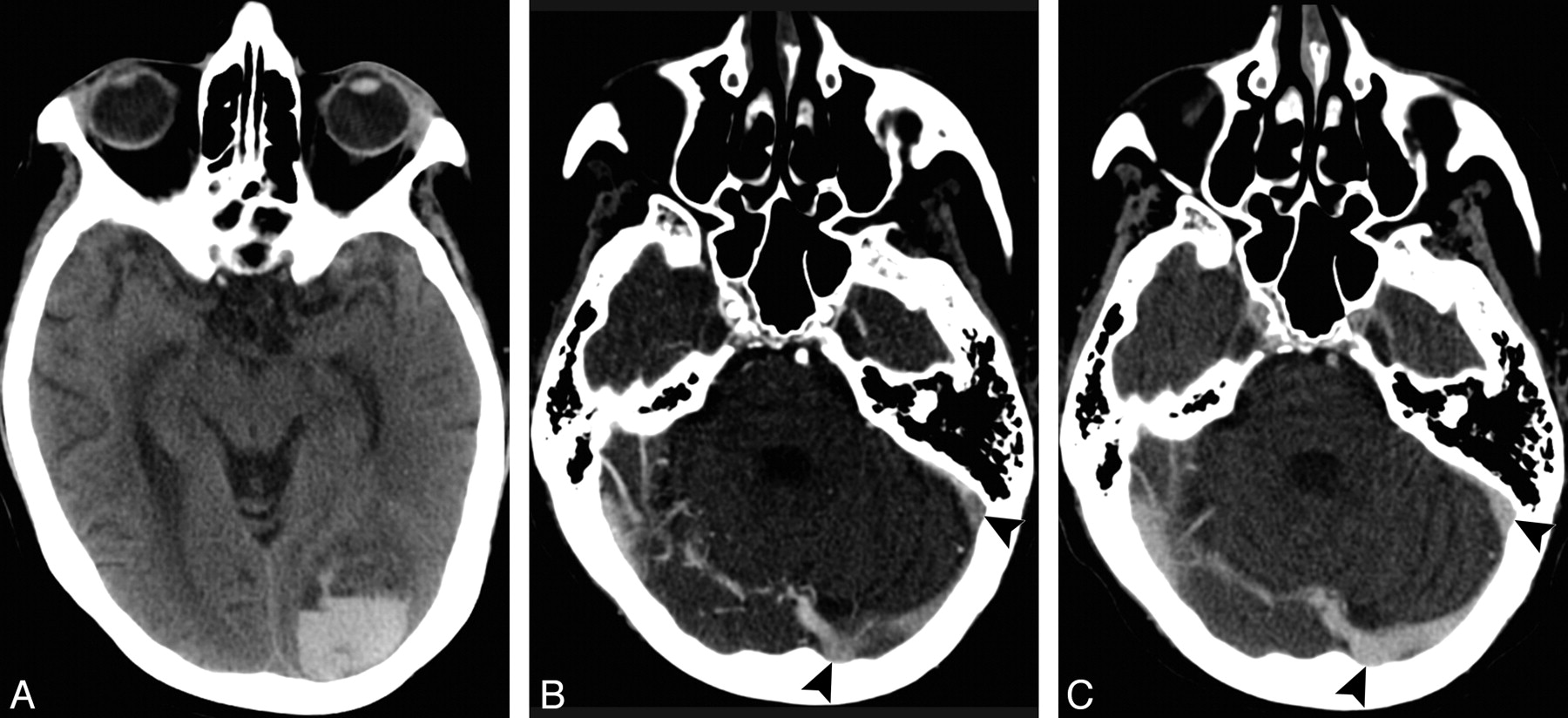
- Fig 1.
A 57-year-old woman who presented with headache and a right visual field deficit. A, NCCT demonstrates a left occipital ICH. B, Axial source image of a first-pass CTA performed in a 64-section CT scanner demonstrates inhomogeneous opacification of the left transverse and sigmoid sinuses (arrowheads), which may be related to scan timing or partial DVST. C, Axial source image of a delayed CTA acquisition performed 110 seconds after the first-pass scan demonstrates homogeneous opacification of the left transverse and sigmoid sinuses (arrowheads), which excludes DVST as the ICH etiology.
- Fig 2.
A 65-year-old woman who presented with worsening aphasia and confusion. A, NCCT demonstrates a large left temporo-occipital ICH with intraventricular extension. B, Axial source image of a first-pass CTA performed in a 64-section CT scanner demonstrates nonopacification of the left transverse and sigmoid sinuses (arrowheads), which may be related to scan timing or DVST. C, Axial source image of a delayed CTA acquisition performed 32 seconds after the first-pass scan demonstrates homogeneous opacification of the left transverse and sigmoid sinuses (arrowheads), which excludes DVST as the ICH etiology.


Tables
ICH locationb Presumed Venous Drainage Frontal lobes Superior sagittal sinus Superior parietal lobes Superior sagittal sinus Inferior parietal lobes Transverse or sigmoid sinusc Occipital lobes Superior sagittal sinus Temporal lobes Transverse or sigmoid sinusc Peri-Sylvian cortex Cavernous sinusc Insular cortex Cavernous sinusc Basal ganglia Internal cerebral veins,c vein of Galen, straight sinus Thalami Internal cerebral veins,c vein of Galen, straight sinus Cerebellar vermis Vein of Galen, straight sinus Cerebellar hemispheres Transverse or sigmoid sinusc Brain stem Superior petrosal sinusc -
a Adapted from Meder et al13 for the supratentorial brain and Jinkins14 for the infratentorial brain. Note that there may be considerable variability in the venous drainage pattern of the cortical supratentorial brain.
-
b The frontal, temporal, and parietal lobes exclude the peri-Sylvian cortex within these lobes.
-
c Ipsilateral to the ICH location.
-
No. % Sex Male 90 53 Female 80 47 Age (yr) 18–45 25 15 46–70 74 43 71–92 71 42 History of hypertension Yes 107 63 No 63 37 Antiplatelet therapy Yes 60 35 No 110 65 Admission INR <1.5 148 87 1.5–2.5 13 8 >2.5 9 5 ICH location Frontal 24 14 Superior parietal 16 9 Inferior parietal 10 6 Occipital 6 4 Temporal 11 7 Peri-Sylvian 2 1 Insular 1 1 Multilobar 24 14 Basal ganglia 43 25 Thalamic 8 5 Vermian 2 1 Cerebellar 16 9 Brain stem 7 4 ICH volume (mL) 0.2–29.9 105 62 30–59.9 39 23 ≥60 26 15 IVH volume (mL) 0 81 48 0.1–4.9 55 32 5–14.9 15 9 ≥15 19 11 - Table 3.
Frequency of adequate contrast opacification of the major intracranial venous structures in first-pass CTAs performed for evaluation of intracerebral hemorrhagea
Venous Structure All CTAs (%) (n = 170) 16-Section CTAs (%) (n = 58) 64-Section CTAs (%) (n = 112) P Valueb Deep cerebral veinsc 88 95 84 .03 Straight sinus 85 93 80 .03 Superior sagittal sinus 81 97 72 <.001 Right transverse sinus 66 83 58 .001 Left transverse sinus 63 81 54 <.001 Right sigmoid sinus 49 62 42 .012 Left sigmoid sinus 48 71 36 <.001 Right cavernous sinus 11 16 8 .13 Left cavernous sinus 7 12 4 .11 All noncavernous venous structures 42 60 33 <.001 -
a Adequate contrast opacification defined as homogeneous contrast opacification within the venous structure examined.
-
b P value is for the difference between 16- and 64-section CTAs using the Pearson χ2 test.
-
c Includes internal cerebral veins and the vein of Galen.
-
In this issue


Jump to section
Related Articles
Cited By...
- No citing articles found.