Advertisement

Main menu

  • Home
  • Content
    • Current Issue
    • Accepted Manuscripts
    • Article Preview
    • Past Issue Archive
    • Video Articles
    • AJNR Case Collection
    • Case of the Week Archive
    • Case of the Month Archive
    • Classic Case Archive
  • Special Collections
    • AJNR Awards
    • Low-Field MRI
    • Alzheimer Disease
    • ASNR Foundation Special Collection
    • Photon-Counting CT
    • View All
  • Multimedia
    • AJNR Podcasts
    • AJNR SCANtastic
    • Trainee Corner
    • MRI Safety Corner
    • Imaging Protocols
  • For Authors
    • Submit a Manuscript
    • Submit a Video Article
    • Submit an eLetter to the Editor/Response
    • Manuscript Submission Guidelines
    • Statistical Tips
    • Fast Publishing of Accepted Manuscripts
    • Graphical Abstract Preparation
    • Imaging Protocol Submission
    • Author Policies
  • About Us
    • About AJNR
    • Editorial Board
    • Editorial Board Alumni
  • More
    • Become a Reviewer/Academy of Reviewers
    • Subscribers
    • Permissions
    • Alerts
    • Feedback
    • Advertisers
    • ASNR Home

User menu

  • Alerts
  • Log in

Search

  • Advanced search
American Journal of Neuroradiology

American Journal of Neuroradiology

ASHNR American Society of Functional Neuroradiology ASHNR American Society of Pediatric Neuroradiology ASSR
  • Alerts
  • Log in

Advanced Search

  • Home
  • Content
    • Current Issue
    • Accepted Manuscripts
    • Article Preview
    • Past Issue Archive
    • Video Articles
    • AJNR Case Collection
    • Case of the Week Archive
    • Case of the Month Archive
    • Classic Case Archive
  • Special Collections
    • AJNR Awards
    • Low-Field MRI
    • Alzheimer Disease
    • ASNR Foundation Special Collection
    • Photon-Counting CT
    • View All
  • Multimedia
    • AJNR Podcasts
    • AJNR SCANtastic
    • Trainee Corner
    • MRI Safety Corner
    • Imaging Protocols
  • For Authors
    • Submit a Manuscript
    • Submit a Video Article
    • Submit an eLetter to the Editor/Response
    • Manuscript Submission Guidelines
    • Statistical Tips
    • Fast Publishing of Accepted Manuscripts
    • Graphical Abstract Preparation
    • Imaging Protocol Submission
    • Author Policies
  • About Us
    • About AJNR
    • Editorial Board
    • Editorial Board Alumni
  • More
    • Become a Reviewer/Academy of Reviewers
    • Subscribers
    • Permissions
    • Alerts
    • Feedback
    • Advertisers
    • ASNR Home
  • Follow AJNR on Twitter
  • Visit AJNR on Facebook
  • Follow AJNR on Instagram
  • Join AJNR on LinkedIn
  • RSS Feeds

AJNR Awards, New Junior Editors, and more. Read the latest AJNR updates

Research ArticleSpine
Open Access

CT-Guided Epidural Blood Patching of Directly Observed or Potential Leak Sites for the Targeted Treatment of Spontaneous Intracranial Hypotension

P.G. Kranz, L. Gray and J.N. Taylor
American Journal of Neuroradiology May 2011, 32 (5) 832-838; DOI: https://doi.org/10.3174/ajnr.A2384
P.G. Kranz
  • Find this author on Google Scholar
  • Find this author on PubMed
  • Search for this author on this site
L. Gray
  • Find this author on Google Scholar
  • Find this author on PubMed
  • Search for this author on this site
J.N. Taylor
  • Find this author on Google Scholar
  • Find this author on PubMed
  • Search for this author on this site
  • Article
  • Figures & Data
  • Supplemental
  • Info & Metrics
  • Responses
  • References
  • PDF
Loading

Article Figures & Data

Figures

  • Tables
  • Fig 1.
    • Download figure
    • Open in new tab
    • Download powerpoint
    Fig 1.

    Technique for CT-guided transforaminal blood patching. A, Needle placement before patching. The needle tip is located in the posterior third of the neural foramen, adjacent to a diverticulum of the nerve root (arrow). B, Contrast injected before blood patching confirms the location in the epidural space. C, Image obtained during injection of autologous blood demonstrates displacement of the contrast by the injected blood and mild mass effect on the diverticulum.
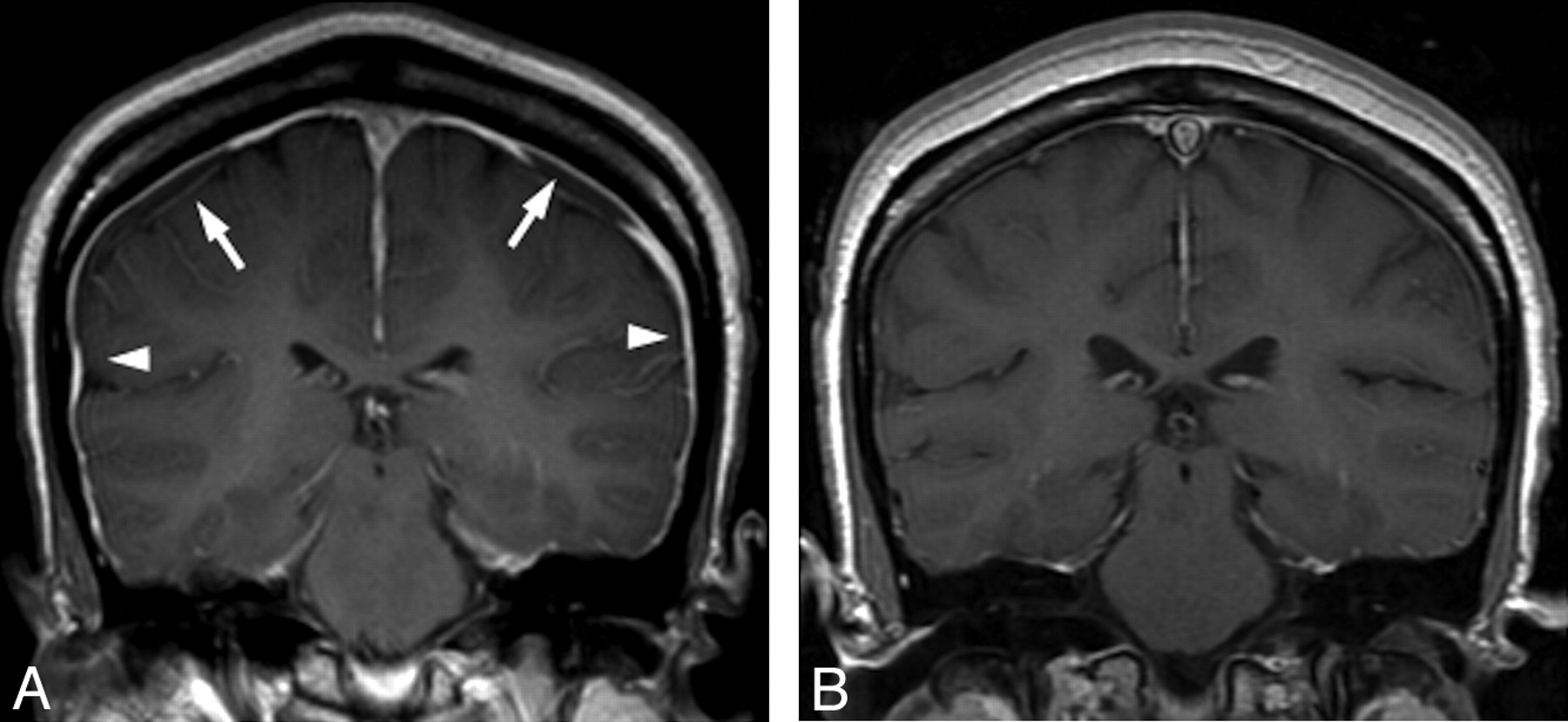
  • Fig 2.
    • Download figure
    • Open in new tab
    • Download powerpoint
    Fig 2.

    Resolution of imaging changes of intracranial hypotension. A, Pretreatment MR image shows typical signs of intracranial hypotension, including smooth dural thickening and enhancement (arrowheads) and subdural collections (arrows). B, Follow-up brain MR image after CT-guided targeted blood patching demonstrates resolution of dural enhancement and subdural collections.
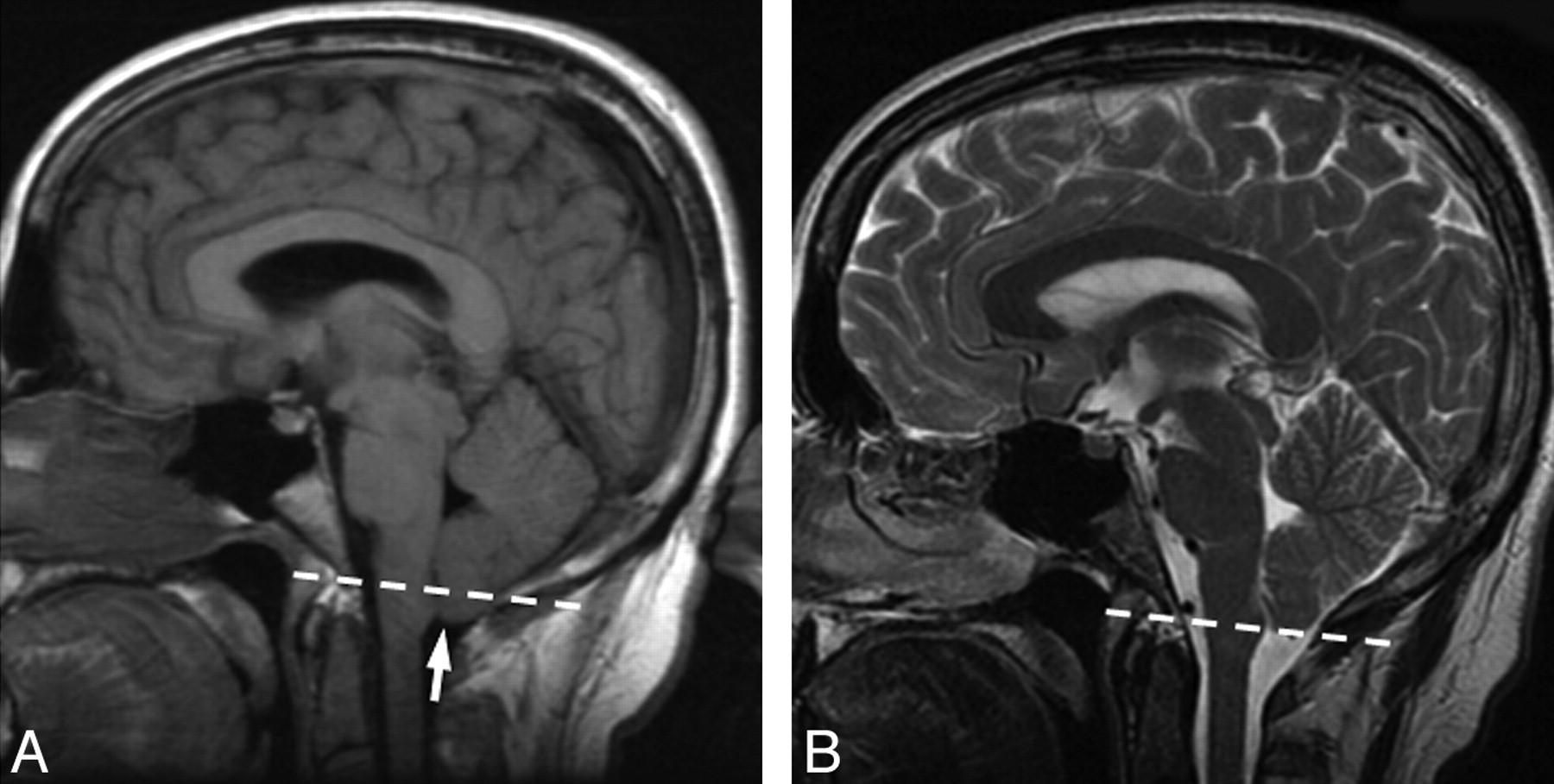
  • Fig 3.
    • Download figure
    • Open in new tab
    • Download powerpoint
    Fig 3.

    Pre- and posttreatment MR imaging from patient 2. Dashed line indicates the foramen magnum. A, Pretreatment MR image demonstrates cerebellar tonsillar ectopia (arrow). The dashed line indicates the plane of the foramen magnum. Midline sagging of the brain with inferior displacement of the mammillary bodies is seen. In this patient, there was no abnormal dural enhancement on pretreatment MR imaging (not shown). B, Follow-up brain MR image after CT-guided blood patching demonstrates resolution of the cerebellar tonsillar ectopia and midline sag.
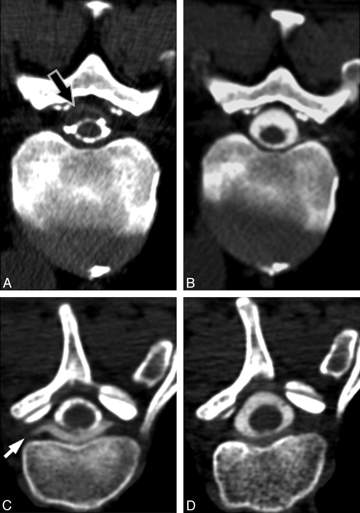
  • Fig 4.
    • Download figure
    • Open in new tab
    • Download powerpoint
    Fig 4.

    Pre- and posttreatment appearance of spinal fluid collections. A, Pretreatment CT myelogram from patient 6 demonstrates a subdural fluid collection (arrow) in the spinal canal, which narrows the CSF space. B, Follow-up CT myelogram 15 days after CT-guided blood patching at the site of leak demonstrates resolution of the subdural collection. C, Pretreatment CT myelogram from patient 3 demonstrates an extradural collection ventral to the thecal sac, extending into the nerve root sleeves (arrow). D, Follow-up CT myelogram 17 days after CT-guided blood patching of the nerve root sleeves demonstrates near-complete resolution of the collection.

  • Fig 5.
    • Download figure
    • Open in new tab
    • Download powerpoint
    Fig 5.

    Representative examples of targets for blood patching. A, CT myelogram demonstrates extravasation of contrast from a nerve root sleeve (white arrow) due to a high-flow leak. Note the presence of an extradural collection surrounding the thecal sac (black arrow). B, CT myelogram in a different patient demonstrates an enlarged diverticulum of the nerve root sleeve (arrowhead). Although no direct myelographic evidence of leak is seen, this site was chosen for empiric blood patching on the basis of the potential for an intermittent or low-flow leak.

Tables

  • Figures
    • View popup
    Table

    Findings of CT myelographya

    Patient No.Opening Pressure (cm H20)Spinal Fluid CollectionIrregular DiverticulaDirect Visualization of LeakLocation of Diverticula
    CervicalUpper Thoracic (T1–6)Lower Thoracic (T6–12)Lumbar
    10+++0400
    215–+–0120
    30+–+0500
    4N/A–+–0070
    5N/A–+–0020
    60+++0010
    70–+–0251
    812–+–0145
    • Note: a + indicates presence of the finding; –, absence of the finding.

PreviousNext
Back to top

In this issue

American Journal of Neuroradiology: 32 (5)
American Journal of Neuroradiology
Vol. 32, Issue 5
1 May 2011
  • Table of Contents
  • Index by author
Advertisement
Print
Download PDF
Email Article

Thank you for your interest in spreading the word on American Journal of Neuroradiology.

NOTE: We only request your email address so that the person you are recommending the page to knows that you wanted them to see it, and that it is not junk mail. We do not capture any email address.

Enter multiple addresses on separate lines or separate them with commas.
CT-Guided Epidural Blood Patching of Directly Observed or Potential Leak Sites for the Targeted Treatment of Spontaneous Intracranial Hypotension
(Your Name) has sent you a message from American Journal of Neuroradiology
(Your Name) thought you would like to see the American Journal of Neuroradiology web site.
CAPTCHA
This question is for testing whether or not you are a human visitor and to prevent automated spam submissions.
Cite this article
P.G. Kranz, L. Gray, J.N. Taylor
CT-Guided Epidural Blood Patching of Directly Observed or Potential Leak Sites for the Targeted Treatment of Spontaneous Intracranial Hypotension
American Journal of Neuroradiology May 2011, 32 (5) 832-838; DOI: 10.3174/ajnr.A2384

Citation Manager Formats

  • BibTeX
  • Bookends
  • EasyBib
  • EndNote (tagged)
  • EndNote 8 (xml)
  • Medlars
  • Mendeley
  • Papers
  • RefWorks Tagged
  • Ref Manager
  • RIS
  • Zotero
0 Responses
Respond to this article
Share
Bookmark this article
CT-Guided Epidural Blood Patching of Directly Observed or Potential Leak Sites for the Targeted Treatment of Spontaneous Intracranial Hypotension
P.G. Kranz, L. Gray, J.N. Taylor
American Journal of Neuroradiology May 2011, 32 (5) 832-838; DOI: 10.3174/ajnr.A2384
del.icio.us logo Twitter logo Facebook logo Mendeley logo
  • Tweet Widget
  • Facebook Like
  • Google Plus One
Purchase

Jump to section

  • Article
    • Abstract
    • Abbreviations
    • Materials and Methods
    • Results
    • Discussion
    • Conclusions
    • Footnotes
    • References
  • Figures & Data
  • Supplemental
  • Info & Metrics
  • Responses
  • References
  • PDF

Related Articles

  • PubMed
  • Google Scholar

Cited By...

  • A classification system of spontaneous spinal CSF leaks
  • Imaging Signs in Spontaneous Intracranial Hypotension: Prevalence and Relationship to CSF Pressure
  • MR Myelography for Identification of Spinal CSF Leak in Spontaneous Intracranial Hypotension
  • Large-Volume Blood Patch to Multiple Sites in the Epidural Space through a Single-Catheter Access Site for Treatment of Spontaneous Intracranial Hypotension
  • Spinal Meningeal Diverticula in Spontaneous Intracranial Hypotension: Analysis of Prevalence and Myelographic Appearance
  • CT Myelography for the Planning and Guidance of Targeted Epidural Blood Patches in Patients with Persistent Spinal CSF Leakage
  • Crossref (53)
  • Google Scholar

This article has been cited by the following articles in journals that are participating in Crossref Cited-by Linking.

  • A classification system of spontaneous spinal CSF leaks
    Wouter I. Schievink, M. Marcel Maya, Stacey Jean-Pierre, Miriam Nuño, Ravi S. Prasad, Franklin G. Moser
    Neurology 2016 87 7
  • CSF Venous Fistulas in Spontaneous Intracranial Hypotension: Imaging Characteristics on Dynamic and CT Myelography
    Peter G. Kranz, Timothy J. Amrhein, Linda Gray
    American Journal of Roentgenology 2017 209 6
  • Myelographic Techniques for the Detection of Spinal CSF Leaks in Spontaneous Intracranial Hypotension
    Peter G. Kranz, Patrick H. Luetmer, Felix E. Diehn, Timothy J. Amrhein, Teerath Peter Tanpitukpongse, Linda Gray
    American Journal of Roentgenology 2016 206 1
  • Spontaneous Intracranial Hypotension: 10 Myths and Misperceptions
    Peter G. Kranz, Linda Gray, Timothy J. Amrhein
    Headache: The Journal of Head and Face Pain 2018 58 7
  • Imaging Signs in Spontaneous Intracranial Hypotension: Prevalence and Relationship to CSF Pressure
    P. G. Kranz, T. P. Tanpitukpongse, K. R. Choudhury, T. J. Amrhein, L. Gray
    American Journal of Neuroradiology 2016 37 7
  • Spontaneous Intracranial Hypotension
    Peter G. Kranz, Linda Gray, Michael D. Malinzak, Timothy J. Amrhein
    Neuroimaging Clinics of North America 2019 29 4
  • The status of diagnosis and treatment to intracranial hypotension, including SIH
    Jin-ping Lin, Shu-dong Zhang, Fei-fang He, Min-jun Liu, Xiao-xu Ma
    The Journal of Headache and Pain 2017 18 1
  • MR Myelography for Identification of Spinal CSF Leak in Spontaneous Intracranial Hypotension
    J.L. Chazen, J.F. Talbott, J.E. Lantos, W.P. Dillon
    American Journal of Neuroradiology 2014 35 10
  • Spinal Meningeal Diverticula in Spontaneous Intracranial Hypotension: Analysis of Prevalence and Myelographic Appearance
    P.G. Kranz, S.S. Stinnett, K.T. Huang, L. Gray
    American Journal of Neuroradiology 2013 34 6
  • Intracranial hypotension
    Horst Urbach
    Current Opinion in Neurology 2014 27 4

More in this TOC Section

  • Bern Score Validity for SIH
  • MP2RAGE 7T in MS Lesions of the Cervical Spine
  • Resisted Inspiration for CSF-Venous Fistula
Show more Spine

Similar Articles

Advertisement

Indexed Content

  • Current Issue
  • Accepted Manuscripts
  • Article Preview
  • Past Issues
  • Editorials
  • Editor's Choice
  • Fellows' Journal Club
  • Letters to the Editor
  • Video Articles

Cases

  • Case Collection
  • Archive - Case of the Week
  • Archive - Case of the Month
  • Archive - Classic Case

More from AJNR

  • Trainee Corner
  • Imaging Protocols
  • MRI Safety Corner
  • Book Reviews

Multimedia

  • AJNR Podcasts
  • AJNR Scantastics

Resources

  • Turnaround Time
  • Submit a Manuscript
  • Submit a Video Article
  • Submit an eLetter to the Editor/Response
  • Manuscript Submission Guidelines
  • Statistical Tips
  • Fast Publishing of Accepted Manuscripts
  • Graphical Abstract Preparation
  • Imaging Protocol Submission
  • Evidence-Based Medicine Level Guide
  • Publishing Checklists
  • Author Policies
  • Become a Reviewer/Academy of Reviewers
  • News and Updates

About Us

  • About AJNR
  • Editorial Board
  • Editorial Board Alumni
  • Alerts
  • Permissions
  • Not an AJNR Subscriber? Join Now
  • Advertise with Us
  • Librarian Resources
  • Feedback
  • Terms and Conditions
  • AJNR Editorial Board Alumni

American Society of Neuroradiology

  • Not an ASNR Member? Join Now

© 2025 by the American Society of Neuroradiology All rights, including for text and data mining, AI training, and similar technologies, are reserved.
Print ISSN: 0195-6108 Online ISSN: 1936-959X

Powered by HighWire