
Article Figures & Data
Figures
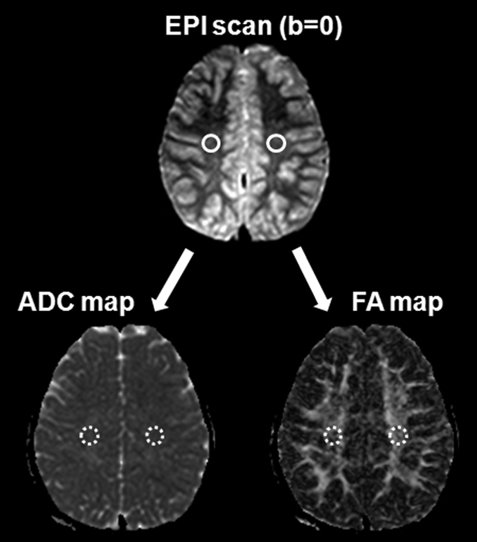
- Fig 1.
ROI method for ADCCS and FACS measurement. Round ROIs (60 mm3) were drawn on the EPI scan of the b = 0 step at the level of the centrum semiovale, followed by automatic overlaying onto the coregistered FA and ADC images.
- Fig 2.
Primary MMD in a 6-year-old girl. A–C, MRA images showed asymmetric MRA cores and stages between both hemispheres (Rt., stage III; Lt., stage II). TTP (D) and CBV (E) maps demonstrated asymmetric hemodynamic status, in which the Rt. hemisphere showed more delayed TTP and increased CBV. Diffusion-weighted image (b = 1000; F) and a FLAIR image (G) did not show any abnormalities. H, ADC map, right side ADCCS (836.3 mm2/s) was higher than left side ADCCS (779.8 mm2/s). I, FA map, right side FACS (0.438 × 10⁻³) was lower than left side FACS (0.487 × 10⁻³).


Tables
- Table 1:
Clinical information and findings for TTP status, degree of supraclinoid ICA or MCA stenosis, and rCBV pattern
No. Sex Age (yr) Clinical Symptom H-TTPdelayed MRA Scorea rCBV in H-TTPdelayed Rt. Lt. 1 F 5 TIA (Lt. HP) Rt. 2 1 Increased 2 M 6 TIA (Rt. HP) Lt. 3 4 Increased 3 F 6 TIA (Rt. HP) Lt. 1 4 Increased 4 F 6 Headache Rt. 4 3 Increased 5 F 6 TIA (Rt. HP) Rt. 3 1 Increased 6 F 7 TIA (Rt. HP) Lt. 3 4 Increased 7 F 7 Seizure Lt. 1 2 Increased 8 F 8 TIA (Rt. HP) Lt. 4 5 Increased 9 F 8 Headache Lt. 2 3 Increased 10 F 8 Family history Lt. 3 4 Increased 11 M 10 TIA (Lt. HP) Lt. 4 5 Increased 12 F 10 TIA (Rt. HP) Lt. 2 3 Increased 13 M 11 TIA (Rt. HP) Lt. 2 3 Increased 14 M 12 Headache Lt. 1 2 Increased 15 F 12 TIA (Lt. HP) Rt. 5 1 Increased 16 M 13 Hypogonadism Lt. 1 3 Increased 17 F 13 TIA (Lt. HP) Lt. 2 3 Increased 18 F 13 TIA (Rt. HP) Lt. 3 5 Increased 19 F 15 TIA (Rt. HP) Lt. 3 5 Increased 20 F 15 TIA (Lt. HP) Rt. 4 2 Increased -
Note:—HP indicates hemisphere
-
↵a MRA score is presented as the sum of anterior cerebral artery, MCA, posterior cerebral artery, and distal ICA using the MRA scoring system proposed by Houkin et al.21
-
Patient Patient Group Control Group H-TTPdelayed H-TTPshorter H-TTPdelayed H-TTPshorter FACS ADCCS FACS ADCCS FACS ADCCS FACS ADCCS 1 438 837.5 486 831.7 420.7 797.6 415.7 788.2 2 445.4 783.8 408 814.7 431.3 951.5 426 829 3 438 836.3 487 779.8 464.2 781.1 454.7 807.9 4 505.6 754.4 445.1 795.5 513.3 749.4 475.6 769.3 5 458.4 934.1 528.2 814.3 454.5 830.9 448.2 804.5 6 388 864 399.9 862.6 486.4 738.8 478.9 743.4 7 406.4 860.8 391.3 863.4 506.4 719.2 484.3 709.1 8 463.8 767 425.4 765.9 472.2 754.7 476 730.2 9 464.2 759.1 425.9 792.9 468.5 773.3 488.4 765.3 10 502.6 748.2 471.2 741.6 501.2 728.8 505.1 717.4 11 521.9 723.9 490.2 707.2 556.2 703.1 512.3 702 12 554.5 774.6 517.6 801.2 522.9 785.6 543.9 765.6 13 492.2 802.5 433 806.9 550.4 722.7 524.9 687.4 14 541.2 713.3 511.7 756.2 504.1 737.1 495.1 705.3 15 439.3 801.3 496.6 741.6 555 728.2 526.8 716.6 16 458 759.1 425 733.3 518.8 846.1 576.5 799.4 17 516.4 742.4 485.9 802.4 462.2 750.8 487.5 686.2 18 484.8 707 465.3 756.8 452.7 830.1 469.1 775.4 19 427.4 794.4 417.4 782.9 584.3 685.1 583.1 666.1 20 462.4 747.4 418.8 798.2 555.3 869.6 589.7 820.3 -
Note:—FA values (×10−3); ADC values (mm2/s).
-
Patient Group Control Group H-TTPdelayed H-TTPshorter Rt. Lt. In each group Mean FACS (× 10−3) 445.77 ± 35.09 481.13 ± 43.93 499.03 ± 45.85 498.09 ± 48.46 P <.0001a .8682 Mean ADCCS (mm2/s) 800.12 ± 51.52 772.89 ± 43.86 753.68 ± 64.75 749.43 ± 49.47 P .0022a .5709 Asymmetric index AIFA 0.075 ± 0.04 0.0023 ± 0.04 P <.0001a AIADC 0.034 ± 0.04 0.004 ± 0.04 P .0294a -
Note:—Values are presented as the mean ± SD.
-
↵a Significant difference between groups (P <.05).
-


