
Article Figures & Data
Figures
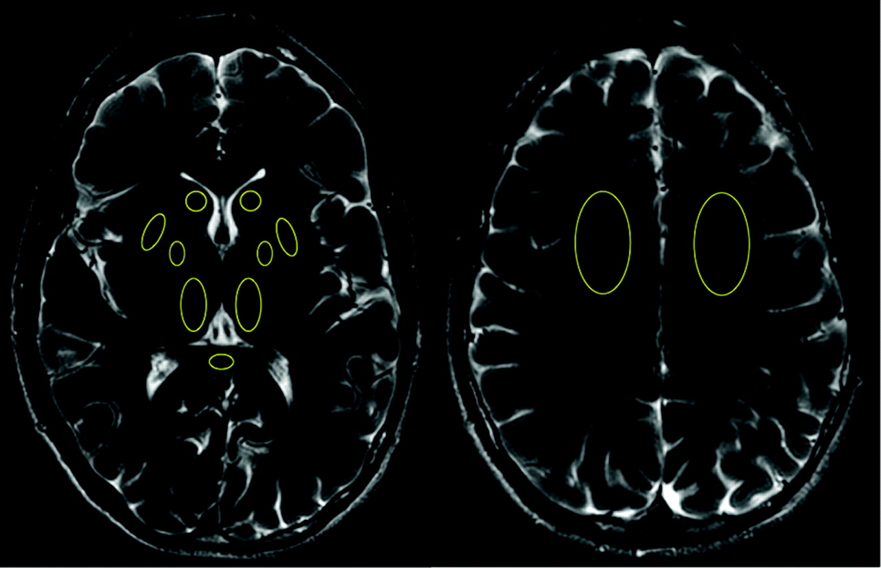
- Fig 1.
Representative of the ROIs drawn on 2 sample T2 images at the level of basal ganglia and centrum semiovale.
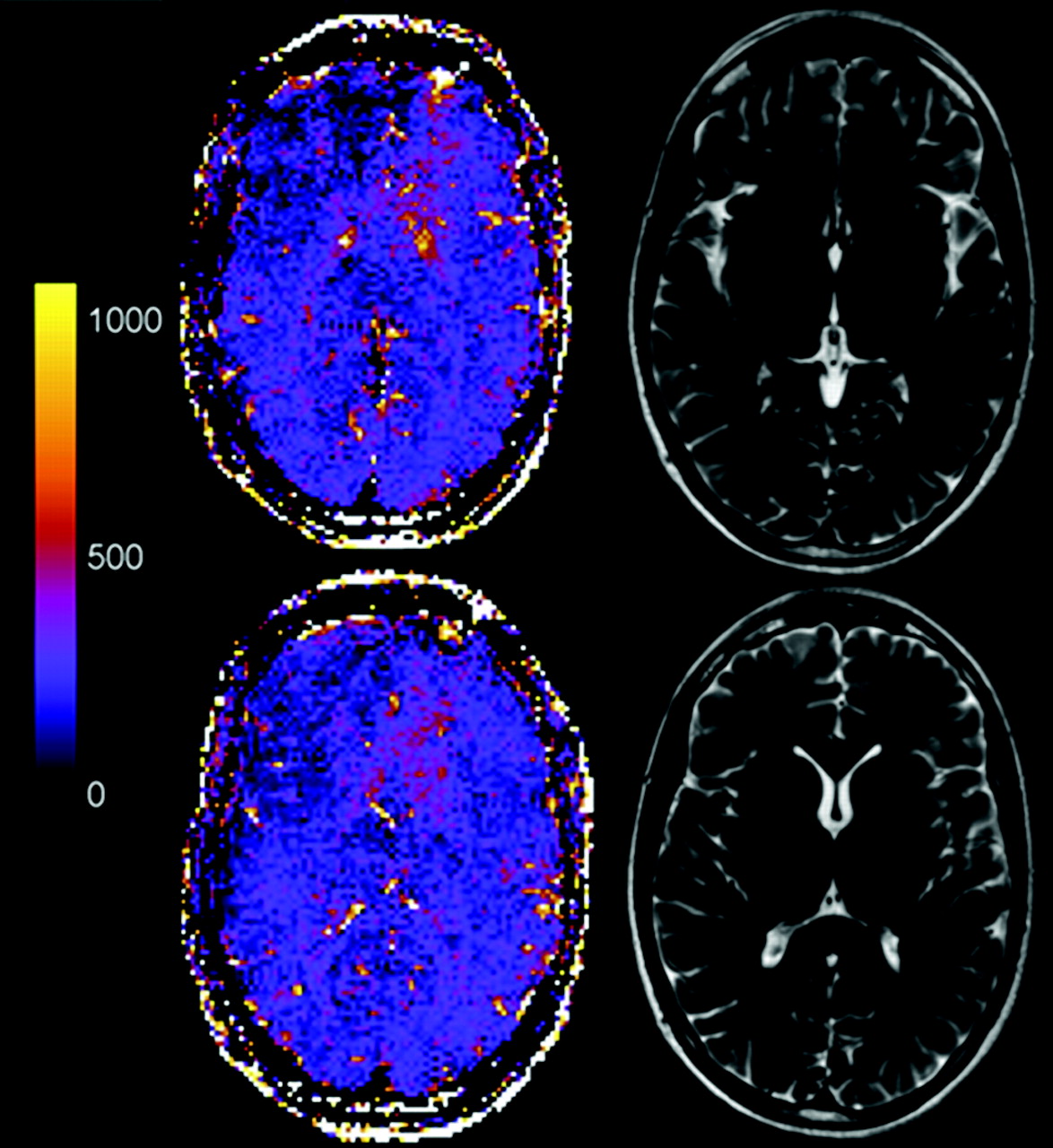
- Fig 2.
Representative MFC color maps and correspondent T2 images of 2 brain sections at the level of globus pallidus and thalamus in a 42-year-old male patient with mTBI. The patient complained of neck stiffness; his GCS score was 15, and the conventional MR imaging scan was normal. Note the clearly increased MFC in the deep gray matter and the correspondent normal T2 images. MFC values are reported in s−2.
- Fig 3.
Representative MFC color maps and correspondent T2 images of 2 brain sections at the level of globus pallidus and thalamus in a 25-year-old male patient with mTBI. The patient complained of headache, neck stiffness, photophobia, and nausea; his GCS score was 13, and the conventional MR imaging scan was normal. Note the clearly increased MFC in the deep gray matter and the correspondent normal T2 images. MFC values are reported in s−2.
- Fig 4.
Representative MFC color maps and correspondent T2 images of 2 brain sections at the level of globus pallidus and thalamus in a 32-year-old male healthy volunteer. MFC values are reported in s−2.


Tables
Symptom No. of Patients Blurry vision 1 Nausea 9 Memory loss 8 Dizziness 7 Photophobia 6 Neck stiffness 5 Loss of smell 4 Sleep disturbance 3 Headaches 2 Pain 1 Numbness 1 Tingling 1 - Table 2:
Average MFC values (s−2) ± SDs in healthy controls and patients with mild traumatic brain injury
Brain Region Controls mTBI Patients P Caudate 269 ± 146 326 ± 147 .111 Thalamus 149 ± 59 181 ± 65 .036* Globus pallidus 689 ± 252 873 ± 327 .002* Putamen 430 ± 166 493 ± 234 .331 Splenium 243 ± 190 210 ± 140 .638 Frontal lobe white matter 273 ± 176 203 ± 153 .130 -
Note:—Asterisk indicates statistically significant comparisons.
-
In this issue


Jump to section
Related Articles
Cited By...
- Individualised quantitative susceptibility mapping reveals abnormal hippocampal iron markers in acute mild traumatic brain injury
- Magnetic susceptibility of the hippocampal subfields and basal ganglia in acute mild traumatic brain injury
- Cortical iron-related markers are elevated in mild Traumatic Brain Injury: An individual-level quantitative susceptibility mapping study
- Distribution of paramagnetic and diamagnetic cortical substrates following mild Traumatic Brain Injury: A depth- and curvature-based quantitative susceptibility mapping study
- Altered oligodendroglia and astroglia in chronic traumatic encephalopathy
- Single Cell Molecular Alterations Reveal Pathogenesis and Targets of Concussive Brain Injury
- Brain iron overload following intracranial haemorrhage
- Classification algorithms using multiple MRI features in mild traumatic brain injury