
Article Figures & Data
Figures
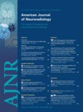
- Fig 1.
The position of the key ROIs on (A) b = 0, (B) b = 1000, and (C) b = 3000 ADC map.
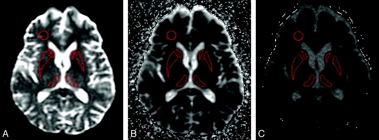
- Fig 2.
Differences in SIs in the basal ganglia in sCJD at (A) b = 1000 and (B) b = 3000 and in vCJD at (C) b = 1000 and (D) b = 3000.
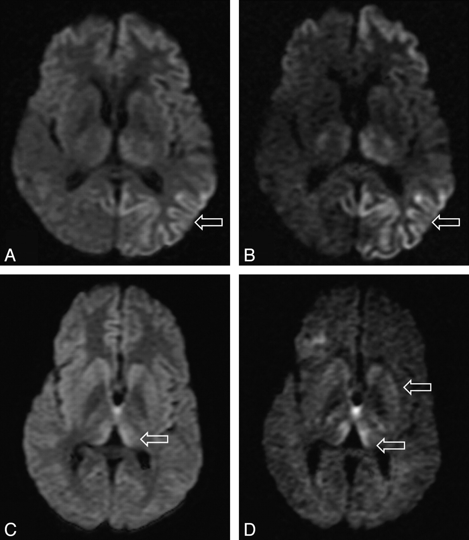
- Fig 3.
Bar charts showing ROI ADC values in sCJD (hashed bars) and controls (solid bars) at (A) b = 1000 s/mm2 and (B) b = 3000 s/mm2.


Tables
- Table 1:
Visual assessment of SI findings in vCJD and sCJD on FLAIR and DWI (b = 1000 s/mm2)
C P DM Cortex SP FWM vCJD 1 + + + − − − 2 + + + − − − 3 − − + − − − 4 + − + − − − 5 − − + − − − 6 + + + − − − 7 + − + − − − 8 + + + − − − sCJD 1 + + + + − − 2 + + − + − − 3 + + + + − − 4 + + + + − − 5 + + + + − − 6 + + − + − − 7 + − − + − − 8 + + + + − − 9 + + + + − − Note:—+ signifies hyperintense to gray matter; −, isointense to gray matter.
- Table 2:
Summary of mean diffusivity values (in mm2/s) measured in vCJD patients, sCJD patients, and controls for each ROI at b = 1000 s/mm2 with P values from post hoc comparisons
Group SP C P DM Pu FWM (A) vCJD 741.6 (104.0) 687.9 (70.6) 670.9 (56.2) 834.1 (51.3) 837.6 (33.0) 820.7 (44.6) (B) sCJD 753.8 (44.3) 587.3 (84.6) 603.3 (98.7) 691.8 (85.7) — 830.9 (89.1) (C) Control 754.8 (64.9) 722.7 (16.6) 727.8 (24.4) 763.7 (17.1) 748.0 (17.4) 773.7 (43.7) A versus C P = .949 P = .655 P = .382 P = .159 P < .001 P = .450 A versus B P = .941 P = .021 P = .167 P = .001 — P = .947 B versus C P = 1.000 P = .007 P = .018 P = .137 — P = .299 Note:—b = 1000 s/mm2 (group averages for 8 vCJD and 9 sCJD patients). All data are expressed as mean values with SD in parentheses. Significant P values are in bold.
- Table 3:
Summary of mean diffusivity values (in mm2/s) measured in vCJD patients, sCJD patients, and controls for each ROI at b = 3000 s/mm2 with P values from post hoc comparisons
Group SP C P DM Pu FWM (A) vCJD 524.5 (87.7) 554.8 (69.4) 530.3 (49.7) 584.7 (48.1) 603.2 (59.0) 556.6 (23.7) (B) sCJD 493.9 (87.9) 478.4 (27.5) 477.1 (77.8) 485.8 (87.4) — 539.5 (39.9) (C) Control 484.1 (19.9) 628.3 (15.4) 594.8 (8.8) 627.3 (13.1) 625.4 (10.6) 528.3 (41.4) A versus C P = .652 P = .063 P = .156 P = .460 P = .432 P = .444 A versus B P = .800 P = .068 P = .302 P = .050 — P = .759 B versus C P = .977 P = .001 P = .014 P = .007 — P = .885 Note:—b = 3000 s/mm2 (group averages for 4 vCJD and 5 sCJD patients). All data are expressed as mean values with SD in parentheses. Significant P values are in bold.


