
Article Figures & Data
Figures
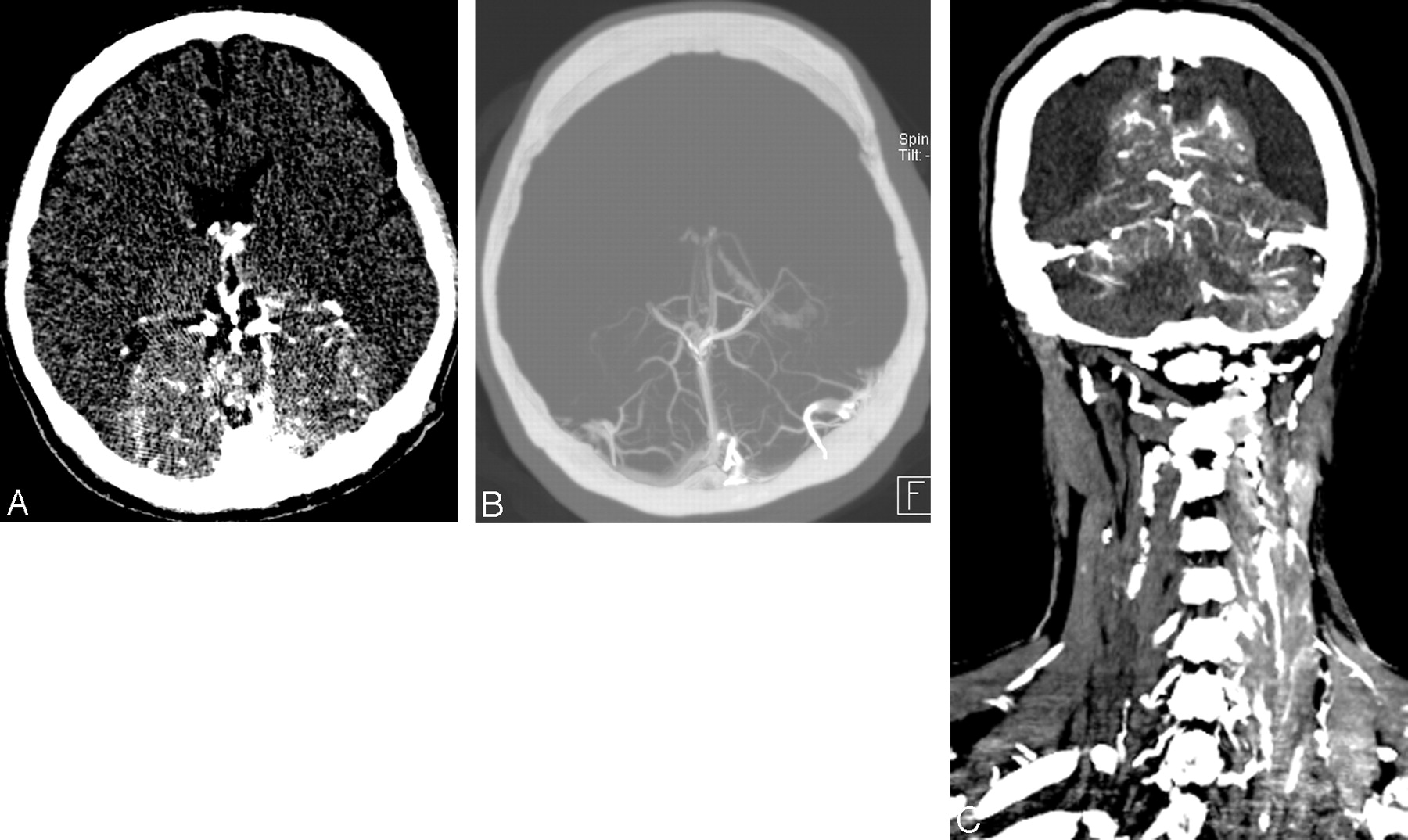
- Fig 1.
A, Source image from the CT angiogram of the brain demonstrates abnormal parenchymal enhancement corresponding to the posterior cerebral circulation territories. B, Maximum-intensity-projection image of the CT angiogram of brain demonstrates abnormal parenchymal enhancement and attenuated opacification of the deep cerebral veins and straight and transverse sinuses. C, Coronal reconstruction from the CT angiogram of the head and neck demonstrates abnormal parenchymal and venous enhancement corresponding to posterior circulation territories. There is also enhancement in the left paraspinal muscles and veins.
- Fig 2.
Axial source image from the CT angiogram of the neck demonstrates narrowing of the brachiocephalic vein between the brachiocephalic artery and the sternum, with a wire from a prior sternotomy. Retrosternal distance measures 2 mm at this level.


