Advertisement

Main menu

  • Home
  • Content
    • Current Issue
    • Accepted Manuscripts
    • Article Preview
    • Past Issue Archive
    • Video Articles
    • AJNR Case Collection
    • Case of the Week Archive
    • Case of the Month Archive
    • Classic Case Archive
  • Special Collections
    • AJNR Awards
    • Low-Field MRI
    • Alzheimer Disease
    • ASNR Foundation Special Collection
    • Photon-Counting CT
    • View All
  • Multimedia
    • AJNR Podcasts
    • AJNR SCANtastic
    • Trainee Corner
    • MRI Safety Corner
    • Imaging Protocols
  • For Authors
    • Submit a Manuscript
    • Submit a Video Article
    • Submit an eLetter to the Editor/Response
    • Manuscript Submission Guidelines
    • Statistical Tips
    • Fast Publishing of Accepted Manuscripts
    • Graphical Abstract Preparation
    • Imaging Protocol Submission
    • Author Policies
  • About Us
    • About AJNR
    • Editorial Board
    • Editorial Board Alumni
  • More
    • Become a Reviewer/Academy of Reviewers
    • Subscribers
    • Permissions
    • Alerts
    • Feedback
    • Advertisers
    • ASNR Home

User menu

  • Alerts
  • Log in

Search

  • Advanced search
American Journal of Neuroradiology

American Journal of Neuroradiology

ASHNR American Society of Functional Neuroradiology ASHNR American Society of Pediatric Neuroradiology ASSR
  • Alerts
  • Log in

Advanced Search

  • Home
  • Content
    • Current Issue
    • Accepted Manuscripts
    • Article Preview
    • Past Issue Archive
    • Video Articles
    • AJNR Case Collection
    • Case of the Week Archive
    • Case of the Month Archive
    • Classic Case Archive
  • Special Collections
    • AJNR Awards
    • Low-Field MRI
    • Alzheimer Disease
    • ASNR Foundation Special Collection
    • Photon-Counting CT
    • View All
  • Multimedia
    • AJNR Podcasts
    • AJNR SCANtastic
    • Trainee Corner
    • MRI Safety Corner
    • Imaging Protocols
  • For Authors
    • Submit a Manuscript
    • Submit a Video Article
    • Submit an eLetter to the Editor/Response
    • Manuscript Submission Guidelines
    • Statistical Tips
    • Fast Publishing of Accepted Manuscripts
    • Graphical Abstract Preparation
    • Imaging Protocol Submission
    • Author Policies
  • About Us
    • About AJNR
    • Editorial Board
    • Editorial Board Alumni
  • More
    • Become a Reviewer/Academy of Reviewers
    • Subscribers
    • Permissions
    • Alerts
    • Feedback
    • Advertisers
    • ASNR Home
  • Follow AJNR on Twitter
  • Visit AJNR on Facebook
  • Follow AJNR on Instagram
  • Join AJNR on LinkedIn
  • RSS Feeds

AJNR Awards, New Junior Editors, and more. Read the latest AJNR updates

Case ReportBrain

Pseudopathologic Brain Parenchymal Enhancement due to Venous Reflux from Left-Sided Injection and Brachiocephalic Vein Narrowing

J.Y. Chen, A.C. Mamourian, S.R. Messe and R.L. Wolf
American Journal of Neuroradiology January 2010, 31 (1) 86-87; DOI: https://doi.org/10.3174/ajnr.A1688
J.Y. Chen
  • Find this author on Google Scholar
  • Find this author on PubMed
  • Search for this author on this site
A.C. Mamourian
  • Find this author on Google Scholar
  • Find this author on PubMed
  • Search for this author on this site
S.R. Messe
  • Find this author on Google Scholar
  • Find this author on PubMed
  • Search for this author on this site
R.L. Wolf
  • Find this author on Google Scholar
  • Find this author on PubMed
  • Search for this author on this site
  • Article
  • Figures & Data
  • Info & Metrics
  • Responses
  • References
  • PDF
Loading

Abstract

SUMMARY: In this report, we present a case of a patient with CT angiographic artifacts related to left-sided venous injection resulting in a striking pattern of enhancement simulating vascular abnormalities, which prompted additional diagnostic imaging. To our knowledge, no similar case has been reported in the published literature to date.

With the development of high-speed multidetector CT units, CT angiography is increasingly becoming a primary tool for evaluation of the vasculature of the head and neck. Although a major advantage is its relative noninvasiveness compared with conventional catheter-based vascular imaging, disadvantages and pitfalls do exist. Failure to recognize these at the time of acquisition and interpretation can lead to poor-quality examinations or, worse, incorrect diagnoses and additional potentially unnecessary imaging or other work-up.

We present a case of a patient with artifacts related to left-sided venous injection resulting in a striking pattern of enhancement simulating vascular abnormalities, which prompted additional diagnostic imaging. To our knowledge, no similar case has been reported to date.

Case Report

A 59-year-old man with a prior sternotomy for mitral valve repair presented to the emergency department with acutely worsened intermittent vertigo. The possibility of vertebrobasilar ischemia was raised, and a CT angiogram of the neck and head was obtained on a 64-row multidetector CT scanner (Definition; Siemens Medical Systems, Erlangen, Germany) by using a timing bolus triggered from the left internal carotid artery. In this case, venous access was in the left antecubital vein. Contrast injection was 100 mL of iohexol (Omnipaque 350; GE Healthcare, Giles, UK) at 5 mL per second with a 27-second scanning delay.

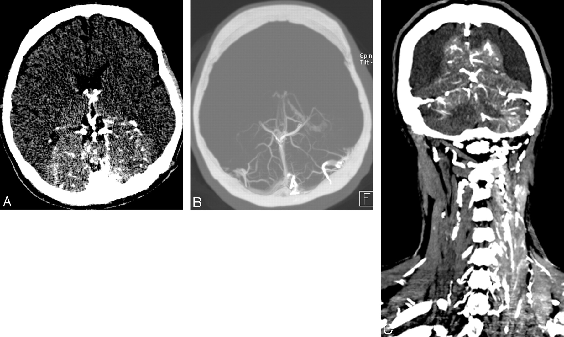
The CT angiogram demonstrated marked parenchymal enhancement in the posterior fossa, medial occipital, and parietal lobes; and opacification of the transverse and sigmoid sinuses and deep venous structures, generally corresponding to posterior circulation territories (Fig 1 A–C). Venous enhancement was also prominent in the left neck more than the right, including the bilateral internal jugular, subclavian, and brachiocephalic veins; the superior vena cava; and the epidural venous plexus and cervical venous plexus, obscuring vertebral artery enhancement. Parenchymal enhancement was evident in the dorsal cervical spinal cord and the left lobe of the thyroid gland. Arterial enhancement in the head and neck was suboptimal, generally less than that of the venous structures detailed above.

Fig 1.
  • Download figure
  • Open in new tab
  • Download powerpoint
Fig 1.

A, Source image from the CT angiogram of the brain demonstrates abnormal parenchymal enhancement corresponding to the posterior cerebral circulation territories. B, Maximum-intensity-projection image of the CT angiogram of brain demonstrates abnormal parenchymal enhancement and attenuated opacification of the deep cerebral veins and straight and transverse sinuses. C, Coronal reconstruction from the CT angiogram of the head and neck demonstrates abnormal parenchymal and venous enhancement corresponding to posterior circulation territories. There is also enhancement in the left paraspinal muscles and veins.

To evaluate further for structural or vascular abnormalities, we performed an MR imaging of the brain with MR angiography. Contrast-enhanced 3D MR angiography of the neck demonstrated prominent enhancement of the cervical venous plexus on arterial and delayed phases, without a clear vascular malformation or arterial feeder. Findings of brain MR imaging, MR angiography, and MR venography were unremarkable. Findings of conventional angiography were normal. CT angiography of the head and neck was ultimately repeated by using a right-sided venous injection, the findings of which were within normal limits.

Discussion

Both original CT and MR angiograms were obtained with left-sided venous injections. Further review of the initial CT angiogram revealed narrowing of the retrosternal distance (Fig 2) to 0.2 cm and narrowing of the brachiocephalic vein ipsilateral to the side of injection, which was redemonstrated on MR angiography.

Fig 2.
  • Download figure
  • Open in new tab
  • Download powerpoint
Fig 2.

Axial source image from the CT angiogram of the neck demonstrates narrowing of the brachiocephalic vein between the brachiocephalic artery and the sternum, with a wire from a prior sternotomy. Retrosternal distance measures 2 mm at this level.

With normal conventional angiography and CT angiography by using a right-sided injection, the most likely explanation for the abnormal pattern of enhancement on the initial studies was functional and/or structural narrowing of the left brachiocephalic vein related to a narrowed substernal distance. We propose that this resulted in preferential retrograde flow into the left jugular vein and intracranial venous system with reflux through the transverse and sigmoid sinuses to the right intracranial and cervical venous system, in this case overwhelming vascular autoregulation such that parenchymal enhancement occurred. The mechanism for the left thyroid gland enhancement is less clear, though elevated venous pressures may be an explanation.

Artifacts related to left-sided venous injection for cross-sectional angiographic studies are well-documented in the literature. These may include attenuated contrast in the brachiocephalic vein obscuring adjacent arteries,1 reflux of contrast into the jugular veins simulating filling defects, and simple reflux into the internal jugular vein and/or vertebral veins or vertebral venous plexus2 reaching the dural venous sinuses.3,4 None, however, have demonstrated retrograde brain parenchymal enhancement.

There are multiple suggested etiologies for the left-sided predominance of venous reflux. Anatomic differences in the course of the brachiocephalic veins may be a factor.5 The right brachiocephalic vein is more parallel to the ascending aorta, whereas the course of the left crosses the aorta and is, therefore, more susceptible to anatomic compression. An aberrant right subclavian artery may compress the left brachiocephalic vein.4 Hypertension or age-related ectasia and tortuosity of the aorta may also compress the brachiocephalic vein.

Retrosternal narrowing was evident in this case, which has been found to predispose to jugular venous reflux with left-sided injections. Tseng et al3 found that patients who were scanned serially with injections on both sides had no reflux when the retrosternal distance was 1.3 cm but did reflux with retrosternal distances of 0.65 cm. Our patient's retrosternal distance was 0.2 cm. The phase of respiration during contrast injection may also affect the retrosternal distance inversely with thoracic volume.4 Also, the jugular venous valves may be absent, more often on the left than the right in autopsy series.6 When present, venous valves may become incompetent, especially in patients with long-standing severe tricuspid regurgitation7 or after thoracoabdominal trauma.8 In addition to left-sided reflux, general venous return from injection of the upper extremities can be decreased by increased intrathoracic pressure, mediastinal masses, occlusion or stenosis of the superior vena cava, arm positioning, or congestive cardiac failure.9–12 Any of these concomitant conditions may contribute to venous reflux or delay of the arterial phase of enhancement.

The best explanation for this unusual pattern of enhancement on our patient's initial CT and MR angiographic studies is retrograde flow into the cervicocranial venous system, due to left-sided injection with impaired venous flow at the level of the left brachiocephalic vein related to narrowing of the retrosternal distance. This explanation confirms and reinforces existing recommendations that CT or MR angiography be performed with right-sided injection when possible, to minimize venous reflux.

References

  1. 1.↵
    1. Takhtani D
    . CT neuroangiography: a glance at the common pitfalls and their prevention. AJR Am J Roentgenol 2005; 185: 772–83
    PubMed
  2. 2.↵
    1. Lee YJ,
    2. Chung TS,
    3. Joo JY,
    4. et al
    . Suboptimal contrast-enhanced carotid MR angiography from the left brachiocephalic venous stasis. J Magn Reson Imaging 1999; 10: 503–09
    CrossRefPubMed
  3. 3.↵
    1. Tseng TC,
    2. Hsu HL,
    3. Lee TH,
    4. et al
    . Venous reflux on carotid computed tomography angiography: relationship with left-arm injection. J Comput Assist Tomogr 2007; 31: 360–64
    CrossRefPubMed
  4. 4.↵
    1. Tanaka T,
    2. Uemura K,
    3. Takahashi M,
    4. et al
    . Compression of the left brachiocephalic vein: cause of high signal intensity of the left sigmoid sinus and internal jugular vein on MR images. Radiology 1993; 188: 355–61
    PubMed
  5. 5.↵
    1. McCarthy J,
    2. Solomon NA
    . The effects of injection site, age, and body position on cervical venous reflux. Radiology 1979; 130: 536–67
    CrossRefPubMed
  6. 6.↵
    1. Harmon JV,
    2. Edwards WD
    . Venous valves in subclavian and internal jugular veins. Am J Cardiovasc Pathol 1986; 1: 51–4
  7. 7.↵
    1. Fisher J,
    2. Vaghaiwalla F,
    3. Tsitlik J,
    4. et al
    . Determinants and clinical significance of jugular venous valve competence. Circulation 1982; 65: 188–96
    Abstract/FREE Full Text
  8. 8.↵
    1. Keith A
    . An account of structures concerned in the production of the jugular pulse. J Anat Physiol 1907; 42 (Pt 1): 1–15
    PubMed
  9. 9.↵
    1. Bok B,
    2. Marsault C,
    3. Aubin ML,
    4. et al
    . Jugular venous reflux in cerebral radionuclide angiography: an explanation. Eur J Nucl Med 1978; 3: 63–65
    PubMed
  10. 10.↵
    1. Silverstein GE,
    2. Burke G,
    3. Goldberg D
    . Superior vena caval system obstruction caused by benign endothoracic goiter. Chest 1969; 56: 519–23
    CrossRefPubMed
  11. 11.↵
    1. Peart RA,
    2. Driedget AA
    . Effect of obstructed mediastinal venous return on dynamic brain blood flow studies: case report. J Nucl Med 1975; 16: 622–25
    Abstract/FREE Full Text
  12. 12.↵
    1. Fred HL,
    2. Wukasch DC,
    3. Petreny P
    . Transient compression of the left innominate vein. Circulation 1964; 29: 758–61
    Abstract/FREE Full Text
  • Received April 15, 2009.
  • Accepted after revision April 20, 2009.
  • Copyright © American Society of Neuroradiology
View Abstract
PreviousNext
Back to top

In this issue

American Journal of Neuroradiology: 31 (1)
American Journal of Neuroradiology
Vol. 31, Issue 1
1 Jan 2010
  • Table of Contents
  • Index by author
Advertisement
Print
Download PDF
Email Article

Thank you for your interest in spreading the word on American Journal of Neuroradiology.

NOTE: We only request your email address so that the person you are recommending the page to knows that you wanted them to see it, and that it is not junk mail. We do not capture any email address.

Enter multiple addresses on separate lines or separate them with commas.
Pseudopathologic Brain Parenchymal Enhancement due to Venous Reflux from Left-Sided Injection and Brachiocephalic Vein Narrowing
(Your Name) has sent you a message from American Journal of Neuroradiology
(Your Name) thought you would like to see the American Journal of Neuroradiology web site.
CAPTCHA
This question is for testing whether or not you are a human visitor and to prevent automated spam submissions.
Cite this article
J.Y. Chen, A.C. Mamourian, S.R. Messe, R.L. Wolf
Pseudopathologic Brain Parenchymal Enhancement due to Venous Reflux from Left-Sided Injection and Brachiocephalic Vein Narrowing
American Journal of Neuroradiology Jan 2010, 31 (1) 86-87; DOI: 10.3174/ajnr.A1688

Citation Manager Formats

  • BibTeX
  • Bookends
  • EasyBib
  • EndNote (tagged)
  • EndNote 8 (xml)
  • Medlars
  • Mendeley
  • Papers
  • RefWorks Tagged
  • Ref Manager
  • RIS
  • Zotero
0 Responses
Respond to this article
Share
Bookmark this article
Pseudopathologic Brain Parenchymal Enhancement due to Venous Reflux from Left-Sided Injection and Brachiocephalic Vein Narrowing
J.Y. Chen, A.C. Mamourian, S.R. Messe, R.L. Wolf
American Journal of Neuroradiology Jan 2010, 31 (1) 86-87; DOI: 10.3174/ajnr.A1688
del.icio.us logo Twitter logo Facebook logo Mendeley logo
  • Tweet Widget
  • Facebook Like
  • Google Plus One
Purchase

Jump to section

  • Article
    • Abstract
    • Case Report
    • Discussion
    • References
  • Figures & Data
  • Info & Metrics
  • Responses
  • References
  • PDF

Related Articles

  • No related articles found.
  • PubMed
  • Google Scholar

Cited By...

  • Pseudopathologic vertebral body enhancement
  • Author Response: Teaching NeuroImages: Pseudopathologic Brain Parenchymal Enhancement due to Vascular Compression in Parotid Tumor
  • Teaching NeuroImages: Pseudopathologic brain parenchymal enhancement due to vascular compression in parotid tumor
  • Suboptimal Contrast Opacification of Dynamic Head and Neck MR Angiography due to Venous Stasis and Reflux: Technical Considerations for Optimization
  • Crossref (8)
  • Google Scholar

This article has been cited by the following articles in journals that are participating in Crossref Cited-by Linking.

  • Pseudopathologic vertebral body enhancement in the presence of superior vena cava obstruction on computed tomography
    Yoon Kyung Kim, Yon Mi Sung, Kyung Hoon Hwang, Eun Kyung Cho, Hye-Young Choi
    The Spine Journal 2015 15 6
  • Suboptimal Contrast Opacification of Dynamic Head and Neck MR Angiography due to Venous Stasis and Reflux: Technical Considerations for Optimization
    D.R. Hingwala, B. Thomas, C. Kesavadas, T.R. Kapilamoorthy
    American Journal of Neuroradiology 2011 32 2
  • Computed Tomography Angiography of the Neurovascular Circulation
    Suyash Mohan, Mohit Agarwal, Bryan Pukenas
    Radiologic Clinics of North America 2016 54 1
  • Prevalence and characteristics of intravertebral enhancement on contrast-enhanced CT scans in cancer patients
    Benjamin Rasselet, Ahmed Larbi, Pierre Viala, Nicolas Molinari, Raphael Tetreau, Marie Faruch-Bilfeld, Patrice Taourel, Catherine Cyteval
    European Journal of Radiology 2017 86
  • Author Response: Teaching NeuroImages: Pseudopathologic Brain Parenchymal Enhancement due to Vascular Compression in Parotid Tumor
    Bernardo C.A. Teixeira, Zeferino Demartini, Debora B. Bertholdo, Luiz O.M. Coelho, Dante L. Escuissato
    Neurology 2021 97 1
  • Diagnostic and Therapeutic Neuroradiology
    Daniel Dias
    2018
  • Intracranial Venous Sinus Reflux on CT Angiography: Benign or Malignant Entity?
    Raghav Seth, Vikas Bhatia, Chirag Jain, Ajay Kumar
    Indian Journal of Radiology and Imaging 2021 31 04
  • Teaching NeuroImages: Pseudopathologic brain parenchymal enhancement due to vascular compression in parotid tumor
    Bernardo Corrêa de Almeida Teixeira, Marianna Cioni, Debora Brighente Bertholdo, Luiz Otávio de Mattos Coelho, Dante Luiz Escuissato
    Neurology 2020 94 16

More in this TOC Section

  • Evaluating the Effects of White Matter Multiple Sclerosis Lesions on the Volume Estimation of 6 Brain Tissue Segmentation Methods
  • Quiet PROPELLER MRI Techniques Match the Quality of Conventional PROPELLER Brain Imaging Techniques
  • Predictors of Reperfusion in Patients with Acute Ischemic Stroke
Show more BRAIN

Similar Articles

Advertisement

Indexed Content

  • Current Issue
  • Accepted Manuscripts
  • Article Preview
  • Past Issues
  • Editorials
  • Editor's Choice
  • Fellows' Journal Club
  • Letters to the Editor
  • Video Articles

Cases

  • Case Collection
  • Archive - Case of the Week
  • Archive - Case of the Month
  • Archive - Classic Case

More from AJNR

  • Trainee Corner
  • Imaging Protocols
  • MRI Safety Corner
  • Book Reviews

Multimedia

  • AJNR Podcasts
  • AJNR Scantastics

Resources

  • Turnaround Time
  • Submit a Manuscript
  • Submit a Video Article
  • Submit an eLetter to the Editor/Response
  • Manuscript Submission Guidelines
  • Statistical Tips
  • Fast Publishing of Accepted Manuscripts
  • Graphical Abstract Preparation
  • Imaging Protocol Submission
  • Evidence-Based Medicine Level Guide
  • Publishing Checklists
  • Author Policies
  • Become a Reviewer/Academy of Reviewers
  • News and Updates

About Us

  • About AJNR
  • Editorial Board
  • Editorial Board Alumni
  • Alerts
  • Permissions
  • Not an AJNR Subscriber? Join Now
  • Advertise with Us
  • Librarian Resources
  • Feedback
  • Terms and Conditions
  • AJNR Editorial Board Alumni

American Society of Neuroradiology

  • Not an ASNR Member? Join Now

© 2025 by the American Society of Neuroradiology All rights, including for text and data mining, AI training, and similar technologies, are reserved.
Print ISSN: 0195-6108 Online ISSN: 1936-959X

Powered by HighWire