Advertisement

Main menu

  • Home
  • Content
    • Current Issue
    • Accepted Manuscripts
    • Article Preview
    • Past Issue Archive
    • Video Articles
    • AJNR Case Collection
    • Case of the Week Archive
    • Case of the Month Archive
    • Classic Case Archive
  • Special Collections
    • AJNR Awards
    • Low-Field MRI
    • Alzheimer Disease
    • ASNR Foundation Special Collection
    • Photon-Counting CT
    • View All
  • Multimedia
    • AJNR Podcasts
    • AJNR SCANtastic
    • Trainee Corner
    • MRI Safety Corner
    • Imaging Protocols
  • For Authors
    • Submit a Manuscript
    • Submit a Video Article
    • Submit an eLetter to the Editor/Response
    • Manuscript Submission Guidelines
    • Statistical Tips
    • Fast Publishing of Accepted Manuscripts
    • Graphical Abstract Preparation
    • Imaging Protocol Submission
    • Author Policies
  • About Us
    • About AJNR
    • Editorial Board
    • Editorial Board Alumni
  • More
    • Become a Reviewer/Academy of Reviewers
    • Subscribers
    • Permissions
    • Alerts
    • Feedback
    • Advertisers
    • ASNR Home

User menu

  • Alerts
  • Log in

Search

  • Advanced search
American Journal of Neuroradiology

American Journal of Neuroradiology

ASHNR American Society of Functional Neuroradiology ASHNR American Society of Pediatric Neuroradiology ASSR
  • Alerts
  • Log in

Advanced Search

  • Home
  • Content
    • Current Issue
    • Accepted Manuscripts
    • Article Preview
    • Past Issue Archive
    • Video Articles
    • AJNR Case Collection
    • Case of the Week Archive
    • Case of the Month Archive
    • Classic Case Archive
  • Special Collections
    • AJNR Awards
    • Low-Field MRI
    • Alzheimer Disease
    • ASNR Foundation Special Collection
    • Photon-Counting CT
    • View All
  • Multimedia
    • AJNR Podcasts
    • AJNR SCANtastic
    • Trainee Corner
    • MRI Safety Corner
    • Imaging Protocols
  • For Authors
    • Submit a Manuscript
    • Submit a Video Article
    • Submit an eLetter to the Editor/Response
    • Manuscript Submission Guidelines
    • Statistical Tips
    • Fast Publishing of Accepted Manuscripts
    • Graphical Abstract Preparation
    • Imaging Protocol Submission
    • Author Policies
  • About Us
    • About AJNR
    • Editorial Board
    • Editorial Board Alumni
  • More
    • Become a Reviewer/Academy of Reviewers
    • Subscribers
    • Permissions
    • Alerts
    • Feedback
    • Advertisers
    • ASNR Home
  • Follow AJNR on Twitter
  • Visit AJNR on Facebook
  • Follow AJNR on Instagram
  • Join AJNR on LinkedIn
  • RSS Feeds

AJNR Awards, New Junior Editors, and more. Read the latest AJNR updates

Research ArticleNeurointervention

Endovascular Treatment of Brain Arteriovenous Fistulas

X. Lv, Y. Li, C. Jiang and Z. Wu
American Journal of Neuroradiology April 2009, 30 (4) 851-856; DOI: https://doi.org/10.3174/ajnr.A1436
X. Lv
  • Find this author on Google Scholar
  • Find this author on PubMed
  • Search for this author on this site
Y. Li
  • Find this author on Google Scholar
  • Find this author on PubMed
  • Search for this author on this site
C. Jiang
  • Find this author on Google Scholar
  • Find this author on PubMed
  • Search for this author on this site
Z. Wu
  • Find this author on Google Scholar
  • Find this author on PubMed
  • Search for this author on this site
  • Article
  • Figures & Data
  • Info & Metrics
  • Responses
  • References
  • PDF
Loading

Article Figures & Data

Figures

  • Tables
  • Fig 1.
    • Download figure
    • Open in new tab
    • Download powerpoint
    Fig 1.

    Patient 1. A, Angiogram of right internal carotid injection, anteroposterior view, shows a conglomerate of abnormally dilated vessels at the A1/A2 junction of the right ACA with a venous aneurysm (arrow). The lesion drains into the left sigmoid sinus and sphenoparietal sinus contralaterally via the right frontal basal vein and left tentorial sinus. B, Superselective angiography shows an arteriovenous fistula. C, Control angiogram of the right internal carotid injection after Onyx embolization, anteroposterior view, shows that the fistula was obliterated completely.
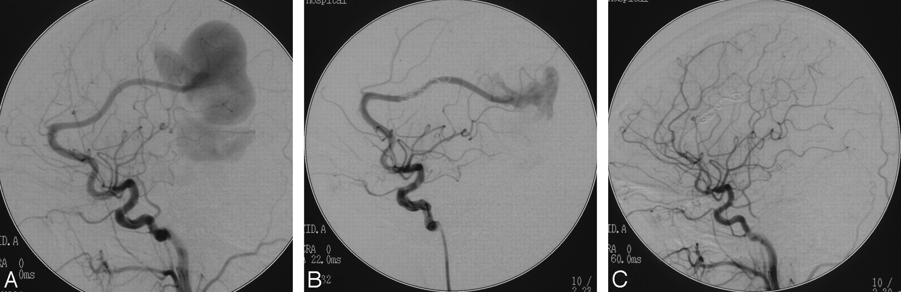
  • Fig 2.
    • Download figure
    • Open in new tab
    • Download powerpoint
    Fig 2.

    Patient 2. A, Angiogram of left internal carotid injection shows a pial AVF supplied by the left distal ACA with a giant venous aneurysm. B, Angiogram of the left internal carotid injection after the procedure shows that the fistula is occluded partially. C, Control angiogram at 4-month follow-up shows the fistula thrombosis spontaneously.
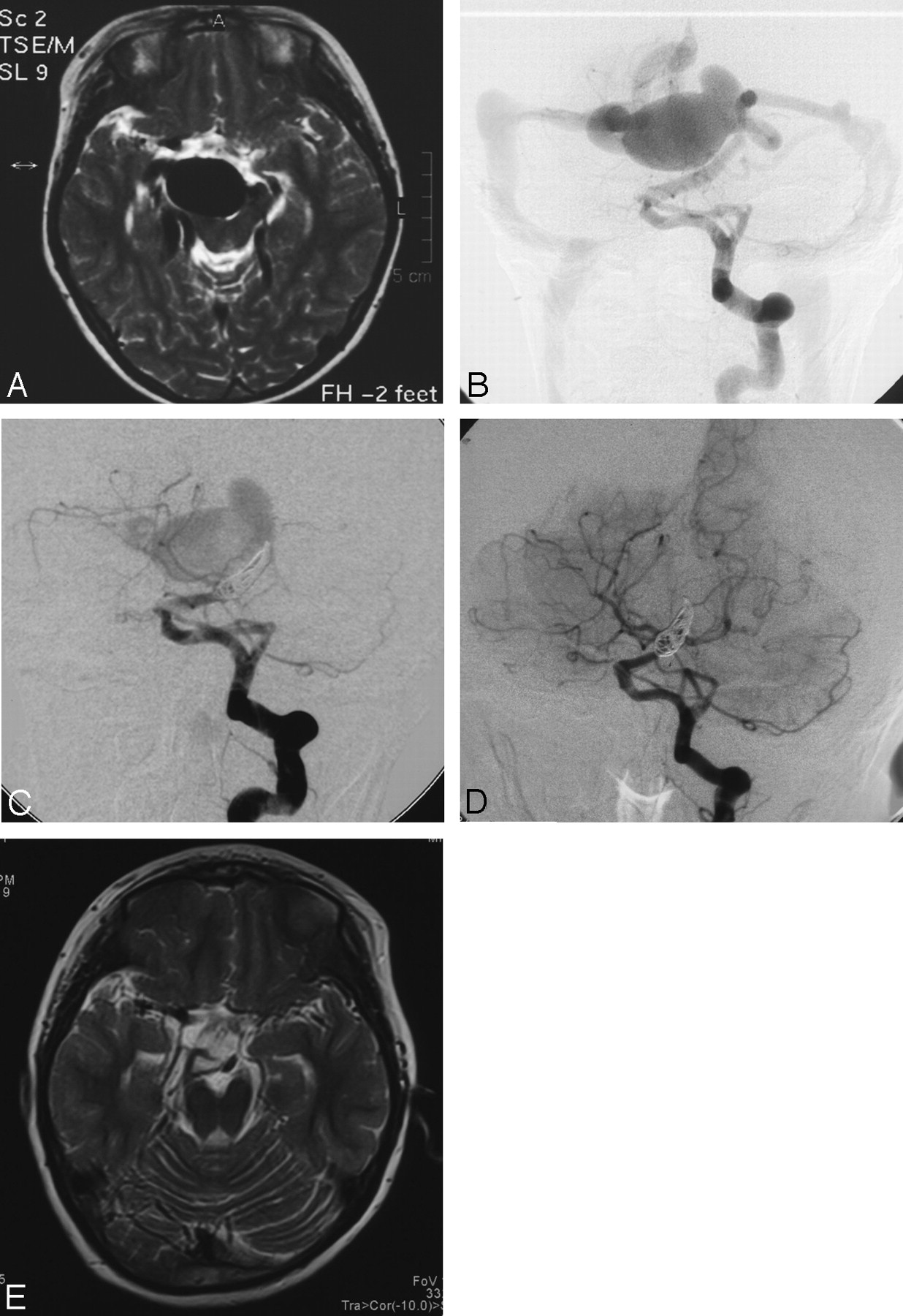
  • Fig 3.
    • Download figure
    • Open in new tab
    • Download powerpoint
    Fig 3.

    Patient 3. A, T2-weighted MR imaging of the head at the level of the midbrain shows giant flow-void signal intensity in the interpeduncular cistern. B, Anteroposterior view of the left vertebral artery injection demonstrates that the fistula is supplied by the primitive trigeminal artery and drained into the basal vein of Rosenthal, with an associated venous varix. C, After coil embolization, anteroposterior view of the left vertebral artery injection demonstrates incomplete disconnection of the fistula by the coils. D, Follow-up angiogram obtained at 7 months shows spontaneous occlusion of the fistula with preserved patency of the basilar artery. E, Disappearance of the venous varix on follow-up T2-weighted MR imaging.

Tables

  • Figures
  • Patients with BAVFs treated with endovascular techniques

    Patient No.Age/SexLocationPresentationFeeding ArteriesTreatment and Immediate Outcome*Follow-up
    1n7/MGyrus rectus regionICHJunction of A1 and A2Onyx-34, complete3
    226/FPosterior aspect of interhemispheric fissureHeadachesDistal ACADetachable coils, incomplete4
    37/MInterpeduncular cisternIncidentalPrimitive trigeminal arteryDetachable coils, incomplete7
    440/MSylvian fissureICHMCA, 3 arterial connections(IV), detachable coils, complete7
    514/MAnterior aspect of interhemispheric fissureSeizureA2 segment of ACA(II), detachable coils, complete3
    67/MPosterior aspect of interhemispheric fissureHeadachesP4 segment of PCADetachable coils, complete12
    735/MThird ventricleICHPosterior medial choroidal arteries of PCAsDetachable coils and 34% n-BCA, complete6
    822/FQuadrigeminal cisternICHP3–P4 segment of PCAFiber coils, incomplete3
    92/MAnterior aspect of interhemispheric fissureIncidentalA2 segment of ACADetachable coils, complete6
    • Note:—BAVFs indicate brain arteriovenous fistulas; R, right; L, left; F, female; M, male; MCA, middle cerebral artery; PCA, posterior cerebral artery; ACA, anterior cerebral artery; n-BCA, n-butyl cyanoacrylate; ICH, intracranial hemorrhage; II, IV, 2 times and 4 times.

    • * All patients were cured on follow-up.

PreviousNext
Back to top

In this issue

American Journal of Neuroradiology: 30 (4)
American Journal of Neuroradiology
Vol. 30, Issue 4
April 2009
  • Table of Contents
  • Index by author
Advertisement
Print
Download PDF
Email Article

Thank you for your interest in spreading the word on American Journal of Neuroradiology.

NOTE: We only request your email address so that the person you are recommending the page to knows that you wanted them to see it, and that it is not junk mail. We do not capture any email address.

Enter multiple addresses on separate lines or separate them with commas.
Endovascular Treatment of Brain Arteriovenous Fistulas
(Your Name) has sent you a message from American Journal of Neuroradiology
(Your Name) thought you would like to see the American Journal of Neuroradiology web site.
CAPTCHA
This question is for testing whether or not you are a human visitor and to prevent automated spam submissions.
Cite this article
X. Lv, Y. Li, C. Jiang, Z. Wu
Endovascular Treatment of Brain Arteriovenous Fistulas
American Journal of Neuroradiology Apr 2009, 30 (4) 851-856; DOI: 10.3174/ajnr.A1436

Citation Manager Formats

  • BibTeX
  • Bookends
  • EasyBib
  • EndNote (tagged)
  • EndNote 8 (xml)
  • Medlars
  • Mendeley
  • Papers
  • RefWorks Tagged
  • Ref Manager
  • RIS
  • Zotero
0 Responses
Respond to this article
Share
Bookmark this article
Endovascular Treatment of Brain Arteriovenous Fistulas
X. Lv, Y. Li, C. Jiang, Z. Wu
American Journal of Neuroradiology Apr 2009, 30 (4) 851-856; DOI: 10.3174/ajnr.A1436
del.icio.us logo Twitter logo Facebook logo Mendeley logo
  • Tweet Widget
  • Facebook Like
  • Google Plus One
Purchase

Jump to section

  • Article
    • Abstract
    • Materials and Methods
    • Results
    • Discussion
    • Conclusions
    • References
  • Figures & Data
  • Info & Metrics
  • Responses
  • References
  • PDF

Related Articles

  • No related articles found.
  • PubMed
  • Google Scholar

Cited By...

  • Non-galenic arteriovenous fistulas in adults: transarterial embolization and literature review
  • Development, clinical presentation and endovascular management of congenital intracranial pial arteriovenous fistulas
  • Pediatric Intracranial Nongalenic Pial Arteriovenous Fistulas: Clinical Features, Angioarchitecture, and Outcomes
  • Crossref (38)
  • Google Scholar

This article has been cited by the following articles in journals that are participating in Crossref Cited-by Linking.

  • Pediatric Intracranial Nongalenic Pial Arteriovenous Fistulas: Clinical Features, Angioarchitecture, and Outcomes
    S.W. Hetts, K. Keenan, H.J. Fullerton, W.L. Young, J.D. English, N. Gupta, C.F. Dowd, R.T. Higashida, M.T. Lawton, V.V. Halbach
    American Journal of Neuroradiology 2012 33 9
  • An institutional series and literature review of pial arteriovenous fistulas in the pediatric population
    Peter J. Madsen, Shih-Shan Lang, Jared M. Pisapia, Phillip B. Storm, Robert W. Hurst, Gregory G. Heuer
    Journal of Neurosurgery: Pediatrics 2013 12 4
  • Intracranial non-galenic pial arteriovenous fistula: A review of the literature
    Jinlu Yu, Lei Shi, Xianli Lv, Zhongxue Wu, Hongfa Yang
    Interventional Neuroradiology 2016 22 5
  • Development, clinical presentation and endovascular management of congenital intracranial pial arteriovenous fistulas
    Srinivasan Paramasivam, Naoki Toma, Yasunari Niimi, Alejandro Berenstein
    Journal of NeuroInterventional Surgery 2013 5 3
  • Neuroendovascular Management of Dural Arteriovenous Malformations
    Kathleen A. McConnell, Stavropoula I. Tjoumakaris, Jason Allen, Maksim Shapiro, Tibor Bescke, Pascal M. Jabbour, Robert H. Rosenwasser, Peter K. Nelson
    Neurosurgery Clinics of North America 2009 20 4
  • Multimodal treatment strategies for complex pediatric cerebral arteriovenous fistulas: contemporary case series at Barrow Neurological Institute
    Hasan A. Zaidi, M. Yashar S. Kalani, Robert F. Spetzler, Cameron G. McDougall, Felipe C. Albuquerque
    Journal of Neurosurgery: Pediatrics 2015 15 6
  • Clinical Outcomes of Endovascular Treatment for Intracranial Pial Arteriovenous Fistulas
    Xianli Lv, Chuhan Jiang, Youxiang Li, Xinjian Yang, Zhongxue Wu
    World Neurosurgery 2010 73 4
  • De novo multiple dural arteriovenous fistulas and arteriovenous malformation after embolization of cerebral arteriovenous fistula: case report
    Yahui Bai, Chuan He, Hongqi Zhang, Feng Ling
    Child's Nervous System 2012 28 11
  • Intracranial Pial Arteriovenous Fistulae: Diagnosis and Treatment Techniques in Pediatric Patients with Review of Literature
    Anand Alurkar, Lakshmi Sudha Prasanna Karanam, Suresh Nayak, Rajesh Kumar Ghanta
    Journal of Clinical Imaging Science 2016 6
  • Stereotactic Radiosurgery for Cavernous Sinus Versus Noncavernous Sinus Dural Arteriovenous Fistulas: Outcomes and Outcome Predictors
    Yi-Chieh Hung, Nasser Mohammed, Kathryn N Kearns, Ching-Jen Chen, Robert M Starke, Hideyuki Kano, John Lee, David Mathieu, Anthony M Kaufmann, Wei Gang Wang, Inga S Grills, Christopher P Cifarelli, John Vargo, Tomas Chytka, Ladislava Janouskova, Caleb E Feliciano, Rafael Rodriguez-Mercado, L Dade Lunsford, Jason P Sheehan
    Neurosurgery 2020 86 5

More in this TOC Section

  • Factors Associated with Major Re-Recanalization following Second Coiling for Recanalized Aneurysms: A Multicenter Experience over 20 Years during Long-Term Follow-up
  • A Key Factor Shapes LS-DAVFs EVT Outcome
  • Optimizing Voxel Size in 3D Rotational Angiography
Show more NEUROINTERVENTION

Similar Articles

Advertisement

Indexed Content

  • Current Issue
  • Accepted Manuscripts
  • Article Preview
  • Past Issues
  • Editorials
  • Editor's Choice
  • Fellows' Journal Club
  • Letters to the Editor
  • Video Articles

Cases

  • Case Collection
  • Archive - Case of the Week
  • Archive - Case of the Month
  • Archive - Classic Case

More from AJNR

  • Trainee Corner
  • Imaging Protocols
  • MRI Safety Corner

Multimedia

  • AJNR Podcasts
  • AJNR Scantastics

Resources

  • Turnaround Time
  • Submit a Manuscript
  • Submit a Video Article
  • Submit an eLetter to the Editor/Response
  • Manuscript Submission Guidelines
  • Statistical Tips
  • Fast Publishing of Accepted Manuscripts
  • Graphical Abstract Preparation
  • Imaging Protocol Submission
  • Evidence-Based Medicine Level Guide
  • Publishing Checklists
  • Author Policies
  • Become a Reviewer/Academy of Reviewers
  • News and Updates

About Us

  • About AJNR
  • Editorial Board
  • Editorial Board Alumni
  • Alerts
  • Permissions
  • Not an AJNR Subscriber? Join Now
  • Advertise with Us
  • Librarian Resources
  • Feedback
  • Terms and Conditions
  • AJNR Editorial Board Alumni

American Society of Neuroradiology

  • Not an ASNR Member? Join Now

© 2025 by the American Society of Neuroradiology All rights, including for text and data mining, AI training, and similar technologies, are reserved.
Print ISSN: 0195-6108 Online ISSN: 1936-959X

Powered by HighWire