
Article Figures & Data
Figures
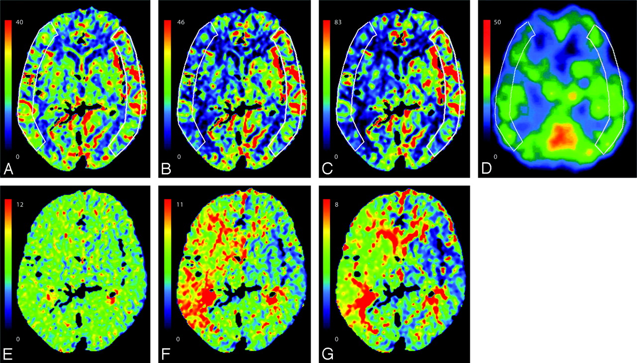
- Fig 1.
CBF and MTT maps of a 70-year-old woman with occlusion of the right MCA. A and E, CTP with bSVD. B and F, CTP with sSVD. C and G, CTP with bMTF. D, Quantitative SPECT with 123I-IMP. A–D, CBF. E–G, MTT. AIF was obtained from the second portion of the left MCA. A–C, E–G, CBF and MTT color maps on CTP appear different among the different deconvolution algorithms. B–D, Images obtained by using sSVD (B) and bMTF (C) overestimate the decrease in CBF of the right MCA territory, compared with that generated by SPECT (D), whereas images using bSVD (A) correspond well with those using SPECT (D).
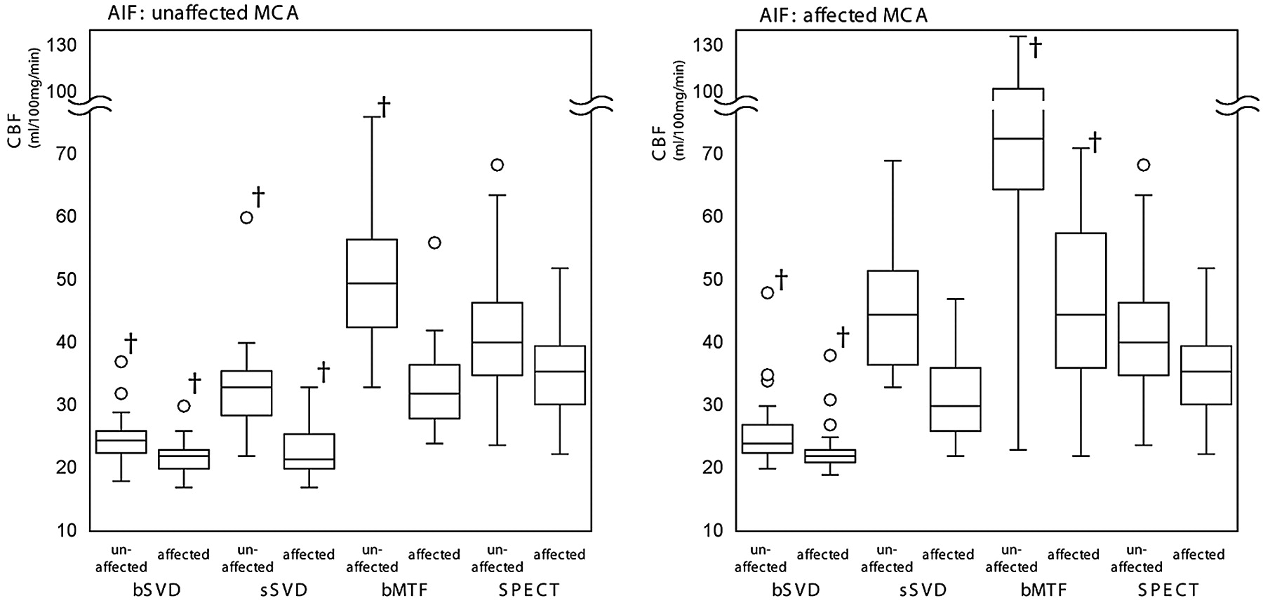
- Fig 2.
Absolute CBF values of the unaffected and affected MCA territories with different algorithms and different locations of AIFs. The ranges of the CBF obtained by bSVD in any conditions and those obtained by sSVD with the AIF on the unaffected MCA are significantly smaller than the corresponding ranges obtained by quantitative SPECT. However, those obtained by bMTF are significantly larger than those obtained by SPECT, particularly when the AIF is on the affected MCA. The CBF ranges obtained by sSVD with the AIF on the affected MCA are compatible with those generated by SPECT. †P < .01.
- Fig 3.
The CBF ratios of the affected MCA territory to the contralateral corresponding area with different algorithms and different locations of AIFs. When comparing the CBF ratios with SPECT, we observed that the CBF ratios were underestimated in many patients on CTP with sSVD and bMTF but not with bSVD, regardless of the location (affected or unaffected side) of the AIF on the MCA.


Tables
Correlation of the CBF values between CTP and SPECT
CBF AIF Unaffected Side AIF Affected Side Unaffected Side Affected Side Ratio Unaffected Side Affected Side Ratio bSVD r2 0.58* 0.46† 0.70‡ 0.44 0.36 0.60* ICC −0.31 −0.42 0.64‡ −0.22 −0.30 0.58* sSVD r2 0.52† 0.22 0.50† 0.46† 0.08 0.40 ICC 0.25 −0.36 0.02 0.43† 0.02 0.03 bMTF r2 0.40 0.25 0.57* 0.22 0.22 0.11 ICC 0.21 0.26 −0.04 −0.31 −0.30 −0.23 Note:—CBF indicates cerebral blood flow; CTP, CT perfusion; SPECT, single-photon emission CT; AIF, arterial input function; bSVD, block-circulant singular value decomposition; SSVD, standard SVD; bMTF, box-modulation transfer function: ICC, intraclass correlation coefficient.
* P < .01.
† P < .05.
‡ P < .001.
In this issue


Jump to section
Related Articles
Cited By...
- Predicting Impaired Cerebrovascular Reactivity and Hyperperfusion Syndrome with BeamSAT MRI in Carotid Artery Stenosis
- CT Perfusion in Acute Lacunar Stroke: Detection Capabilities Based on Infarct Location
- Using Quantitative CT Perfusion for Evaluation of Delayed Cerebral Ischemia Following Aneurysmal Subarachnoid Hemorrhage
- CT Cerebral Blood Flow Maps Optimally Correlate With Admission Diffusion-Weighted Imaging in Acute Stroke but Thresholds Vary by Postprocessing Platform