Advertisement

Main menu

  • Home
  • Content
    • Current Issue
    • Accepted Manuscripts
    • Article Preview
    • Past Issue Archive
    • Video Articles
    • AJNR Case Collection
    • Case of the Week Archive
    • Case of the Month Archive
    • Classic Case Archive
  • Special Collections
    • AJNR Awards
    • Low-Field MRI
    • Alzheimer Disease
    • ASNR Foundation Special Collection
    • Photon-Counting CT
    • View All
  • Multimedia
    • AJNR Podcasts
    • AJNR SCANtastic
    • Trainee Corner
    • MRI Safety Corner
    • Imaging Protocols
  • For Authors
    • Submit a Manuscript
    • Submit a Video Article
    • Submit an eLetter to the Editor/Response
    • Manuscript Submission Guidelines
    • Statistical Tips
    • Fast Publishing of Accepted Manuscripts
    • Graphical Abstract Preparation
    • Imaging Protocol Submission
    • Author Policies
  • About Us
    • About AJNR
    • Editorial Board
    • Editorial Board Alumni
  • More
    • Become a Reviewer/Academy of Reviewers
    • Subscribers
    • Permissions
    • Alerts
    • Feedback
    • Advertisers
    • ASNR Home

User menu

  • Alerts
  • Log in

Search

  • Advanced search
American Journal of Neuroradiology

American Journal of Neuroradiology

ASHNR American Society of Functional Neuroradiology ASHNR American Society of Pediatric Neuroradiology ASSR
  • Alerts
  • Log in

Advanced Search

  • Home
  • Content
    • Current Issue
    • Accepted Manuscripts
    • Article Preview
    • Past Issue Archive
    • Video Articles
    • AJNR Case Collection
    • Case of the Week Archive
    • Case of the Month Archive
    • Classic Case Archive
  • Special Collections
    • AJNR Awards
    • Low-Field MRI
    • Alzheimer Disease
    • ASNR Foundation Special Collection
    • Photon-Counting CT
    • View All
  • Multimedia
    • AJNR Podcasts
    • AJNR SCANtastic
    • Trainee Corner
    • MRI Safety Corner
    • Imaging Protocols
  • For Authors
    • Submit a Manuscript
    • Submit a Video Article
    • Submit an eLetter to the Editor/Response
    • Manuscript Submission Guidelines
    • Statistical Tips
    • Fast Publishing of Accepted Manuscripts
    • Graphical Abstract Preparation
    • Imaging Protocol Submission
    • Author Policies
  • About Us
    • About AJNR
    • Editorial Board
    • Editorial Board Alumni
  • More
    • Become a Reviewer/Academy of Reviewers
    • Subscribers
    • Permissions
    • Alerts
    • Feedback
    • Advertisers
    • ASNR Home
  • Follow AJNR on Twitter
  • Visit AJNR on Facebook
  • Follow AJNR on Instagram
  • Join AJNR on LinkedIn
  • RSS Feeds

AJNR Awards, New Junior Editors, and more. Read the latest AJNR updates

Research ArticleBRAIN

Arterial Spin-Labeling MR Imaging Measurements of Timing Parameters in Patients with a Carotid Artery Occlusion

R.P.H. Bokkers, P.J. van Laar, K.C.C. van de Ven, L.J. Kapelle, C.J.M. Klijn and J. Hendrikse
American Journal of Neuroradiology October 2008, 29 (9) 1698-1703; DOI: https://doi.org/10.3174/ajnr.A1232
R.P.H. Bokkers
  • Find this author on Google Scholar
  • Find this author on PubMed
  • Search for this author on this site
P.J. van Laar
  • Find this author on Google Scholar
  • Find this author on PubMed
  • Search for this author on this site
K.C.C. van de Ven
  • Find this author on Google Scholar
  • Find this author on PubMed
  • Search for this author on this site
L.J. Kapelle
  • Find this author on Google Scholar
  • Find this author on PubMed
  • Search for this author on this site
C.J.M. Klijn
  • Find this author on Google Scholar
  • Find this author on PubMed
  • Search for this author on this site
J. Hendrikse
  • Find this author on Google Scholar
  • Find this author on PubMed
  • Search for this author on this site
  • Article
  • Figures & Data
  • Info & Metrics
  • Responses
  • References
  • PDF
Loading

Article Figures & Data

Figures

  • Tables
  • Fig 1.
    • Download figure
    • Open in new tab
    • Download powerpoint
    Fig 1.

    A, sagittal orientation image illustrating the planning of the imaging section (1) and the arterial spin-labeling slab (2) parallel to the orbitomeatal angle. B, regions of interest used for quantification of the hemodynamic parameters. In each hemisphere, 2 regions of interests were drawn in the frontal lobe, and 1, in the frontal parietal, parietal occipital, and occipital regions.

  • Fig 2.
    • Download figure
    • Open in new tab
    • Download powerpoint
    Fig 2.

    Measured perfusion images of a 53-year-old man with a unilateral left-sided ICA occlusion. The images show the absolute cerebral blood flow (A), transit time (B), and trailing edge (C) maps. Decreased cerebral blood flow, increased transit time, and increased trailing edge can be appreciated in the left hemisphere.
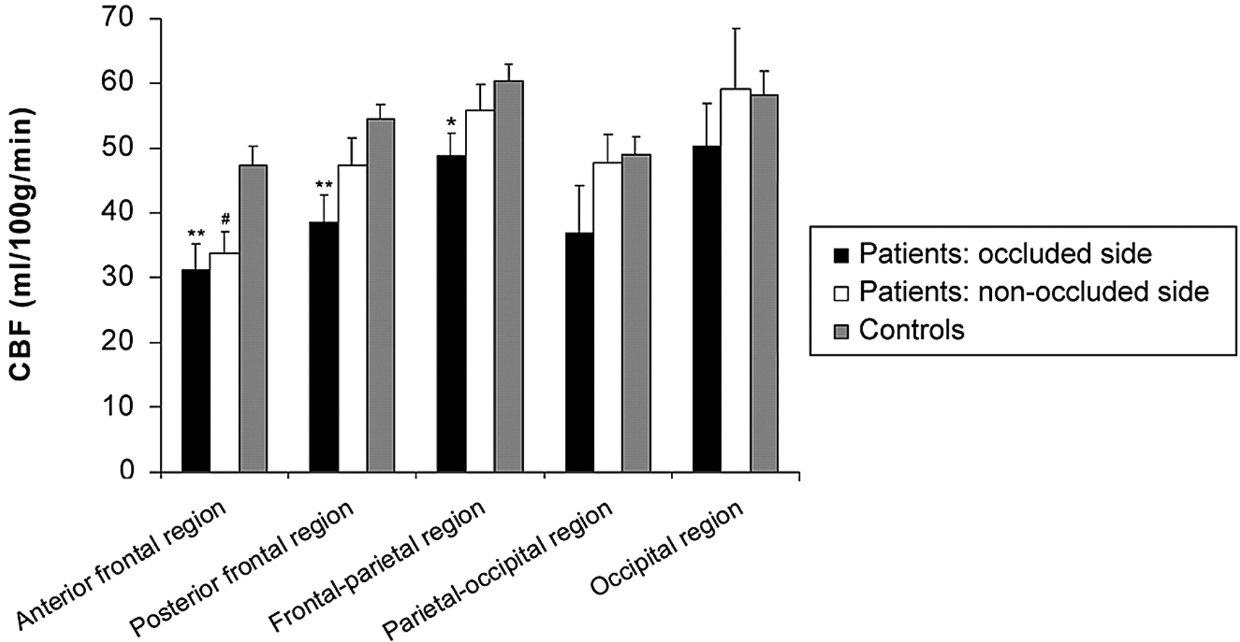
  • Fig 3.
    • Download figure
    • Open in new tab
    • Download powerpoint
    Fig 3.

    Cerebral blood flow (mean ± SEM) in the regions of the hemisphere ipsi- and contralateral to the occlusion and of the control group. Asterisk indicates P < .05; double asterisks, P < .01, a significant difference between the hemisphere ipsilateral to the ICA occlusion and the control subjects; number sign, a significant (P < .05) difference between the hemisphere contralateral to the ICA occlusion and the control subjects.

  • Fig 4.
    • Download figure
    • Open in new tab
    • Download powerpoint
    Fig 4.

    Transit time and trailing edge time (mean ± SEM) in the regions of the hemisphere ipsi- and contralateral to the occlusion and of the control group. Asterisk indicates P < .05; double asterisks, P < .01, a significant difference between the hemisphere ipsilateral to the ICA occlusion and the control subjects; number sign, a significant (P < .05) asymmetry between the hemisphere contralateral to the ICA occlusion and the control subjects; dagger, a significant (P < .01) difference between the hemisphere ipsi- and contralateral to the ICA occlusion.

Tables

  • Figures
    • View popup
    Table 1:

    Patient characteristics

    Patient No.Age (yr)SexSymptomsSide of OcclusionContralateral SideCollateral Flow Pathways
    144FStrokeRight30%P + L
    255MTIARight–P
    355MStrokeRight–A + P
    448MTIARight–A + P + L
    553MStrokeLeft–A + P
    665MStrokeRight–A + L
    777MAFRight–A + P
    856MTIALeft–A + P
    947FStrokeRight–P
    1055MStrokeLeft90%A + L
    1168MTIALeft70%L
    1258MStrokeRight30%A
    1353MTIARight30%A + P + L
    1465MTIARight–A + L
    1560MStrokeLeft30%A + P
    1653MStrokeRight–A + L
    1758MStrokeLeft50%P + L
    • Note:—TIA indicates transient ischemic attack; AF, amaurosis fugax; A, anterior collateral pathway; P, posterior collateral pathway; L, leptomeningeal vessels; –, no stenosis.

    • View popup
    Table 2:

    Hemodynamic values for the hemisphere ipsilateral to the occlusion in patients with (n = 9) and with leptomeningeal collateral vessels

    Leptomeningeal Vessels PresentNo Leptomeningeal Vessels
    CBF*Transit TimeTrailing EdgeCBFTransit TimeTrailing Edge
    Anterior frontal31 ± 983 ± 462436 ± 275†31 ± 225 ± 151648 ± 201
    Posterior frontal29 ± 5†196 ± 1032700 ± 46947 ± 685 ± 321598 ± 348
    Frontal parietal45 ± 4228 ± 532362 ± 20852 ± 697 ± 762103 ± 259
    Parietal occipital41 ± 11529 ± 1632089 ± 28533 ± 10411 ± 1062185 ± 271
    Occipital54 ± 13381 ± 1381815 ± 128†47 ± 5359 ± 942388 ± 203
    • * Cerebral blood flow values are given in milliliters/minute/100 g, and transit and trailing edge times, in milliseconds.

    • † A statistically significant difference between the patients with and without leptomeningeal vessels (t test, P < .05).

PreviousNext
Back to top

In this issue

American Journal of Neuroradiology: 29 (9)
American Journal of Neuroradiology
Vol. 29, Issue 9
October 2008
  • Table of Contents
  • Index by author
Advertisement
Print
Download PDF
Email Article

Thank you for your interest in spreading the word on American Journal of Neuroradiology.

NOTE: We only request your email address so that the person you are recommending the page to knows that you wanted them to see it, and that it is not junk mail. We do not capture any email address.

Enter multiple addresses on separate lines or separate them with commas.
Arterial Spin-Labeling MR Imaging Measurements of Timing Parameters in Patients with a Carotid Artery Occlusion
(Your Name) has sent you a message from American Journal of Neuroradiology
(Your Name) thought you would like to see the American Journal of Neuroradiology web site.
CAPTCHA
This question is for testing whether or not you are a human visitor and to prevent automated spam submissions.
Cite this article
R.P.H. Bokkers, P.J. van Laar, K.C.C. van de Ven, L.J. Kapelle, C.J.M. Klijn, J. Hendrikse
Arterial Spin-Labeling MR Imaging Measurements of Timing Parameters in Patients with a Carotid Artery Occlusion
American Journal of Neuroradiology Oct 2008, 29 (9) 1698-1703; DOI: 10.3174/ajnr.A1232

Citation Manager Formats

  • BibTeX
  • Bookends
  • EasyBib
  • EndNote (tagged)
  • EndNote 8 (xml)
  • Medlars
  • Mendeley
  • Papers
  • RefWorks Tagged
  • Ref Manager
  • RIS
  • Zotero
0 Responses
Respond to this article
Share
Bookmark this article
Arterial Spin-Labeling MR Imaging Measurements of Timing Parameters in Patients with a Carotid Artery Occlusion
R.P.H. Bokkers, P.J. van Laar, K.C.C. van de Ven, L.J. Kapelle, C.J.M. Klijn, J. Hendrikse
American Journal of Neuroradiology Oct 2008, 29 (9) 1698-1703; DOI: 10.3174/ajnr.A1232
del.icio.us logo Twitter logo Facebook logo Mendeley logo
  • Tweet Widget
  • Facebook Like
  • Google Plus One
Purchase

Jump to section

  • Article
    • Abstract
    • Materials and Methods
    • Results
    • Discussion
    • Conclusion
    • Footnotes
    • References
  • Figures & Data
  • Info & Metrics
  • Responses
  • References
  • PDF

Related Articles

  • No related articles found.
  • PubMed
  • Google Scholar

Cited By...

  • Cerebral Perfusion After Repair of Congenital Diaphragmatic Hernia with Common Carotid Artery Occlusion After ECMO Therapy
  • Noninvasive Evaluation of CBF and Perfusion Delay of Moyamoya Disease Using Arterial Spin-Labeling MRI with Multiple Postlabeling Delays: Comparison with 15O-Gas PET and DSC-MRI
  • Arterial Spin Labeling Magnetic Resonance Imaging Estimation of Antegrade and Collateral Flow in Unilateral Middle Cerebral Artery Stenosis
  • Regional Cerebral Arterial Transit Time Hemodynamics Correlate with Vascular Risk Factors and Cognitive Function in Men with Coronary Artery Disease
  • Cerebrovascular Collaterals Correlate with Disease Severity in Adult North American Patients with Moyamoya Disease
  • Hemodynamic Alterations in Vertebrobasilar Large Artery Disease Assessed by Arterial Spin-Labeling MR Imaging
  • Assessment of Cortical Hemodynamics by Multichannel Near-Infrared Spectroscopy in Steno-Occlusive Disease of the Middle Cerebral Artery
  • Systematic Review of Methods for Assessing Leptomeningeal Collateral Flow
  • Cerebral Perfusion Long Term after Therapeutic Occlusion of the Internal Carotid Artery in Patients Who Tolerated Angiographic Balloon Test Occlusion
  • Multiple Inflow Pulsed Arterial Spin-Labeling Reveals Delays in the Arterial Arrival Time in Minor Stroke and Transient Ischemic Attack
  • Noninvasive MR imaging of cerebral perfusion in patients with a carotid artery stenosis
  • Simultaneous Measurement of Arterial Transit Time, Arterial Blood Volume, and Cerebral Blood Flow Using Arterial Spin-Labeling in Patients with Alzheimer Disease
  • Crossref (49)
  • Google Scholar

This article has been cited by the following articles in journals that are participating in Crossref Cited-by Linking.

  • Applications of arterial spin labeled MRI in the brain
    John A. Detre, Hengyi Rao, Danny J.J. Wang, Yu Fen Chen, Ze Wang
    Journal of Magnetic Resonance Imaging 2012 35 5
  • Systematic Review of Methods for Assessing Leptomeningeal Collateral Flow
    F. McVerry, D.S. Liebeskind, K.W. Muir
    American Journal of Neuroradiology 2012 33 3
  • Arterial spin labeling in neuroimaging
    Sasitorn Petcharunpaisan
    World Journal of Radiology 2010 2 10
  • Assessment of arterial arrival times derived from multiple inversion time pulsed arterial spin labeling MRI
    Bradley J. MacIntosh, Nicola Filippini, Michael A. Chappell, Mark W. Woolrich, Clare E. Mackay, Peter Jezzard
    Magnetic Resonance in Medicine 2010 63 3
  • Separation of macrovascular signal in multi‐inversion time arterial spin labelling MRI
    Michael A. Chappell, Bradley J. MacIntosh, Manus J. Donahue, Matthias Günther, Peter Jezzard, Mark W. Woolrich
    Magnetic Resonance in Medicine 2010 63 5
  • Multiple Inflow Pulsed Arterial Spin-Labeling Reveals Delays in the Arterial Arrival Time in Minor Stroke and Transient Ischemic Attack
    B.J. MacIntosh, A.C. Lindsay, I. Kylintireas, W. Kuker, M. Günther, M.D. Robson, J. Kennedy, R.P. Choudhury, P. Jezzard
    American Journal of Neuroradiology 2010 31 10
  • Simultaneous Measurement of Arterial Transit Time, Arterial Blood Volume, and Cerebral Blood Flow Using Arterial Spin-Labeling in Patients with Alzheimer Disease
    T. Yoshiura, A. Hiwatashi, K. Yamashita, Y. Ohyagi, A. Monji, Y. Takayama, E. Nagao, H. Kamano, T. Noguchi, H. Honda
    American Journal of Neuroradiology 2009 30 7
  • State-of-the-art MRI techniques in neuroradiology: principles, pitfalls, and clinical applications
    Magalie Viallon, Victor Cuvinciuc, Benedicte Delattre, Laura Merlini, Isabelle Barnaure-Nachbar, Seema Toso-Patel, Minerva Becker, Karl-Olof Lovblad, Sven Haller
    Neuroradiology 2015 57 5
  • Noninvasive MR imaging of cerebral perfusion in patients with a carotid artery stenosis
    R.P.H. Bokkers, H. B. van der Worp, W. P.T.M. Mali, J. Hendrikse
    Neurology 2009 73 11
  • Intravoxel incoherent motion perfusion imaging in acute stroke: initial clinical experience
    C. Federau, S. Sumer, F. Becce, P. Maeder, K. O’Brien, R. Meuli, M. Wintermark
    Neuroradiology 2014 56 8

More in this TOC Section

  • Usefulness of Quantitative Susceptibility Mapping for the Diagnosis of Parkinson Disease
  • White Matter Alterations in the Brains of Patients with Active, Remitted, and Cured Cushing Syndrome: A DTI Study
  • Qualitative and Quantitative Analysis of MR Imaging Findings in Patients with Middle Cerebral Artery Stroke Implanted with Mesenchymal Stem Cells
Show more BRAIN

Similar Articles

Advertisement

Indexed Content

  • Current Issue
  • Accepted Manuscripts
  • Article Preview
  • Past Issues
  • Editorials
  • Editor's Choice
  • Fellows' Journal Club
  • Letters to the Editor
  • Video Articles

Cases

  • Case Collection
  • Archive - Case of the Week
  • Archive - Case of the Month
  • Archive - Classic Case

More from AJNR

  • Trainee Corner
  • Imaging Protocols
  • MRI Safety Corner
  • Book Reviews

Multimedia

  • AJNR Podcasts
  • AJNR Scantastics

Resources

  • Turnaround Time
  • Submit a Manuscript
  • Submit a Video Article
  • Submit an eLetter to the Editor/Response
  • Manuscript Submission Guidelines
  • Statistical Tips
  • Fast Publishing of Accepted Manuscripts
  • Graphical Abstract Preparation
  • Imaging Protocol Submission
  • Evidence-Based Medicine Level Guide
  • Publishing Checklists
  • Author Policies
  • Become a Reviewer/Academy of Reviewers
  • News and Updates

About Us

  • About AJNR
  • Editorial Board
  • Editorial Board Alumni
  • Alerts
  • Permissions
  • Not an AJNR Subscriber? Join Now
  • Advertise with Us
  • Librarian Resources
  • Feedback
  • Terms and Conditions
  • AJNR Editorial Board Alumni

American Society of Neuroradiology

  • Not an ASNR Member? Join Now

© 2025 by the American Society of Neuroradiology All rights, including for text and data mining, AI training, and similar technologies, are reserved.
Print ISSN: 0195-6108 Online ISSN: 1936-959X

Powered by HighWire