Advertisement

Main menu

  • Home
  • Content
    • Current Issue
    • Accepted Manuscripts
    • Article Preview
    • Past Issue Archive
    • Video Articles
    • AJNR Case Collection
    • Case of the Week Archive
    • Case of the Month Archive
    • Classic Case Archive
  • Special Collections
    • AJNR Awards
    • Low-Field MRI
    • Alzheimer Disease
    • ASNR Foundation Special Collection
    • Photon-Counting CT
    • View All
  • Multimedia
    • AJNR Podcasts
    • AJNR SCANtastic
    • Trainee Corner
    • MRI Safety Corner
    • Imaging Protocols
  • For Authors
    • Submit a Manuscript
    • Submit a Video Article
    • Submit an eLetter to the Editor/Response
    • Manuscript Submission Guidelines
    • Statistical Tips
    • Fast Publishing of Accepted Manuscripts
    • Graphical Abstract Preparation
    • Imaging Protocol Submission
    • Author Policies
  • About Us
    • About AJNR
    • Editorial Board
    • Editorial Board Alumni
  • More
    • Become a Reviewer/Academy of Reviewers
    • Subscribers
    • Permissions
    • Alerts
    • Feedback
    • Advertisers
    • ASNR Home

User menu

  • Alerts
  • Log in

Search

  • Advanced search
American Journal of Neuroradiology

American Journal of Neuroradiology

ASHNR American Society of Functional Neuroradiology ASHNR American Society of Pediatric Neuroradiology ASSR
  • Alerts
  • Log in

Advanced Search

  • Home
  • Content
    • Current Issue
    • Accepted Manuscripts
    • Article Preview
    • Past Issue Archive
    • Video Articles
    • AJNR Case Collection
    • Case of the Week Archive
    • Case of the Month Archive
    • Classic Case Archive
  • Special Collections
    • AJNR Awards
    • Low-Field MRI
    • Alzheimer Disease
    • ASNR Foundation Special Collection
    • Photon-Counting CT
    • View All
  • Multimedia
    • AJNR Podcasts
    • AJNR SCANtastic
    • Trainee Corner
    • MRI Safety Corner
    • Imaging Protocols
  • For Authors
    • Submit a Manuscript
    • Submit a Video Article
    • Submit an eLetter to the Editor/Response
    • Manuscript Submission Guidelines
    • Statistical Tips
    • Fast Publishing of Accepted Manuscripts
    • Graphical Abstract Preparation
    • Imaging Protocol Submission
    • Author Policies
  • About Us
    • About AJNR
    • Editorial Board
    • Editorial Board Alumni
  • More
    • Become a Reviewer/Academy of Reviewers
    • Subscribers
    • Permissions
    • Alerts
    • Feedback
    • Advertisers
    • ASNR Home
  • Follow AJNR on Twitter
  • Visit AJNR on Facebook
  • Follow AJNR on Instagram
  • Join AJNR on LinkedIn
  • RSS Feeds

AJNR Awards, New Junior Editors, and more. Read the latest AJNR updates

OtherNeurointervention

Endovascular Treatment of Giant Serpentine Aneurysms

W.J. van Rooij, M. Sluzewski and G.N. Beute
American Journal of Neuroradiology August 2008, 29 (7) 1418-1419; DOI: https://doi.org/10.3174/ajnr.A1071
W.J. van Rooij
  • Find this author on Google Scholar
  • Find this author on PubMed
  • Search for this author on this site
M. Sluzewski
  • Find this author on Google Scholar
  • Find this author on PubMed
  • Search for this author on this site
G.N. Beute
  • Find this author on Google Scholar
  • Find this author on PubMed
  • Search for this author on this site
  • Article
  • Figures & Data
  • Supplemental
  • Info & Metrics
  • Responses
  • References
  • PDF
Loading

Abstract

SUMMARY: Giant serpentine aneurysms are fusiform partially thrombosed aneurysms with a separate outflow tract to normal distal cerebral vessels. Three patients with giant serpentine aneurysms of the anterior and middle cerebral arteries were treated with endovascular occlusion of the aneurysmal lumen with coils or glue after balloon test occlusion of the involved vessel. In all 3 patients, leptomeningeal collateral circulation was sufficient to prevent distal ischemia.

Serpentine aneurysms are uncommon fusiform arterial dilations in which the lumen extends longitudinally along the axis and curves of the original artery, creating a serpentine pathway with a separate entrance and outflow tract. Repeating dissection with intramural thrombus formation is proposed as the underlying cause.1–3 Most serpentine aneurysms are giant partially thrombosed lesions with a preferential location in the posterior circulation and on the middle cerebral artery. Clinical presentation is usually by mass effect, seizures, or ischemic symptoms; hemorrhage is rare. Surgical treatment of serpentine aneurysms is technically challenging because distal revascularization is considered required.1–4

In this article, we present 3 patients with giant serpentine aneurysms treated with endovascular intraluminal trapping with coils or glue after selective balloon test occlusion of the involved vessel.

Case Reports

Case 1

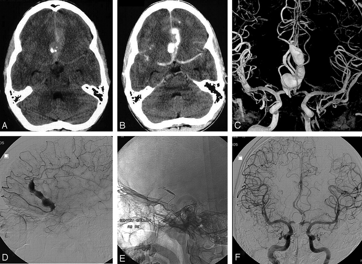
A 16-year-old boy presented with acute severe headache (Fig 1). Apart from drowsiness, no neurologic symptoms were apparent. Native and contrast-enhanced CT scans showed a subarachnoid hemorrhage from a calcified partially thrombosed fusiform aneurysm in the frontal interhemispheric fissure. Bilateral internal carotid artery angiography demonstrated a serpentine aneurysm from the A2 segment of the right anterior cerebral artery. Additional 3D rotational angiography revealed a duplicated right A2 segment with the aneurysm located on the lateral branch. Selective angiography of this segment demonstrated the fusiform nature of the aneurysm, with branches arising both proximal and distal to the dilated segment. Under full heparinization, a nondetachable microballoon (Sentry 15 mm; Boston Scientific, Fremont, Calif) was navigated just proximal to the aneurysm and inflated for test occlusion. For 30 minutes, the patient was repeatedly examined by a senior neurology resident. No clinical symptoms developed, and subsequently the proximal part of the aneurysm was completely occluded with coils (Guglielmi Detachable Coil 18; Boston Scientific). The patient made an uneventful recovery and went back to high school 2 weeks later. A follow-up MR imaging 2 months later showed no infarctions.

  Fig 1.
  • Download figure
  • Open in new tab
  • Download powerpoint
Fig 1.

A 16-year-old boy presented with acute subarachnoid hemorrhage from a giant serpentine anterior cerebral artery aneurysm. A and B, Native (A) and contrast-enhanced (B) CT scans show subarachnoid hemorrhage from a calcified partially thrombosed fusiform aneurysm in the frontal interhemispheric fissure. C, Bilateral internal carotid artery 3D angiography reveals a duplicated right A2 segment with the aneurysm located on the lateral branch. D, Selective angiography of the aneurysm-bearing segment demonstrates branches arising both proximal and distal from the fusiform dilated segment. E, Microballoon for test occlusion. F, Complete exclusion from the circulation after coil occlusion of the proximal part of the lumen. No infarction developed.

Case 2

A 68-year-old woman presented with several episodes of dysphasia that lasted 5–10 minutes during the previous 2 months (On-line Fig 1). CT showed a multilobular hyperattenuated structure in the left Sylvian fissure. Additional MR imaging suggested a partially thrombosed serpentine left middle cerebral artery aneurysm with various stages of clot formation, confirmed at 2D and 3D angiography. A microballoon (HyperForm; ev3, Irvine, Calif) was positioned just proximal to the dilated segment of the middle cerebral artery branch and inflated for test occlusion. No symptoms developed during the 30-minute test occlusion. The next day, with the patient under general anesthesia, the aneurysm lumen was completely occluded with coils (Trufill Orbit; Cordis Neurovascular, Miami Lakes, Fla). Several hours later, she gradually developed slurred speech that recovered after rapid volume expansion. Repeat T2-weighted MR imaging 2 days later showed several new punctuate white matter hyperintensities in the left parietal region. However, during 3 months of follow-up, she has been free of symptoms with no new episodes of dysphasia.

Case 3

A 51-year-old woman presented with a frontal syndrome, which gradually progressed for 2 years (On-line Fig 2). MR imaging demonstrated a 5-cm giant serpentine aneurysm in the frontal midline with surrounding edema. Left internal carotid angiography showed the aneurysm to originate from the left anterior cerebral artery with a tortuous luminal channel. With steroid treatment, her functional status improved. A microballoon (Endeavor; Boston Scientific) was positioned in the proximal involved A2 and inflated to occlude its lumen. No symptoms developed during the 30-minute test occlusion. Then, a microcatheter was advanced into the aneurysm, and the luminal channel was occluded with glue (Histoacryl; Braun, Melsungen, Germany). No infarction in the distal anterior cerebral artery territory developed on follow-up MR imaging. Steroid treatment was discontinued without aggravation of symptoms. Ten months later, the thrombosed aneurysm was surgically partially removed.

Discussion

Giant serpentine aneurysms are extremely rare, with an incidence of <0.1%.3 Pathogenesis is assumed by repeated dissection of an intrinsic abnormal vessel wall with intramural hemorrhages.1 Giant serpentine aneurysms may develop from smaller fusiform aneurysms and represent a spectrum of the same pathologic entity.1,2 Clinical presentation is mostly related to mass effect and sometimes to distal ischemia by distal emboli, occlusion of side branches, or direct flow impairment. Hemorrhage is rare. Most giant serpentine aneurysms are located in the posterior circulation and the middle cerebral artery; other locations, including the anterior cerebral artery as in 2 of our patients, are very uncommon.

Characteristic of serpentine aneurysms is the involvement of a length of blood vessel with separate inflow and outflow sites. To prevent distal ischemia, surgical treatment usually includes a bypass procedure with inherent technical difficulties.1–4 However, distal revascularization is not necessary in patients with adequate leptomeningeal collateral supply. With endovascular techniques, it is now possible to perform a selective temporary balloon test occlusion of the involved vessel in the awake patient. If no neurologic deficit develops, it is safe to occlude the aneurysmal lumen with coils,5 balloons, or glue in an endovascular procedure. All 3 of our patients tolerated simple endovascular occlusion well. In our opinion, this is the therapy of choice. Only in patients who do not tolerate test occlusion may sophisticated bypass procedures be considered.

Footnotes

  • indicates article with supplemental on-line figures.

References

  1. ↵
    Day AL, Gaposchkin CG, Yu CJ, et al. Spontaneous fusiform middle cerebral artery aneurysms: characteristics and a proposed mechanism of formation. J Neurosurg 2003;99:228–40
    PubMed
  2. ↵
    Vishteh AG, Spetzler RF. Evolution of a dolichoectatic aneurysm into a giant serpentine aneurysm during long-term follow-up: case illustration. J Neurosurg 1999;91:346
    PubMed
  3. ↵
    Anson JA, Lawton MT, Spetzler RF. Characteristics and surgical treatment of dolichoectatic and fusiform aneurysms. J Neurosurg 1996;84:185–93
    PubMed
  4. ↵
    Anson JA. Treatment strategies for intracranial fusiform aneurysms. Neurosurg Clin N Am 1998;9:743
    PubMed
  5. ↵
    Coley SC, Hodgson TJ, Jakubowski J. Coil embolization of giant serpentine aneurysms: report of two cases arising from the posterior cerebral artery. Br J Neurosurg 2002;16:43–47
    CrossRefPubMed
  • Received December 12, 2007.
  • Accepted after revision January 10, 2008.
  • Copyright © American Society of Neuroradiology
View Abstract
PreviousNext
Back to top

In this issue

American Journal of Neuroradiology: 29 (7)
American Journal of Neuroradiology
Vol. 29, Issue 7
August 2008
  • Table of Contents
  • Index by author
Advertisement
Print
Download PDF
Email Article

Thank you for your interest in spreading the word on American Journal of Neuroradiology.

NOTE: We only request your email address so that the person you are recommending the page to knows that you wanted them to see it, and that it is not junk mail. We do not capture any email address.

Enter multiple addresses on separate lines or separate them with commas.
Endovascular Treatment of Giant Serpentine Aneurysms
(Your Name) has sent you a message from American Journal of Neuroradiology
(Your Name) thought you would like to see the American Journal of Neuroradiology web site.
CAPTCHA
This question is for testing whether or not you are a human visitor and to prevent automated spam submissions.
Cite this article
W.J. van Rooij, M. Sluzewski, G.N. Beute
Endovascular Treatment of Giant Serpentine Aneurysms
American Journal of Neuroradiology Aug 2008, 29 (7) 1418-1419; DOI: 10.3174/ajnr.A1071

Citation Manager Formats

  • BibTeX
  • Bookends
  • EasyBib
  • EndNote (tagged)
  • EndNote 8 (xml)
  • Medlars
  • Mendeley
  • Papers
  • RefWorks Tagged
  • Ref Manager
  • RIS
  • Zotero
0 Responses
Respond to this article
Share
Bookmark this article
Endovascular Treatment of Giant Serpentine Aneurysms
W.J. van Rooij, M. Sluzewski, G.N. Beute
American Journal of Neuroradiology Aug 2008, 29 (7) 1418-1419; DOI: 10.3174/ajnr.A1071
del.icio.us logo Twitter logo Facebook logo Mendeley logo
  • Tweet Widget
  • Facebook Like
  • Google Plus One
Purchase

Jump to section

  • Article
    • Abstract
    • Case Reports
    • Discussion
    • Footnotes
    • References
  • Figures & Data
  • Supplemental
  • Info & Metrics
  • Responses
  • References
  • PDF

Related Articles

  • PubMed
  • Google Scholar

Cited By...

  • Intracranial Serpentine Aneurysms: Spontaneous Changes of Angiographic Filling Pattern
  • Balloon test occlusion and endosurgical parent artery sacrifice for the evaluation and management of complex intracranial aneurysmal disease
  • Endovascular Treatment of Large and Giant Aneurysms
  • Crossref (27)
  • Google Scholar

This article has been cited by the following articles in journals that are participating in Crossref Cited-by Linking.

  • Endovascular Treatment of Large and Giant Aneurysms
    W.J. van Rooij, M. Sluzewski
    American Journal of Neuroradiology 2009 30 1
  • Current Imaging Assessment and Treatment of Intracranial Aneurysms
    Lotfi Hacein-Bey, James M. Provenzale
    American Journal of Roentgenology 2011 196 1
  • Giant serpentine aneurysms
    Lana D. Christiano, Gaurav Gupta, Charles J. Prestigiacomo, Chirag D. Gandhi
    Neurosurgical Focus 2009 26 5
  • Flow reversal bypass surgery: a treatment option for giant serpentine and dolichoectatic aneurysms—internal maxillary artery bypass with an interposed radial artery graft followed by parent artery occlusion
    Long Wang, Xiang’en Shi, Hai Qian
    Neurosurgical Review 2017 40 2
  • Balloon test occlusion and endosurgical parent artery sacrifice for the evaluation and management of complex intracranial aneurysmal disease
    W S Lesley, R Rangaswamy
    Journal of NeuroInterventional Surgery 2009 1 2
  • Intracranial Serpentine Aneurysms: Spontaneous Changes of Angiographic Filling Pattern
    S.G. Kandemirli, S. Cekirge, I. Oran, I. Saatci, O. Kizilkilic, C. Cinar, C. Islak, N. Kocer
    American Journal of Neuroradiology 2018 39 9
  • Giant Serpentine Aneurysm of the Distal Anterior Cerebral Artery
    Nobuo Senbokuya, Kazuya Kanemaru, Hiroyuki Kinouchi, Toru Horikoshi
    Journal of Stroke and Cerebrovascular Diseases 2012 21 8
  • Dolichoektazja tętnic koła tętniczego Willisa i tętniak wrzecionowaty tętnicy podstawnej mózgu – opis przypadku i przegląd piśmiennictwa
    Bogusława Baran, Olga Kornafel, Maciej Guziński, Marek Sąsiadek
    Polish Journal of Radiology 2012 77 2
  • Intraoperative Functional and Perfusion Monitoring During Surgery for Giant Serpentine Middle Cerebral Artery Aneurysms
    Giacomo Pavesi, Stavros Dimitriadis, Stefano Baroni, Stefano Vallone, Franco Valzania, Giovanni Battista Costella, Alberto Feletti
    World Neurosurgery 2015 84 2
  • Imaging of giant cerebral aneurysms
    E. Tollard, G. Perot, E. Clavier, E. Gerardin
    Neurochirurgie 2015 61 6

More in this TOC Section

  • Optimizing Voxel Size in 3D Rotational Angiography
  • Neuroform Atlas Stent for Intracranial Aneurysms
  • Neurologic Complication in Transradial Angiography
Show more NEUROINTERVENTION

Similar Articles

Advertisement

Indexed Content

  • Current Issue
  • Accepted Manuscripts
  • Article Preview
  • Past Issues
  • Editorials
  • Editor's Choice
  • Fellows' Journal Club
  • Letters to the Editor
  • Video Articles

Cases

  • Case Collection
  • Archive - Case of the Week
  • Archive - Case of the Month
  • Archive - Classic Case

More from AJNR

  • Trainee Corner
  • Imaging Protocols
  • MRI Safety Corner

Multimedia

  • AJNR Podcasts
  • AJNR Scantastics

Resources

  • Turnaround Time
  • Submit a Manuscript
  • Submit a Video Article
  • Submit an eLetter to the Editor/Response
  • Manuscript Submission Guidelines
  • Statistical Tips
  • Fast Publishing of Accepted Manuscripts
  • Graphical Abstract Preparation
  • Imaging Protocol Submission
  • Evidence-Based Medicine Level Guide
  • Publishing Checklists
  • Author Policies
  • Become a Reviewer/Academy of Reviewers
  • News and Updates

About Us

  • About AJNR
  • Editorial Board
  • Editorial Board Alumni
  • Alerts
  • Permissions
  • Not an AJNR Subscriber? Join Now
  • Advertise with Us
  • Librarian Resources
  • Feedback
  • Terms and Conditions
  • AJNR Editorial Board Alumni

American Society of Neuroradiology

  • Not an ASNR Member? Join Now

© 2025 by the American Society of Neuroradiology All rights, including for text and data mining, AI training, and similar technologies, are reserved.
Print ISSN: 0195-6108 Online ISSN: 1936-959X

Powered by HighWire