Advertisement

Main menu

  • Home
  • Content
    • Current Issue
    • Accepted Manuscripts
    • Article Preview
    • Past Issue Archive
    • Video Articles
    • AJNR Case Collection
    • Case of the Week Archive
    • Case of the Month Archive
    • Classic Case Archive
  • Special Collections
    • AJNR Awards
    • Low-Field MRI
    • Alzheimer Disease
    • ASNR Foundation Special Collection
    • Photon-Counting CT
    • View All
  • Multimedia
    • AJNR Podcasts
    • AJNR SCANtastic
    • Trainee Corner
    • MRI Safety Corner
    • Imaging Protocols
  • For Authors
    • Submit a Manuscript
    • Submit a Video Article
    • Submit an eLetter to the Editor/Response
    • Manuscript Submission Guidelines
    • Statistical Tips
    • Fast Publishing of Accepted Manuscripts
    • Graphical Abstract Preparation
    • Imaging Protocol Submission
    • Author Policies
  • About Us
    • About AJNR
    • Editorial Board
    • Editorial Board Alumni
  • More
    • Become a Reviewer/Academy of Reviewers
    • Subscribers
    • Permissions
    • Alerts
    • Feedback
    • Advertisers
    • ASNR Home

User menu

  • Alerts
  • Log in

Search

  • Advanced search
American Journal of Neuroradiology

American Journal of Neuroradiology

ASHNR American Society of Functional Neuroradiology ASHNR American Society of Pediatric Neuroradiology ASSR
  • Alerts
  • Log in

Advanced Search

  • Home
  • Content
    • Current Issue
    • Accepted Manuscripts
    • Article Preview
    • Past Issue Archive
    • Video Articles
    • AJNR Case Collection
    • Case of the Week Archive
    • Case of the Month Archive
    • Classic Case Archive
  • Special Collections
    • AJNR Awards
    • Low-Field MRI
    • Alzheimer Disease
    • ASNR Foundation Special Collection
    • Photon-Counting CT
    • View All
  • Multimedia
    • AJNR Podcasts
    • AJNR SCANtastic
    • Trainee Corner
    • MRI Safety Corner
    • Imaging Protocols
  • For Authors
    • Submit a Manuscript
    • Submit a Video Article
    • Submit an eLetter to the Editor/Response
    • Manuscript Submission Guidelines
    • Statistical Tips
    • Fast Publishing of Accepted Manuscripts
    • Graphical Abstract Preparation
    • Imaging Protocol Submission
    • Author Policies
  • About Us
    • About AJNR
    • Editorial Board
    • Editorial Board Alumni
  • More
    • Become a Reviewer/Academy of Reviewers
    • Subscribers
    • Permissions
    • Alerts
    • Feedback
    • Advertisers
    • ASNR Home
  • Follow AJNR on Twitter
  • Visit AJNR on Facebook
  • Follow AJNR on Instagram
  • Join AJNR on LinkedIn
  • RSS Feeds

AJNR Awards, New Junior Editors, and more. Read the latest AJNR updates

Research ArticleBRAIN

Dynamic 3D-CT Angiography

M. Matsumoto, N. Kodama, Y. Endo, J. Sakuma, Ky. Suzuki, T. Sasaki, K. Murakami, Ke. Suzuki, T. Katakura and F. Shishido
American Journal of Neuroradiology February 2007, 28 (2) 299-304;
M. Matsumoto
  • Find this author on Google Scholar
  • Find this author on PubMed
  • Search for this author on this site
N. Kodama
  • Find this author on Google Scholar
  • Find this author on PubMed
  • Search for this author on this site
Y. Endo
  • Find this author on Google Scholar
  • Find this author on PubMed
  • Search for this author on this site
J. Sakuma
  • Find this author on Google Scholar
  • Find this author on PubMed
  • Search for this author on this site
Ky. Suzuki
  • Find this author on Google Scholar
  • Find this author on PubMed
  • Search for this author on this site
T. Sasaki
  • Find this author on Google Scholar
  • Find this author on PubMed
  • Search for this author on this site
K. Murakami
  • Find this author on Google Scholar
  • Find this author on PubMed
  • Search for this author on this site
Ke. Suzuki
  • Find this author on Google Scholar
  • Find this author on PubMed
  • Search for this author on this site
T. Katakura
  • Find this author on Google Scholar
  • Find this author on PubMed
  • Search for this author on this site
F. Shishido
  • Find this author on Google Scholar
  • Find this author on PubMed
  • Search for this author on this site
  • Article
  • Figures & Data
  • Info & Metrics
  • Responses
  • References
  • PDF
Loading

Article Figures & Data

Figures

  • Tables
  • Fig 1.
    • Download figure
    • Open in new tab
    • Download powerpoint
    Fig 1.

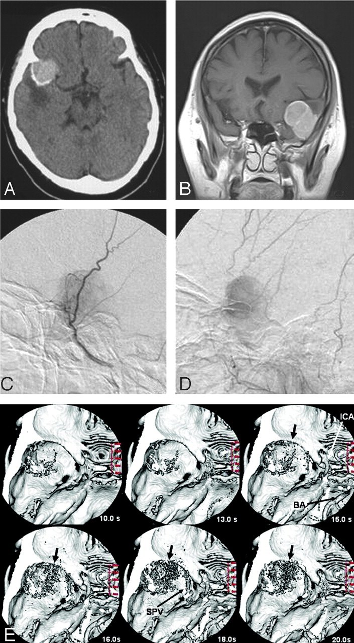
    A 52-year-old woman with a sphenoid ridge meningioma (case 3). A, Plain CT shows a mass with calcification in the left frontotemporal region.

    B, Coronal contrast-enhanced T1-weighted image reveals a homogenous enhanced mass in the left middle fossa.

    C and D, A left external carotid artery angiogram, lateral views of the early (C) and late (D) phase, reveals a tumor stain fed by the middle meningeal artery.

    E, d3D-CTA (superior view). d3D-CT angiographs obtained at 10, 13, 15, 16, 18, and 20 seconds after contrast injection are shown. The tumor stain is first demonstrated in the arterial phase (at 15 seconds). d3D-CTA indicates that the blood supply to the tumor is from the sphenoid ridge. Calcifications within the tumor are shown at 10 seconds. ICA, left internal carotid artery; BA, basilar artery; SPV, sphenoparietal vein.

  • Fig 2.
    • Download figure
    • Open in new tab
    • Download powerpoint
    Fig 2.

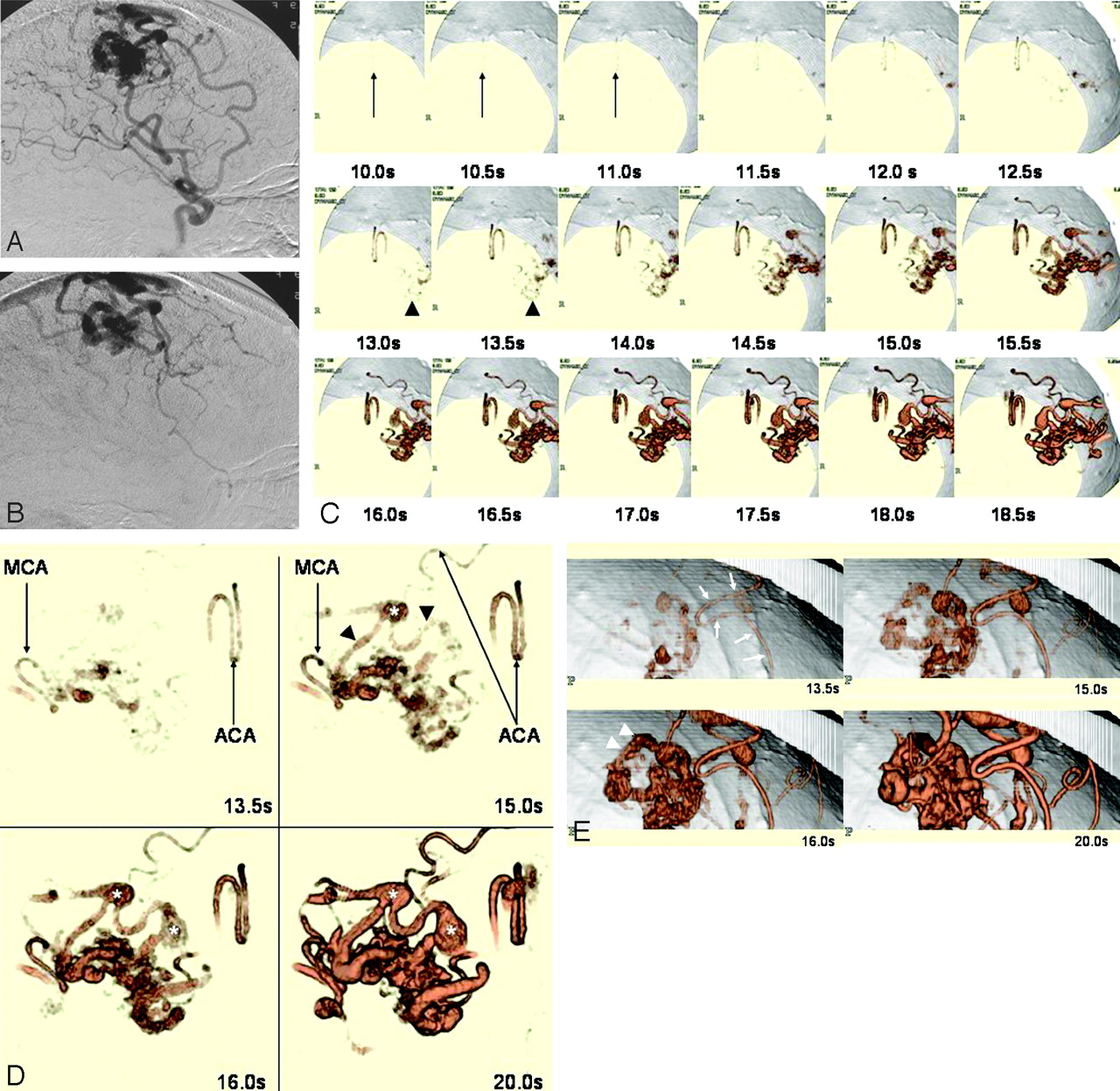
    A 61-year-old woman with an arteriovenous malformation (AVM) in the left frontal lobe (case 8). A and B, Left internal carotid artery angiogram, lateral views of the arterial (A) and venous (B) phase, reveals an AVM fed by branches of the anterior and middle cerebral arteries; drainage is into the frontal ascending vein.

    C, d3D-CTA (inferior view) obtained at 0.5-second intervals after contrast injection. The anterior cerebral artery (feeder, arrow) is demonstrated at 10 seconds; the frontal ascending vein (drainer, arrowhead) is shown in the early arterial phase (at 13 and 13.5 seconds). The d3D-CTA scans demonstrate the hemodynamics of the AVM.

    D, d3D-CTA (superior view) obtained at 13.5, 15, 16, and 20 seconds after contrast injection. The feeding arteries, nidus, and drainer are visualized in the arterial phase (at 13.5 and 15 seconds). MCA, branch of the middle cerebral artery; ACA, branches of the anterior cerebral artery; *, varices.

    E, d3D-CTA (left lateral view) obtained at 13.5, 15, 16, and 20 seconds after contrast injection. In the early arterial phase (at 13.5 seconds) the feeder, a branch of the anterior cerebral artery (arrow), and the nidus are demonstrated. The arterial phase (at 16 seconds) revealed a drainer (arrowheads), indicating that there is an arteriovenous shunt. d3D-CTA provided information on the hemodynamics and the 3D vascular structures of the AVM.

  • Fig 3.
    • Download figure
    • Open in new tab
    • Download powerpoint
    Fig 3.

    A 75-year-old woman with occlusion of the right cervical internal carotid artery (case 12). A, Cervical 3D-CTA showing occlusion of the right internal carotid artery (arrow). Right lateral (left) and left lateral (right) views of the right cervical vessels are shown. CCA, common carotid artery.

    B, d3D-CTA (anterosuperior view). 3D-CTA scans were obtained at 12, 12.5, 13, 13.5, 14, and 14.5 seconds after contrast injection. The right middle cerebral artery (MCA, arrowheads) is visualized later (at 13 or 13.5 seconds) than the left MCA (arrows), which appeared at 12.5 seconds. d3D-CTA demonstrates the right MCA with a 0.5- and 1-second filling delay compared with the left MCA. The smaller caliber of the right compared with the left MCA indicates that the blood flow volume in the right MCA is smaller than in the left MCA. Cal, calcification.

    C, Functional maps of cerebral blood flow (CBF), cerebral blood volume (CBV), and mean transit time (MTT) produced from d3D-CTA data. The maps showed no significant difference between the 2 hemispheres.

Tables

  • Figures
    • View popup
    Table 1:

    Clinical information on 12 patients with brain lesions

    Case No.Age/SexDiagnosisContrast Medium Injection Speed (mL/s)/Total Volume (mL)Pulse Rate/MinArrival Time (s)
    170/FTentorial meningioma6/307712.0
    269/FFalx meningioma6/308211.5
    352/FSphenoid ridge meningioma6/306413.0
    450/FConvexity meningioma6/307713.0
    557/MTrigeminal schwannoma7/356214.0
    654/MTrigeminal schwannoma6/305715.5
    770/MMetastatic brain tumor6/306613.5
    861/FFrontal AVM6/309010.5
    954/FOccipital AVM6/306514.0
    1049/FTemporal AVM6/307312.0
    1117/FParietal AVM6/307812.5
    1275/FCervical carotid artery occlusion6/306712.5
    • Note:—AVM indicates arteriovenous malformation; pulse rate, pulse rate just before scanning; arrival time, arrival of injected contrast medium within the scan range.

PreviousNext
Back to top

In this issue

American Journal of Neuroradiology: 28 (2)
American Journal of Neuroradiology
Vol. 28, Issue 2
February 2007
  • Table of Contents
  • Index by author
Advertisement
Print
Download PDF
Email Article

Thank you for your interest in spreading the word on American Journal of Neuroradiology.

NOTE: We only request your email address so that the person you are recommending the page to knows that you wanted them to see it, and that it is not junk mail. We do not capture any email address.

Enter multiple addresses on separate lines or separate them with commas.
Dynamic 3D-CT Angiography
(Your Name) has sent you a message from American Journal of Neuroradiology
(Your Name) thought you would like to see the American Journal of Neuroradiology web site.
CAPTCHA
This question is for testing whether or not you are a human visitor and to prevent automated spam submissions.
Cite this article
M. Matsumoto, N. Kodama, Y. Endo, J. Sakuma, Ky. Suzuki, T. Sasaki, K. Murakami, Ke. Suzuki, T. Katakura, F. Shishido
Dynamic 3D-CT Angiography
American Journal of Neuroradiology Feb 2007, 28 (2) 299-304;

Citation Manager Formats

  • BibTeX
  • Bookends
  • EasyBib
  • EndNote (tagged)
  • EndNote 8 (xml)
  • Medlars
  • Mendeley
  • Papers
  • RefWorks Tagged
  • Ref Manager
  • RIS
  • Zotero
0 Responses
Respond to this article
Share
Bookmark this article
Dynamic 3D-CT Angiography
M. Matsumoto, N. Kodama, Y. Endo, J. Sakuma, Ky. Suzuki, T. Sasaki, K. Murakami, Ke. Suzuki, T. Katakura, F. Shishido
American Journal of Neuroradiology Feb 2007, 28 (2) 299-304;
del.icio.us logo Twitter logo Facebook logo Mendeley logo
  • Tweet Widget
  • Facebook Like
  • Google Plus One

Jump to section

  • Article
    • Abstract
    • Materials and Methods
    • Results
    • Discussion
    • Conclusion
    • References
  • Figures & Data
  • Info & Metrics
  • Responses
  • References
  • PDF

Related Articles

  • No related articles found.
  • PubMed
  • Google Scholar

Cited By...

  • Multi-Detector Row CT Angiography with Direct Intra-Arterial Contrast Injection for the Evaluation of Neurovascular Disease: Technique, Applications, and Initial Experience
  • Crossref
  • Google Scholar

This article has not yet been cited by articles in journals that are participating in Crossref Cited-by Linking.

More in this TOC Section

  • Statin Therapy Does Not Affect the Radiographic and Clinical Profile of Patients with TIA and Minor Stroke
  • Usefulness of Quantitative Susceptibility Mapping for the Diagnosis of Parkinson Disease
  • White Matter Alterations in the Brains of Patients with Active, Remitted, and Cured Cushing Syndrome: A DTI Study
Show more BRAIN

Similar Articles

Advertisement

Indexed Content

  • Current Issue
  • Accepted Manuscripts
  • Article Preview
  • Past Issues
  • Editorials
  • Editor's Choice
  • Fellows' Journal Club
  • Letters to the Editor
  • Video Articles

Cases

  • Case Collection
  • Archive - Case of the Week
  • Archive - Case of the Month
  • Archive - Classic Case

More from AJNR

  • Trainee Corner
  • Imaging Protocols
  • MRI Safety Corner
  • Book Reviews

Multimedia

  • AJNR Podcasts
  • AJNR Scantastics

Resources

  • Turnaround Time
  • Submit a Manuscript
  • Submit a Video Article
  • Submit an eLetter to the Editor/Response
  • Manuscript Submission Guidelines
  • Statistical Tips
  • Fast Publishing of Accepted Manuscripts
  • Graphical Abstract Preparation
  • Imaging Protocol Submission
  • Evidence-Based Medicine Level Guide
  • Publishing Checklists
  • Author Policies
  • Become a Reviewer/Academy of Reviewers
  • News and Updates

About Us

  • About AJNR
  • Editorial Board
  • Editorial Board Alumni
  • Alerts
  • Permissions
  • Not an AJNR Subscriber? Join Now
  • Advertise with Us
  • Librarian Resources
  • Feedback
  • Terms and Conditions
  • AJNR Editorial Board Alumni

American Society of Neuroradiology

  • Not an ASNR Member? Join Now

© 2025 by the American Society of Neuroradiology All rights, including for text and data mining, AI training, and similar technologies, are reserved.
Print ISSN: 0195-6108 Online ISSN: 1936-959X

Powered by HighWire