Advertisement

Main menu

  • Home
  • Content
    • Current Issue
    • Accepted Manuscripts
    • Article Preview
    • Past Issue Archive
    • Video Articles
    • AJNR Case Collection
    • Case of the Week Archive
    • Case of the Month Archive
    • Classic Case Archive
  • Special Collections
    • AJNR Awards
    • Low-Field MRI
    • Alzheimer Disease
    • ASNR Foundation Special Collection
    • Photon-Counting CT
    • View All
  • Multimedia
    • AJNR Podcasts
    • AJNR SCANtastic
    • Trainee Corner
    • MRI Safety Corner
    • Imaging Protocols
  • For Authors
    • Submit a Manuscript
    • Submit a Video Article
    • Submit an eLetter to the Editor/Response
    • Manuscript Submission Guidelines
    • Statistical Tips
    • Fast Publishing of Accepted Manuscripts
    • Graphical Abstract Preparation
    • Imaging Protocol Submission
    • Author Policies
  • About Us
    • About AJNR
    • Editorial Board
    • Editorial Board Alumni
  • More
    • Become a Reviewer/Academy of Reviewers
    • Subscribers
    • Permissions
    • Alerts
    • Feedback
    • Advertisers
    • ASNR Home

User menu

  • Alerts
  • Log in

Search

  • Advanced search
American Journal of Neuroradiology

American Journal of Neuroradiology

ASHNR American Society of Functional Neuroradiology ASHNR American Society of Pediatric Neuroradiology ASSR
  • Alerts
  • Log in

Advanced Search

  • Home
  • Content
    • Current Issue
    • Accepted Manuscripts
    • Article Preview
    • Past Issue Archive
    • Video Articles
    • AJNR Case Collection
    • Case of the Week Archive
    • Case of the Month Archive
    • Classic Case Archive
  • Special Collections
    • AJNR Awards
    • Low-Field MRI
    • Alzheimer Disease
    • ASNR Foundation Special Collection
    • Photon-Counting CT
    • View All
  • Multimedia
    • AJNR Podcasts
    • AJNR SCANtastic
    • Trainee Corner
    • MRI Safety Corner
    • Imaging Protocols
  • For Authors
    • Submit a Manuscript
    • Submit a Video Article
    • Submit an eLetter to the Editor/Response
    • Manuscript Submission Guidelines
    • Statistical Tips
    • Fast Publishing of Accepted Manuscripts
    • Graphical Abstract Preparation
    • Imaging Protocol Submission
    • Author Policies
  • About Us
    • About AJNR
    • Editorial Board
    • Editorial Board Alumni
  • More
    • Become a Reviewer/Academy of Reviewers
    • Subscribers
    • Permissions
    • Alerts
    • Feedback
    • Advertisers
    • ASNR Home
  • Follow AJNR on Twitter
  • Visit AJNR on Facebook
  • Follow AJNR on Instagram
  • Join AJNR on LinkedIn
  • RSS Feeds

AJNR Awards, New Junior Editors, and more. Read the latest AJNR updates

OtherBRAIN

Subcortical Calcification on CT in Dural Arteriovenous Fistula with Cortical Venous Reflux

T. Metoki, S. Mugikura, S. Higano, M. Ezura, Y. Matsumoto, K. Hirayama and S. Takahashi
American Journal of Neuroradiology May 2006, 27 (5) 1076-1078;
T. Metoki
  • Find this author on Google Scholar
  • Find this author on PubMed
  • Search for this author on this site
S. Mugikura
  • Find this author on Google Scholar
  • Find this author on PubMed
  • Search for this author on this site
S. Higano
  • Find this author on Google Scholar
  • Find this author on PubMed
  • Search for this author on this site
M. Ezura
  • Find this author on Google Scholar
  • Find this author on PubMed
  • Search for this author on this site
Y. Matsumoto
  • Find this author on Google Scholar
  • Find this author on PubMed
  • Search for this author on this site
K. Hirayama
  • Find this author on Google Scholar
  • Find this author on PubMed
  • Search for this author on this site
S. Takahashi
  • Find this author on Google Scholar
  • Find this author on PubMed
  • Search for this author on this site
  • Article
  • Figures & Data
  • Info & Metrics
  • Responses
  • References
  • PDF
Loading

Article Figures & Data

Figures

  • Fig 1.
    • Download figure
    • Open in new tab
    • Download powerpoint
    Fig 1.

    Case 1.

    A, Precontrast CT scan shows curvilinear calcification (arrowhead) in the cortico-medullary junction at the inferior part of the left temporal lobe.

    B, Postcontrast T1-weighted image shows multiple foci of curvilinear enhancement along the gyri, indicating dilated venous channels due to reflux (arrowheads).

    C, Lateral view of left external carotid angiogram shows dAVF at left transverse sinus fed by the middle meningeal artery (arrow) and cortical venous reflux (arrowhead).

    D, SPECT shows markedly reduced blood flow in the left temporal lobe where the cortical venous reflux was found.
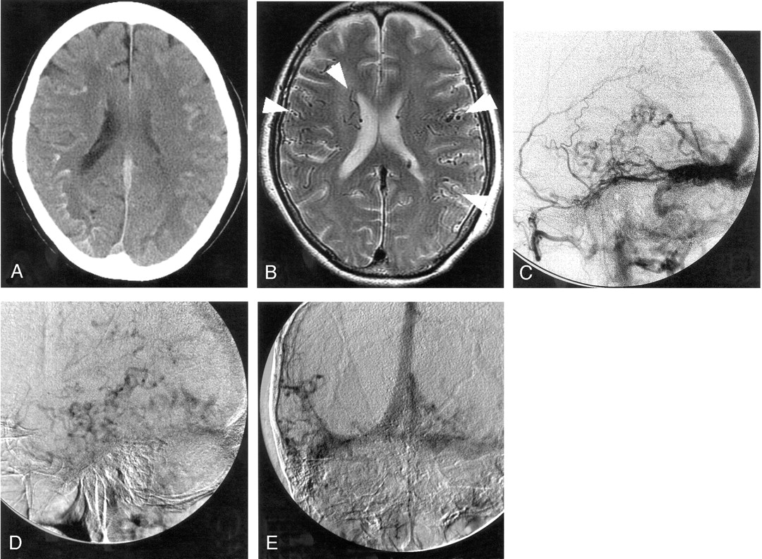
  • Fig 2.
    • Download figure
    • Open in new tab
    • Download powerpoint
    Fig 2.

    Case 2.

    A, CT scan shows curvilinear calcification in the cortico-medullary junction at the bottom of cerebral sulci bilaterally but more predominantly in right cerebral hemisphere.

    B, T2-weighted MR image shows prominent cortical and subependymal veins (arrowheads), which possibly suggests an arteriovenous shunt with cortical venous reflux as well as hyperintensity in deep white matter that was more apparent on the right.

    C–E, Serial right external carotid angiograms (C and D, lateral view; E, frontal view) show enlargement of the middle meningeal artery with early filling of the transverse sinus and retrograde filling of the straight and superior sagittal sinuses. Cortical venous reflux is also evident in the later phase.

  • Fig 3.
    • Download figure
    • Open in new tab
    • Download powerpoint
    Fig 3.

    Case 3.

    A, CT scans shows diffuse atrophy and subtle calcification in the basal ganglia and subcortical region at the bottom of cerebral gyri bilaterally. The subcortical calcification was curvilinear and predominant on the right side.

    B–E, Right external carotid angiograms (B–D, lateral projection; E, frontal projection) show dAVF fed by the enlarged middle meningeal (arrow) and ascending pharyngeal arteries with cortical venous reflux (arrowheads).

    F, Repeated CT scans after 1 year reveal a more advanced degree of diffuse atrophy and denser subcortical calcification.

PreviousNext
Back to top

In this issue

American Journal of Neuroradiology: 27 (5)
American Journal of Neuroradiology
Vol. 27, Issue 5
May 2006
  • Table of Contents
  • Index by author
Advertisement
Print
Download PDF
Email Article

Thank you for your interest in spreading the word on American Journal of Neuroradiology.

NOTE: We only request your email address so that the person you are recommending the page to knows that you wanted them to see it, and that it is not junk mail. We do not capture any email address.

Enter multiple addresses on separate lines or separate them with commas.
Subcortical Calcification on CT in Dural Arteriovenous Fistula with Cortical Venous Reflux
(Your Name) has sent you a message from American Journal of Neuroradiology
(Your Name) thought you would like to see the American Journal of Neuroradiology web site.
CAPTCHA
This question is for testing whether or not you are a human visitor and to prevent automated spam submissions.
Cite this article
T. Metoki, S. Mugikura, S. Higano, M. Ezura, Y. Matsumoto, K. Hirayama, S. Takahashi
Subcortical Calcification on CT in Dural Arteriovenous Fistula with Cortical Venous Reflux
American Journal of Neuroradiology May 2006, 27 (5) 1076-1078;

Citation Manager Formats

  • BibTeX
  • Bookends
  • EasyBib
  • EndNote (tagged)
  • EndNote 8 (xml)
  • Medlars
  • Mendeley
  • Papers
  • RefWorks Tagged
  • Ref Manager
  • RIS
  • Zotero
0 Responses
Respond to this article
Share
Bookmark this article
Subcortical Calcification on CT in Dural Arteriovenous Fistula with Cortical Venous Reflux
T. Metoki, S. Mugikura, S. Higano, M. Ezura, Y. Matsumoto, K. Hirayama, S. Takahashi
American Journal of Neuroradiology May 2006, 27 (5) 1076-1078;
del.icio.us logo Twitter logo Facebook logo Mendeley logo
  • Tweet Widget
  • Facebook Like
  • Google Plus One

Jump to section

  • Article
    • Abstract
    • Case 1
    • Case 2
    • Case 3
    • Discussion
    • References
  • Figures & Data
  • Info & Metrics
  • Responses
  • References
  • PDF

Related Articles

  • No related articles found.
  • PubMed
  • Google Scholar

Cited By...

  • Simultaneous Arteriovenous Shunting and Venous Congestion Identification in Dural Arteriovenous Fistulas Using Susceptibility-Weighted Imaging: Initial Experience
  • Adult-onset leukoencephalopathy with calcifications associated with primary antiphospholipid syndrome
  • Crossref
  • Google Scholar

This article has not yet been cited by articles in journals that are participating in Crossref Cited-by Linking.

More in this TOC Section

  • Predictors of Reperfusion in Patients with Acute Ischemic Stroke
  • Enhanced Axonal Metabolism during Early Natalizumab Treatment in Relapsing-Remitting Multiple Sclerosis
  • Progression of Microstructural Damage in Spinocerebellar Ataxia Type 2: A Longitudinal DTI Study
Show more BRAIN

Similar Articles

Advertisement

Indexed Content

  • Current Issue
  • Accepted Manuscripts
  • Article Preview
  • Past Issues
  • Editorials
  • Editor's Choice
  • Fellows' Journal Club
  • Letters to the Editor
  • Video Articles

Cases

  • Case Collection
  • Archive - Case of the Week
  • Archive - Case of the Month
  • Archive - Classic Case

More from AJNR

  • Trainee Corner
  • Imaging Protocols
  • MRI Safety Corner
  • Book Reviews

Multimedia

  • AJNR Podcasts
  • AJNR Scantastics

Resources

  • Turnaround Time
  • Submit a Manuscript
  • Submit a Video Article
  • Submit an eLetter to the Editor/Response
  • Manuscript Submission Guidelines
  • Statistical Tips
  • Fast Publishing of Accepted Manuscripts
  • Graphical Abstract Preparation
  • Imaging Protocol Submission
  • Evidence-Based Medicine Level Guide
  • Publishing Checklists
  • Author Policies
  • Become a Reviewer/Academy of Reviewers
  • News and Updates

About Us

  • About AJNR
  • Editorial Board
  • Editorial Board Alumni
  • Alerts
  • Permissions
  • Not an AJNR Subscriber? Join Now
  • Advertise with Us
  • Librarian Resources
  • Feedback
  • Terms and Conditions
  • AJNR Editorial Board Alumni

American Society of Neuroradiology

  • Not an ASNR Member? Join Now

© 2025 by the American Society of Neuroradiology All rights, including for text and data mining, AI training, and similar technologies, are reserved.
Print ISSN: 0195-6108 Online ISSN: 1936-959X

Powered by HighWire