Advertisement

Main menu

  • Home
  • Content
    • Current Issue
    • Accepted Manuscripts
    • Article Preview
    • Past Issue Archive
    • Video Articles
    • AJNR Case Collection
    • Case of the Week Archive
    • Case of the Month Archive
    • Classic Case Archive
  • Special Collections
    • AJNR Awards
    • Low-Field MRI
    • Alzheimer Disease
    • ASNR Foundation Special Collection
    • Photon-Counting CT
    • View All
  • Multimedia
    • AJNR Podcasts
    • AJNR SCANtastic
    • Trainee Corner
    • MRI Safety Corner
    • Imaging Protocols
  • For Authors
    • Submit a Manuscript
    • Submit a Video Article
    • Submit an eLetter to the Editor/Response
    • Manuscript Submission Guidelines
    • Statistical Tips
    • Fast Publishing of Accepted Manuscripts
    • Graphical Abstract Preparation
    • Imaging Protocol Submission
    • Author Policies
  • About Us
    • About AJNR
    • Editorial Board
    • Editorial Board Alumni
  • More
    • Become a Reviewer/Academy of Reviewers
    • Subscribers
    • Permissions
    • Alerts
    • Feedback
    • Advertisers
    • ASNR Home

User menu

  • Alerts
  • Log in

Search

  • Advanced search
American Journal of Neuroradiology

American Journal of Neuroradiology

ASHNR American Society of Functional Neuroradiology ASHNR American Society of Pediatric Neuroradiology ASSR
  • Alerts
  • Log in

Advanced Search

  • Home
  • Content
    • Current Issue
    • Accepted Manuscripts
    • Article Preview
    • Past Issue Archive
    • Video Articles
    • AJNR Case Collection
    • Case of the Week Archive
    • Case of the Month Archive
    • Classic Case Archive
  • Special Collections
    • AJNR Awards
    • Low-Field MRI
    • Alzheimer Disease
    • ASNR Foundation Special Collection
    • Photon-Counting CT
    • View All
  • Multimedia
    • AJNR Podcasts
    • AJNR SCANtastic
    • Trainee Corner
    • MRI Safety Corner
    • Imaging Protocols
  • For Authors
    • Submit a Manuscript
    • Submit a Video Article
    • Submit an eLetter to the Editor/Response
    • Manuscript Submission Guidelines
    • Statistical Tips
    • Fast Publishing of Accepted Manuscripts
    • Graphical Abstract Preparation
    • Imaging Protocol Submission
    • Author Policies
  • About Us
    • About AJNR
    • Editorial Board
    • Editorial Board Alumni
  • More
    • Become a Reviewer/Academy of Reviewers
    • Subscribers
    • Permissions
    • Alerts
    • Feedback
    • Advertisers
    • ASNR Home
  • Follow AJNR on Twitter
  • Visit AJNR on Facebook
  • Follow AJNR on Instagram
  • Join AJNR on LinkedIn
  • RSS Feeds

AJNR Awards, New Junior Editors, and more. Read the latest AJNR updates

OtherBRAIN

MR Findings of Decerebrate Rigidity with Preservation of Consciousness

C.-D. Kao, W.-Y. Guo, J.-T. Chen, Z.-A. Wu and K.-K. Liao
American Journal of Neuroradiology May 2006, 27 (5) 1074-1075;
C.-D. Kao
  • Find this author on Google Scholar
  • Find this author on PubMed
  • Search for this author on this site
W.-Y. Guo
  • Find this author on Google Scholar
  • Find this author on PubMed
  • Search for this author on this site
J.-T. Chen
  • Find this author on Google Scholar
  • Find this author on PubMed
  • Search for this author on this site
Z.-A. Wu
  • Find this author on Google Scholar
  • Find this author on PubMed
  • Search for this author on this site
K.-K. Liao
  • Find this author on Google Scholar
  • Find this author on PubMed
  • Search for this author on this site
  • Article
  • Figures & Data
  • Info & Metrics
  • Responses
  • References
  • PDF
Loading

Abstract

SUMMARY: We describe a case of decerebrate rigidity, with preservation of consciousness, caused by a discrete pontine tegmentum lesion identified on MR imaging. Lesions within a certain brain stem region are responsible for decerebrate rigidity in animal studies, but there has been a lack of MR imaging evidence in humans. This report also implies that a discrete lesion was responsible for the decerebrate rigidity, while consciousness was preserved.

The term “decerebrate rigidity” (DR) was first used in 1898 by Sherrington to describe posture after an intercollicular transection,1—ie, the lesion between the red nuclei and the vestibular nuclei found in animal studies. Such lesions result in a loss of flexor modification normally supplied by the rubrospinal tracts.2 The extensor reflexes are then exaggerated, leading to a rigid extension of the limbs accompanied by hyper-reflexia and opisthotonus. Although DR is familiar to most neurologists, it is usually associated with consciousness disturbance and severe motor dysfunction.3 So far, there has been no evidence from human studies to prove a discrete lesion at the same level as in the animal findings of Sherrington,1 along with preserved consciousness with brain stem lesions found on MR imaging. We report the case of a DR patient with a preservation of consciousness.

Case Report

A 48-year-old woman was admitted to our hospital because of a sudden change of consciousness. Beside 2 small low-signal-intensity densities in the basal ganglia, the brain CT showed a rostral pontine hemorrhage with a fourth ventricle extension (Fig 1A). She had mild hypertension, but without a history of stroke. We believed that hemorrhage of the brain stem was the major cause of her loss of consciousness and another 2 small lesions were old, silent strokes. The Glasgow Coma Scale score on admission was 4; the best motor response was 2 (DR posture); the best verbal response was 1 (no response); and the eye-opening response was 1 (no response). Neurologic examination revealed a normal light reflex, an absence of right-eye corneal reflex, and an oculocephalic reflex. A mechanical ventilator was used to support her respiration. Extension of her 4 limbs with internal rotation of bilateral arms and plantar flexion, clenching of the jaws, and opisthotonus had been noted since admission, and these findings were amplified by pain or pressure stimulation on her limbs. She regained consciousness 5 days after the stroke onset and was able to follow simple instructions (such as opening and closing her eyes). Limb movement was limited, and she could only move the right upper limb slightly. Motor-evoked potentials to transcranial magnetic stimulation 1 week after she regained consciousness showed preserved corticospinal tract responses, though the onset latencies were delayed and the peak amplitudes were decreased.

  Fig 1.
  • Download figure
  • Open in new tab
  • Download powerpoint
Fig 1.

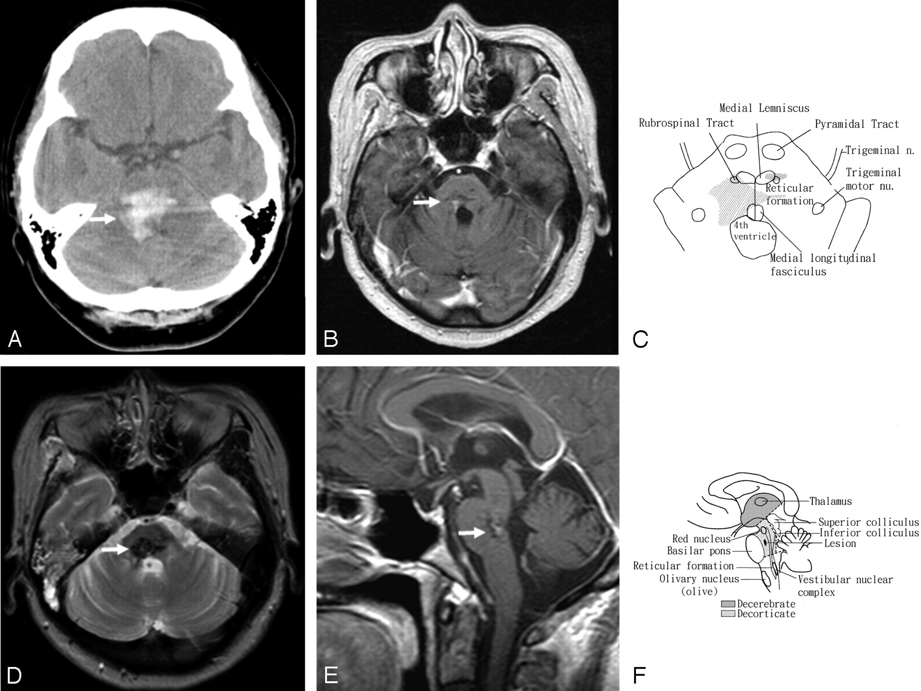
A, Noncontrast CT of a 48-year-old woman who presented with DR and preservation of consciousness shows a right rostral pontine tegmental hemorrhage (arrow) with an extension into the fourth ventricle.

B, T1-weighted axial contrast MR image 3 months after the stroke shows a sequela of the prior insult in the right pontine tegmentum (arrow).

C, Schematic drawing of the brain stem illustrates the lesion and its surrounding structures in an axial view (compared with panel B).

D, T2-weighted axial MR image shows the sequela of old insult in the right pontine tegmentum (arrow).

E, T1-weighted sagittal contrast MR image shows the abnormality in the right rostral pontine tegmentum (arrow).

F, Schematic drawing illustrates the sequela lesion and its surrounding structures in a sagittal view (compared with panel E).

Three months later, there were residual neurologic deficits with right abducens palsy, right facial and crossed left hemibody sensory impairment, poor hand dexterities, and truncal unsteadiness. She was able to follow commands to execute simple tasks, such as moving her shoulders and flexing and extending her wrists or elbows. She could not perform some tasks, such as by using her hands to write or to eat with chopsticks, a fork, or a spoon. She could not maintain a steady sitting position without support or maintain an upright posture even with support. Because the daily record of blood pressure did not show any evidence of hypertension, she did not receive antihypertensive medications after discharge. MR imaging of the brain was performed 3 months after onset. It revealed a discrete sequela of old insult in right pontine tegmentum (Fig 1B, -D, -E ) and a tiny low-signal-intensity abnormality on both T1-weighted and T2-weighted images in the left thalamus. There was no evidence of any other vascular malformation. The muscle response to transcranial magnetic stimulation became larger along with improvement of her muscle strength, though her muscle tone was hypotonic. She remained in flaccid paralysis and did not show significant improvement in motor function on the series of 6-month and 1-year follow-up examinations.

Discussion

DR shows a typical pattern of classic gamma rigidity with the arms in extension, adduction, and hyperpronation and with the feet in extension and plantar flexion. A state of hypertonus in the postural muscles occurred when the brain stems of cats were transected at the intercollicular level.1 The term “decerebrate rigidity” was applied to this condition because it also occurred after removal of the cerebral hemispheres. A series of animal studies revealed that loss of integrity of the reticular, cerebellar, vestibular, and cortical descending neural systems was responsible for the development of DR.4 The rostral pontine tegmental lesion between the red nuclei and vestibular nuclei disrupted the rubrospinal effect on the facilitation of flexor movements in the contralateral upper limbs2 and exaggerated the extensor effect by the removal of the central (cortical) inhibition on pontine reticular formation (medial reticulospinal tract) and Dieter’s nucleus (lateral vestibulospinal tract), the 2 centers of an excitatory influence on extensor muscles5 (Fig 1C, -F).

Imaging studies have been reported in DR patients with preserved consciousness because of ischemic hemispheric damage without a brain stem lesion.6, 7 In Damasceno’s study,7 DR was noted only in the upper limbs, differing from our patient, who had involvement of the 4 limbs, as in Sherrington’s report.1 The case of Damasceno provided another mechanism that a conscious patient with DR might result from a supratentorial lesion affecting the corticoreticular neurons instead of the brain stem involvement of the critical structures mentioned above.7 In contrast with their cases,6, 7 lesions of brain stem were identified by pathologic studies8 and MR imaging in our case. These findings have shown that the mechanism of DR was mainly due to a lesion involving vestibulospinal and rubrospinal pathways and did not arise from the destruction of the corticospinal pathway, as found in animal studies.

References

  1. ↵
    Sherrington CS. Decerebrate rigidity and reflex coordination of movements. J Physiol 1898;22:319–22
  2. ↵
    Jeneskog T, Johansson H. The rubro-bulbospinal path: a descending system known to influence dynamic fusimotor neurons and its interaction with distal cutaneous afferents in the control of flexor reflex afferent pathways. Exp Brain Res 1977;27:161–79
    PubMed
  3. ↵
    Bricolo A, Turazzi S, Alexandre A, et al. Decerebrate rigidity in acute head injury. J Neurosurg 1977;47:680–89
    PubMed
  4. ↵
    Davis RA, Davis L. Decerebrate rigidity in animals. Neurosurg 1981;9:79–89
  5. ↵
    Paul AY, Paul HY. Basic clinical neuroanatomy. Baltimore: Williams & Wilkins;1997 :76–77
  6. ↵
    Kawano H, Suga T, Terasaki T, et al. Decerebrate rigidity after bilateral carotid arteries occlusion. Rinsho Shinkeigaku 2004;44:545–48
    PubMed
  7. ↵
    Damasceno BP. Decerebrate rigidity with preserved cognition and gait: a possible role of anoxic-ischemic brain damage. Int J Neurosci 1991;58:283–87
    PubMed
  8. ↵
    Halsey JH, Downie AW. Decerebrate rigidity with preservation of consciousness. J Neurol Neurosurg Psychiatry 1966;29:350–55
    FREE Full Text
  • Received April 4, 2005.
  • Accepted after revision April 28, 2005.
  • Copyright © American Society of Neuroradiology
View Abstract
PreviousNext
Back to top

In this issue

American Journal of Neuroradiology: 27 (5)
American Journal of Neuroradiology
Vol. 27, Issue 5
May 2006
  • Table of Contents
  • Index by author
Advertisement
Print
Download PDF
Email Article

Thank you for your interest in spreading the word on American Journal of Neuroradiology.

NOTE: We only request your email address so that the person you are recommending the page to knows that you wanted them to see it, and that it is not junk mail. We do not capture any email address.

Enter multiple addresses on separate lines or separate them with commas.
MR Findings of Decerebrate Rigidity with Preservation of Consciousness
(Your Name) has sent you a message from American Journal of Neuroradiology
(Your Name) thought you would like to see the American Journal of Neuroradiology web site.
CAPTCHA
This question is for testing whether or not you are a human visitor and to prevent automated spam submissions.
Cite this article
C.-D. Kao, W.-Y. Guo, J.-T. Chen, Z.-A. Wu, K.-K. Liao
MR Findings of Decerebrate Rigidity with Preservation of Consciousness
American Journal of Neuroradiology May 2006, 27 (5) 1074-1075;

Citation Manager Formats

  • BibTeX
  • Bookends
  • EasyBib
  • EndNote (tagged)
  • EndNote 8 (xml)
  • Medlars
  • Mendeley
  • Papers
  • RefWorks Tagged
  • Ref Manager
  • RIS
  • Zotero
0 Responses
Respond to this article
Share
Bookmark this article
MR Findings of Decerebrate Rigidity with Preservation of Consciousness
C.-D. Kao, W.-Y. Guo, J.-T. Chen, Z.-A. Wu, K.-K. Liao
American Journal of Neuroradiology May 2006, 27 (5) 1074-1075;
del.icio.us logo Twitter logo Facebook logo Mendeley logo
  • Tweet Widget
  • Facebook Like
  • Google Plus One

Jump to section

  • Article
    • Abstract
    • Case Report
    • Discussion
    • References
  • Figures & Data
  • Info & Metrics
  • Responses
  • References
  • PDF

Related Articles

  • No related articles found.
  • PubMed
  • Google Scholar

Cited By...

  • No citing articles found.
  • Crossref
  • Google Scholar

This article has not yet been cited by articles in journals that are participating in Crossref Cited-by Linking.

More in this TOC Section

  • Evaluating the Effects of White Matter Multiple Sclerosis Lesions on the Volume Estimation of 6 Brain Tissue Segmentation Methods
  • Quiet PROPELLER MRI Techniques Match the Quality of Conventional PROPELLER Brain Imaging Techniques
  • Predictors of Reperfusion in Patients with Acute Ischemic Stroke
Show more BRAIN

Similar Articles

Advertisement

Indexed Content

  • Current Issue
  • Accepted Manuscripts
  • Article Preview
  • Past Issues
  • Editorials
  • Editor's Choice
  • Fellows' Journal Club
  • Letters to the Editor
  • Video Articles

Cases

  • Case Collection
  • Archive - Case of the Week
  • Archive - Case of the Month
  • Archive - Classic Case

More from AJNR

  • Trainee Corner
  • Imaging Protocols
  • MRI Safety Corner
  • Book Reviews

Multimedia

  • AJNR Podcasts
  • AJNR Scantastics

Resources

  • Turnaround Time
  • Submit a Manuscript
  • Submit a Video Article
  • Submit an eLetter to the Editor/Response
  • Manuscript Submission Guidelines
  • Statistical Tips
  • Fast Publishing of Accepted Manuscripts
  • Graphical Abstract Preparation
  • Imaging Protocol Submission
  • Evidence-Based Medicine Level Guide
  • Publishing Checklists
  • Author Policies
  • Become a Reviewer/Academy of Reviewers
  • News and Updates

About Us

  • About AJNR
  • Editorial Board
  • Editorial Board Alumni
  • Alerts
  • Permissions
  • Not an AJNR Subscriber? Join Now
  • Advertise with Us
  • Librarian Resources
  • Feedback
  • Terms and Conditions
  • AJNR Editorial Board Alumni

American Society of Neuroradiology

  • Not an ASNR Member? Join Now

© 2025 by the American Society of Neuroradiology All rights, including for text and data mining, AI training, and similar technologies, are reserved.
Print ISSN: 0195-6108 Online ISSN: 1936-959X

Powered by HighWire