Advertisement

Main menu

  • Home
  • Content
    • Current Issue
    • Accepted Manuscripts
    • Article Preview
    • Past Issue Archive
    • Video Articles
    • AJNR Case Collection
    • Case of the Week Archive
    • Case of the Month Archive
    • Classic Case Archive
  • Special Collections
    • Advancing NeuroMRI with High-Relaxivity Contrast Agents
    • AJNR Awards
    • Low-Field MRI
    • Alzheimer Disease
    • ASNR Foundation Special Collection
    • Photon-Counting CT
    • View All
  • Multimedia
    • AJNR Podcasts
    • AJNR SCANtastic
    • Trainee Corner
    • MRI Safety Corner
    • Imaging Protocols
  • For Authors
    • Submit a Manuscript
    • Submit a Video Article
    • Submit an eLetter to the Editor/Response
    • Manuscript Submission Guidelines
    • Statistical Tips
    • Fast Publishing of Accepted Manuscripts
    • Graphical Abstract Preparation
    • Imaging Protocol Submission
    • Author Policies
  • About Us
    • About AJNR
    • Editorial Board
    • Editorial Board Alumni
  • More
    • Become a Reviewer/Academy of Reviewers
    • Subscribers
    • Permissions
    • Alerts
    • Feedback
    • Advertisers
    • ASNR Home

User menu

  • Alerts
  • Log in

Search

  • Advanced search
American Journal of Neuroradiology

American Journal of Neuroradiology

ASHNR American Society of Functional Neuroradiology ASHNR American Society of Pediatric Neuroradiology ASSR
  • Alerts
  • Log in

Advanced Search

  • Home
  • Content
    • Current Issue
    • Accepted Manuscripts
    • Article Preview
    • Past Issue Archive
    • Video Articles
    • AJNR Case Collection
    • Case of the Week Archive
    • Case of the Month Archive
    • Classic Case Archive
  • Special Collections
    • Advancing NeuroMRI with High-Relaxivity Contrast Agents
    • AJNR Awards
    • Low-Field MRI
    • Alzheimer Disease
    • ASNR Foundation Special Collection
    • Photon-Counting CT
    • View All
  • Multimedia
    • AJNR Podcasts
    • AJNR SCANtastic
    • Trainee Corner
    • MRI Safety Corner
    • Imaging Protocols
  • For Authors
    • Submit a Manuscript
    • Submit a Video Article
    • Submit an eLetter to the Editor/Response
    • Manuscript Submission Guidelines
    • Statistical Tips
    • Fast Publishing of Accepted Manuscripts
    • Graphical Abstract Preparation
    • Imaging Protocol Submission
    • Author Policies
  • About Us
    • About AJNR
    • Editorial Board
    • Editorial Board Alumni
  • More
    • Become a Reviewer/Academy of Reviewers
    • Subscribers
    • Permissions
    • Alerts
    • Feedback
    • Advertisers
    • ASNR Home
  • Follow AJNR on Twitter
  • Visit AJNR on Facebook
  • Follow AJNR on Instagram
  • Join AJNR on LinkedIn
  • RSS Feeds

AJNR Awards, New Junior Editors, and more. Read the latest AJNR updates


Improved Turnaround Times | Median time to first decision: 12 days

Research ArticleBrain

Ellipsoid Approximation versus 3D Rotational Angiography in the Volumetric Assessment of Intracranial Aneurysms

M. Piotin, B. Daghman, C. Mounayer, L. Spelle and J. Moret
American Journal of Neuroradiology April 2006, 27 (4) 839-842;
M. Piotin
  • Find this author on Google Scholar
  • Find this author on PubMed
  • Search for this author on this site
B. Daghman
  • Find this author on Google Scholar
  • Find this author on PubMed
  • Search for this author on this site
C. Mounayer
  • Find this author on Google Scholar
  • Find this author on PubMed
  • Search for this author on this site
L. Spelle
  • Find this author on Google Scholar
  • Find this author on PubMed
  • Search for this author on this site
J. Moret
  • Find this author on Google Scholar
  • Find this author on PubMed
  • Search for this author on this site
  • Article
  • Figures & Data
  • Info & Metrics
  • Responses
  • References
  • PDF
Loading

Article Figures & Data

Figures

  • Tables
  • Fig 1.
    • Download figure
    • Open in new tab
    • Download powerpoint
    Fig 1.

    A, Radiograph of the 50-mm-diameter iron ball that served for the calibration of the 3D rotational angiography (3D-RA) system.

    B, Radiograph of the 5-mm-diameter iron ball that also served for the calibration of the 3D-RA system.
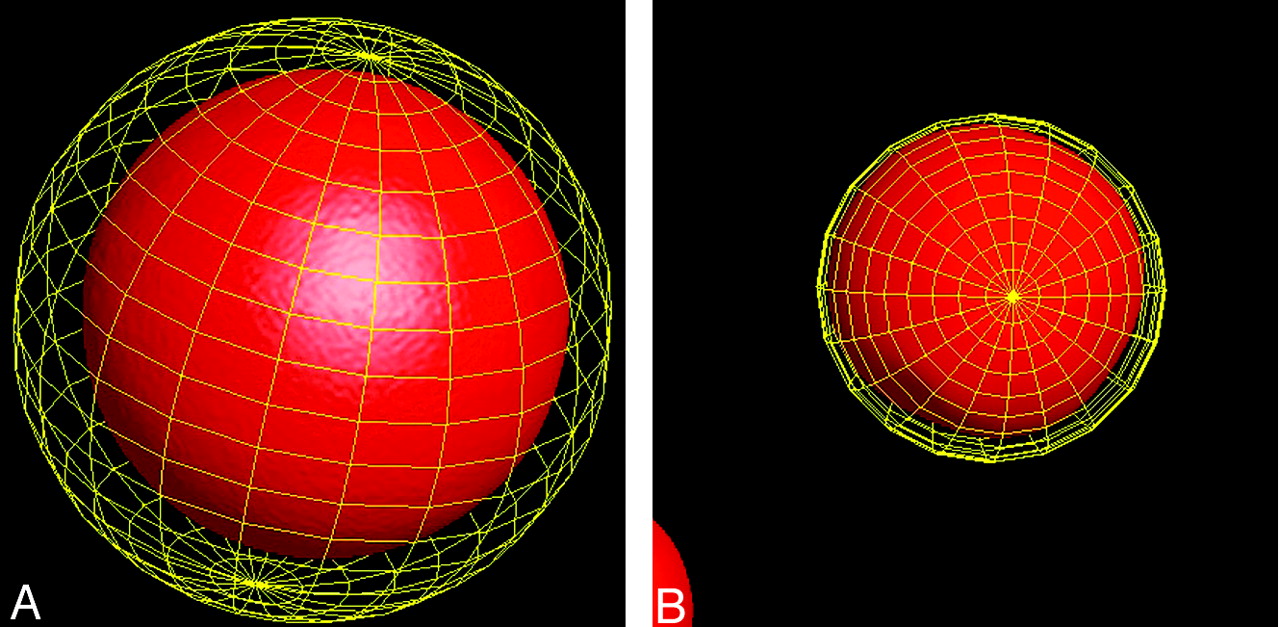
  • Fig 2.
    • Download figure
    • Open in new tab
    • Download powerpoint
    Fig 2.

    A, Volume determination with the 3D-RA software of the 50-mm-diameter iron ball displayed with surface-shaded display. The software is giving a volume of 67.875 mm3, whereas the ellipsoid calculation is giving 65.45 mm3.

    B, Volume determination with the 3D-RA software of the 5-mm-diameter iron ball displayed with surface shaded display. The software is giving a volume of 69 mm3, whereas the ellipsoid calculation is giving 65.5 mm3.
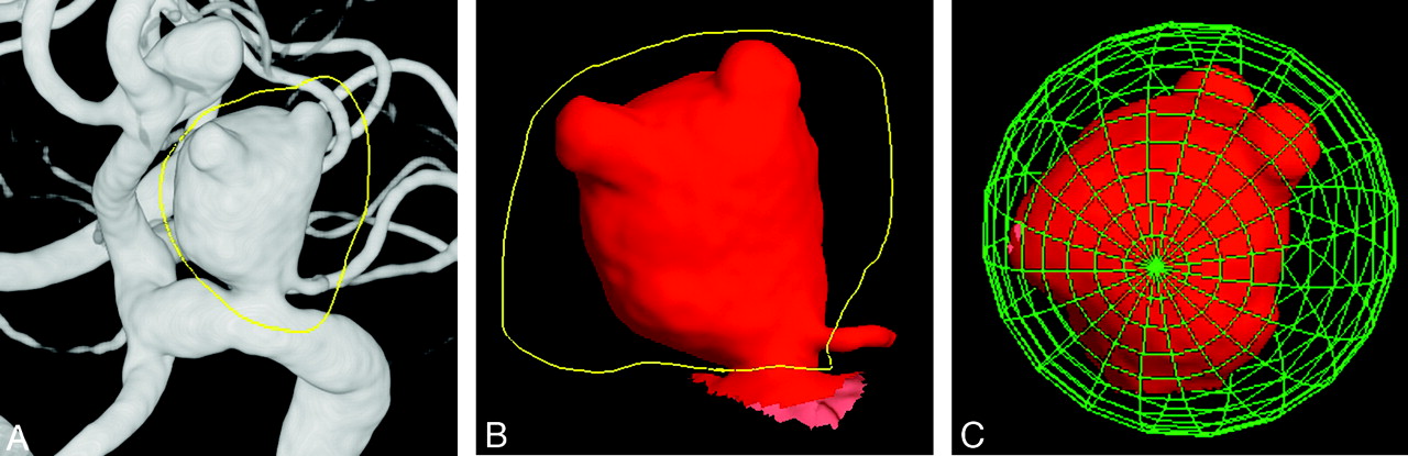
  • Fig 3.
    • Download figure
    • Open in new tab
    • Download powerpoint
    Fig 3.

    Manual segmentation of a 12-mm large carotid ophthalmic artery aneurysm with the 3D rotational angiography (3D-RA) volumetric measurement software.

    A, Volume-rendering display of a 3D-RA representation of the aneurysm obtained after rotational angiography of the right ICA. The gross contour of the aneurysm is first extracted with the cutting graphical tool of the software (yellow line).

    B, Cutting of the aneurysm is refined to extract only the contour of the aneurysm (yellow line).

    C, Finally, the volume is given by placing the segmented aneurysm inside the sphere giving a volume of 591 mm3, which corresponds to a 34% overestimation provided by the ellipsoid approximation.

Tables

  • Figures
  • Volume misestimating of ellipsoid calculation regarding volume provided by 3D measurements

    NumberMean Greater Dimension (mm)Mean Volume Misestimating (%)
    Locations
        MCA1257 ± 3+ 13 ± 41
        AcomA1167 ± 3+ 14 ± 37
        PcomA557 ± 4+ 22 ± 44
        ICA oph429 ± 5+ 16 ± 32
        Basilar tip358 ± 4+ 8 ± 27
        ICA supra339 ± 5+ 17 ± 30
        Cavernous sinus258 ± 4+ 19 ± 46
        ICA bifurcation237 ± 4+ 13 ± 33
        Pericallosal115 ± 2+ 6 ± 22
        Laterobasilar57 ± 4+ 24 ± 52
        Superior cerebellar54 ± 2− 11 ± 27
        Vertebral413 ± 3+ 23 ± 18
        PCA37 ± 3+ 43 ± 59
        PICA25 ± 0+ 10 ± 13
            Kruskal-Wallis test P = .6508
    Sizes
        <10 mm3856 ± 2+ 10 ± 35
        ≥10 mm9913 ± 4+ 32 ± 44
            Mann-Whitney test P < .0001
    All sizes and locations4848 ± 4+ 15 ± 38
    • Note:— MCA indicates cerebral artery; AcomA, anterior communicating artery; PcomA, posterior communicating artery; ICA oph, carotid ophthalmic; ICA supra, supraclinoid internal carotid artery (other than ICA oph and PcomA); PCA, posterior cerebral artery; PICA, posterior-inferior cerebellar artery.

PreviousNext
Back to top

In this issue

American Journal of Neuroradiology: 27 (4)
American Journal of Neuroradiology
Vol. 27, Issue 4
April 2006
  • Table of Contents
  • Index by author
Advertisement
Print
Download PDF
Email Article

Thank you for your interest in spreading the word on American Journal of Neuroradiology.

NOTE: We only request your email address so that the person you are recommending the page to knows that you wanted them to see it, and that it is not junk mail. We do not capture any email address.

Enter multiple addresses on separate lines or separate them with commas.
Ellipsoid Approximation versus 3D Rotational Angiography in the Volumetric Assessment of Intracranial Aneurysms
(Your Name) has sent you a message from American Journal of Neuroradiology
(Your Name) thought you would like to see the American Journal of Neuroradiology web site.
CAPTCHA
This question is for testing whether or not you are a human visitor and to prevent automated spam submissions.
Cite this article
M. Piotin, B. Daghman, C. Mounayer, L. Spelle, J. Moret
Ellipsoid Approximation versus 3D Rotational Angiography in the Volumetric Assessment of Intracranial Aneurysms
American Journal of Neuroradiology Apr 2006, 27 (4) 839-842;

Citation Manager Formats

  • BibTeX
  • Bookends
  • EasyBib
  • EndNote (tagged)
  • EndNote 8 (xml)
  • Medlars
  • Mendeley
  • Papers
  • RefWorks Tagged
  • Ref Manager
  • RIS
  • Zotero
0 Responses
Respond to this article
Share
Bookmark this article
Ellipsoid Approximation versus 3D Rotational Angiography in the Volumetric Assessment of Intracranial Aneurysms
M. Piotin, B. Daghman, C. Mounayer, L. Spelle, J. Moret
American Journal of Neuroradiology Apr 2006, 27 (4) 839-842;
del.icio.us logo Twitter logo Facebook logo Mendeley logo
  • Tweet Widget
  • Facebook Like
  • Google Plus One

Jump to section

  • Article
    • Abstract
    • Methods
    • Results
    • Discussion
    • Potential Pitfalls of 3D-RA
    • Conclusion
    • References
  • Figures & Data
  • Info & Metrics
  • Responses
  • References
  • PDF

Related Articles

  • No related articles found.
  • PubMed
  • Google Scholar

Cited By...

  • Influence of observers, threshold values, and measurement methods on volumetric analysis of cerebral aneurysms with three-dimensional rotational angiography
  • Endovascular treatment of intracranial aneurysms with detachable coils: correlation between aneurysm volume, packing, and angiographic recurrence
  • AngioSuite: an accurate method to calculate aneurysm volumes and packing densities
  • Temporal Evolution of Susceptibility Artifacts from Coiled Aneurysms on MR Angiography: An In Vivo Canine Study
  • Endovascular Treatment of Wide-Neck Intracranial Aneurysms Using a Microcatheter Protective Technique: Results and Outcomes in 75 Aneurysms
  • Durability of Treatment of Intracranial Aneurysms With Hydrocoils Is Not Different From Standard Platinum Coils
  • Crossref
  • Google Scholar

This article has not yet been cited by articles in journals that are participating in Crossref Cited-by Linking.

More in this TOC Section

  • Progression of Microstructural Damage in Spinocerebellar Ataxia Type 2: A Longitudinal DTI Study
  • SWI or T2*: Which MRI Sequence to Use in the Detection of Cerebral Microbleeds? The Karolinska Imaging Dementia Study
  • Statin Therapy Does Not Affect the Radiographic and Clinical Profile of Patients with TIA and Minor Stroke
Show more Brain

Similar Articles

Advertisement

Indexed Content

  • Current Issue
  • Accepted Manuscripts
  • Article Preview
  • Past Issues
  • Editorials
  • Editor's Choice
  • Fellows' Journal Club
  • Letters to the Editor
  • Video Articles

Cases

  • Case Collection
  • Archive - Case of the Week
  • Archive - Case of the Month
  • Archive - Classic Case

More from AJNR

  • Trainee Corner
  • Imaging Protocols
  • MRI Safety Corner
  • Book Reviews

Multimedia

  • AJNR Podcasts
  • AJNR Scantastics

Resources

  • Turnaround Time
  • Submit a Manuscript
  • Submit a Video Article
  • Submit an eLetter to the Editor/Response
  • Manuscript Submission Guidelines
  • Statistical Tips
  • Fast Publishing of Accepted Manuscripts
  • Graphical Abstract Preparation
  • Imaging Protocol Submission
  • Evidence-Based Medicine Level Guide
  • Publishing Checklists
  • Author Policies
  • Become a Reviewer/Academy of Reviewers
  • News and Updates

About Us

  • About AJNR
  • Editorial Board
  • Editorial Board Alumni
  • Alerts
  • Permissions
  • Not an AJNR Subscriber? Join Now
  • Advertise with Us
  • Librarian Resources
  • Feedback
  • Terms and Conditions
  • AJNR Editorial Board Alumni

American Society of Neuroradiology

  • Not an ASNR Member? Join Now

© 2025 by the American Society of Neuroradiology All rights, including for text and data mining, AI training, and similar technologies, are reserved.
Print ISSN: 0195-6108 Online ISSN: 1936-959X

Powered by HighWire