Advertisement

Main menu

  • Home
  • Content
    • Current Issue
    • Accepted Manuscripts
    • Article Preview
    • Past Issue Archive
    • Video Articles
    • AJNR Case Collection
    • Case of the Week Archive
    • Case of the Month Archive
    • Classic Case Archive
  • Special Collections
    • AJNR Awards
    • Low-Field MRI
    • Alzheimer Disease
    • ASNR Foundation Special Collection
    • Photon-Counting CT
    • View All
  • Multimedia
    • AJNR Podcasts
    • AJNR SCANtastic
    • Trainee Corner
    • MRI Safety Corner
    • Imaging Protocols
  • For Authors
    • Submit a Manuscript
    • Submit a Video Article
    • Submit an eLetter to the Editor/Response
    • Manuscript Submission Guidelines
    • Statistical Tips
    • Fast Publishing of Accepted Manuscripts
    • Graphical Abstract Preparation
    • Imaging Protocol Submission
    • Author Policies
  • About Us
    • About AJNR
    • Editorial Board
    • Editorial Board Alumni
  • More
    • Become a Reviewer/Academy of Reviewers
    • Subscribers
    • Permissions
    • Alerts
    • Feedback
    • Advertisers
    • ASNR Home

User menu

  • Alerts
  • Log in

Search

  • Advanced search
American Journal of Neuroradiology

American Journal of Neuroradiology

ASHNR American Society of Functional Neuroradiology ASHNR American Society of Pediatric Neuroradiology ASSR
  • Alerts
  • Log in

Advanced Search

  • Home
  • Content
    • Current Issue
    • Accepted Manuscripts
    • Article Preview
    • Past Issue Archive
    • Video Articles
    • AJNR Case Collection
    • Case of the Week Archive
    • Case of the Month Archive
    • Classic Case Archive
  • Special Collections
    • AJNR Awards
    • Low-Field MRI
    • Alzheimer Disease
    • ASNR Foundation Special Collection
    • Photon-Counting CT
    • View All
  • Multimedia
    • AJNR Podcasts
    • AJNR SCANtastic
    • Trainee Corner
    • MRI Safety Corner
    • Imaging Protocols
  • For Authors
    • Submit a Manuscript
    • Submit a Video Article
    • Submit an eLetter to the Editor/Response
    • Manuscript Submission Guidelines
    • Statistical Tips
    • Fast Publishing of Accepted Manuscripts
    • Graphical Abstract Preparation
    • Imaging Protocol Submission
    • Author Policies
  • About Us
    • About AJNR
    • Editorial Board
    • Editorial Board Alumni
  • More
    • Become a Reviewer/Academy of Reviewers
    • Subscribers
    • Permissions
    • Alerts
    • Feedback
    • Advertisers
    • ASNR Home
  • Follow AJNR on Twitter
  • Visit AJNR on Facebook
  • Follow AJNR on Instagram
  • Join AJNR on LinkedIn
  • RSS Feeds

AJNR Awards, New Junior Editors, and more. Read the latest AJNR updates

OtherHEAD AND NECK

Silent Sinus Syndrome: An Acquired Condition

Roula Hourany, Nafi Aygun, Charley C. Della Santina and S. James Zinreich
American Journal of Neuroradiology October 2005, 26 (9) 2390-2392;
Roula Hourany
  • Find this author on Google Scholar
  • Find this author on PubMed
  • Search for this author on this site
Nafi Aygun
  • Find this author on Google Scholar
  • Find this author on PubMed
  • Search for this author on this site
Charley C. Della Santina
  • Find this author on Google Scholar
  • Find this author on PubMed
  • Search for this author on this site
S. James Zinreich
  • Find this author on Google Scholar
  • Find this author on PubMed
  • Search for this author on this site
  • Article
  • Figures & Data
  • Info & Metrics
  • Responses
  • References
  • PDF
Loading

Abstract

Summary: We present the case of a 30-year-old man with silent sinus syndrome. A CT scan obtained 10 years earlier showed completely normal maxillary sinuses. This case illustrates the acquired nature of this disorder.

The silent sinus syndrome, also known as imploding antrum and chronic maxillary sinus atelectasis, consists of findings of painless enophthalmos and inward retraction of the ipsilateral maxillary sinus walls on imaging studies (1, 2). The resultant volume loss in the maxillary sinus accounts for orbital enlargement and enophthalmos. The distinction between the imaging appearance of hypoplastic maxillary sinus and silent sinus syndrome is not well understood. Some authors believe that a congenital underdevelopment of the maxillary sinus is responsible for the development of silent sinus syndrome, but the acquired nature of this condition is now more readily apparent (3). Obstruction of the maxillary ostium appears to play a critical role in the development of silent sinus syndrome. Events leading to retraction of the walls of the obstructed sinus are not clearly understood.

Case Report

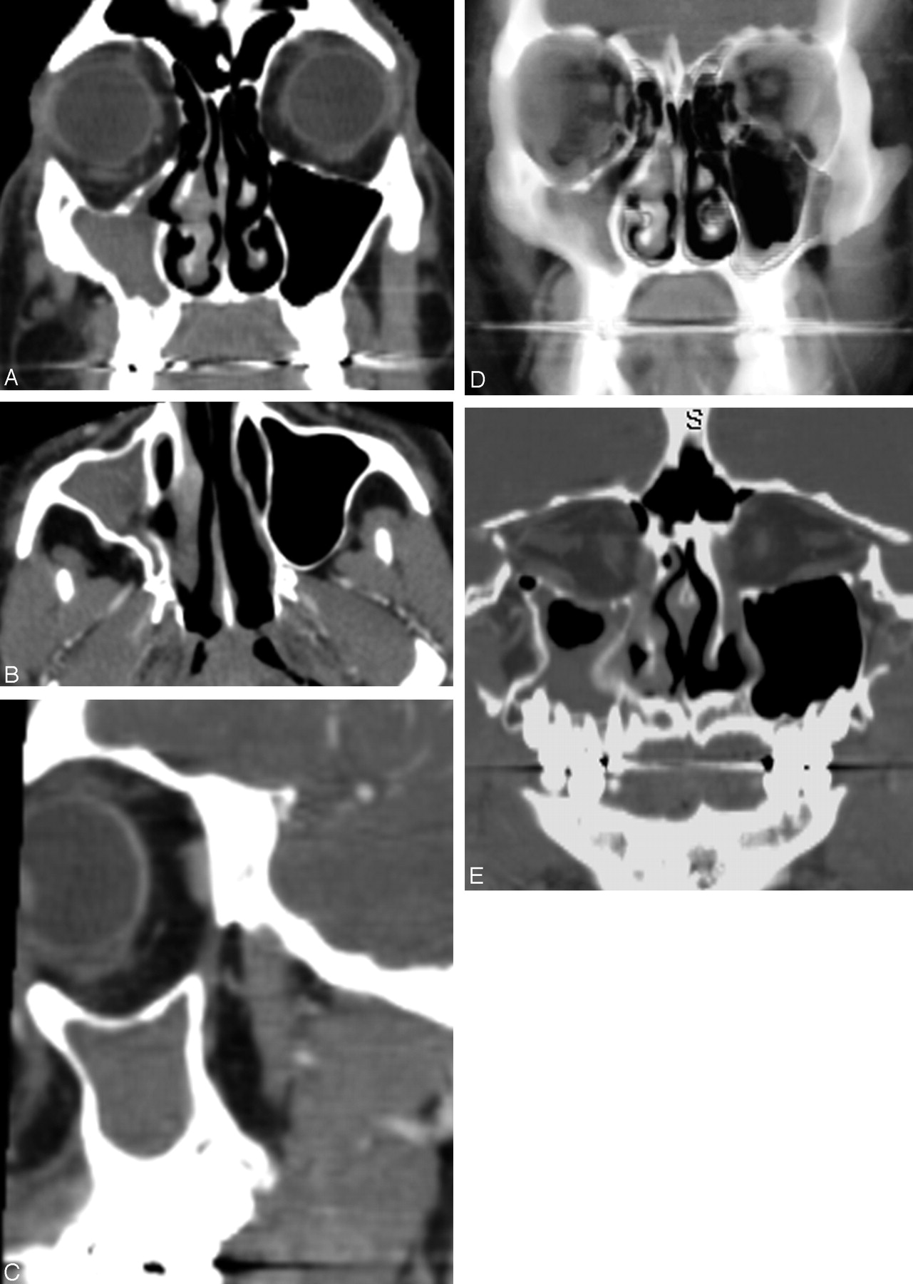
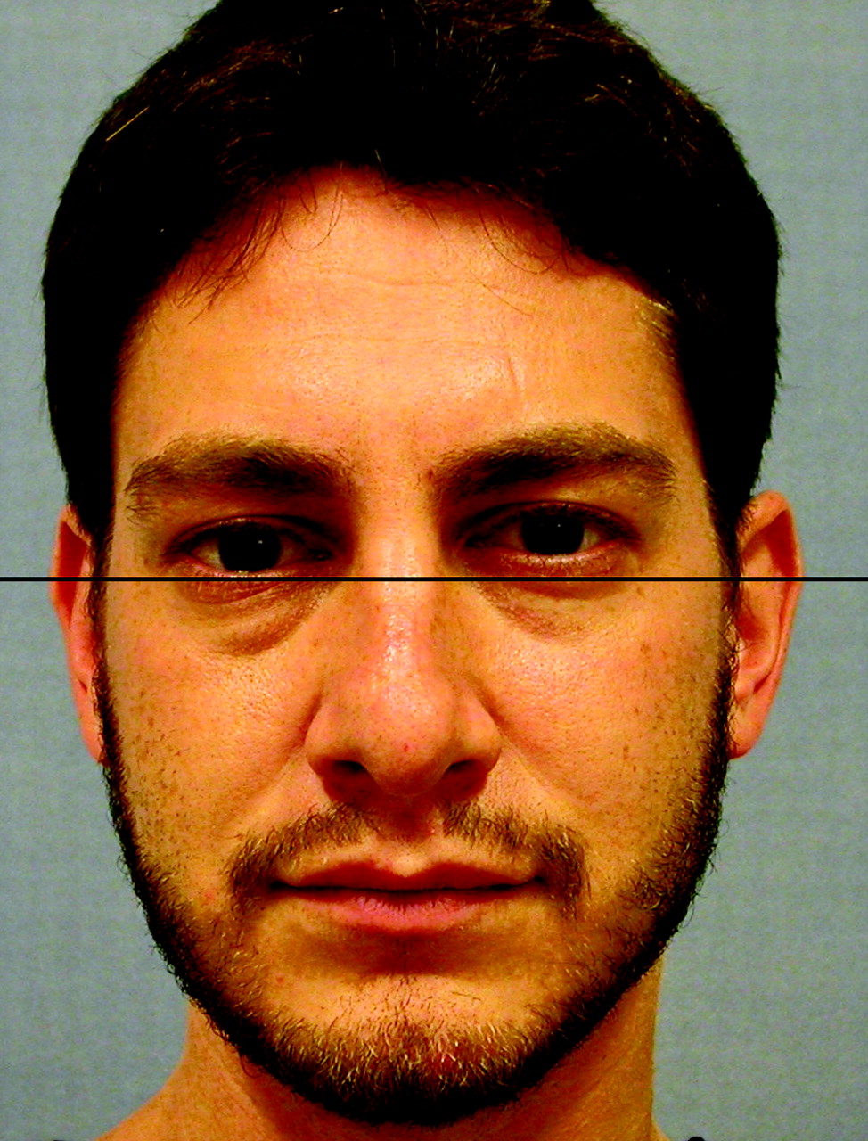
A 30-year-old man was found to have asymmetrically enlarged palatine tonsils, right greater than left, during an upper respiratory infection. The tonsillar asymmetry did not resolve with proper antibiotic treatment. A CT scan of the neck performed for evaluation of a potential underlying mass revealed a completely opacified maxillary sinus on the right. There was significant volume loss of the right maxillary sinus with inward retraction of the slightly thickened walls and consequent enlargement of the right orbit and middle meatus (Fig 1). The uncinate process was thinned and retracted to the orbital wall, obstructing the maxillary sinus infundibulum. Of note, there was no underlying mass to account for tonsillar asymmetry. The patient did not have any symptoms that might be related to sinusitis. He did not notice any enophthalmos but retrospectively recalled a girlfriend several years before noting his right “eye line” being lower than that on the contralateral side. Right enophthalmos and 1–2 mm of hypoglobus were observed on repeat examination (Fig 2).

Fig 1.
  • Download figure
  • Open in new tab
  • Download powerpoint
Fig 1.

(A) Coronal, (B) transverse, and (C) sagittal CT images of the sinuses show inward retraction of all walls of the right maxillary sinus with enlargement of the orbit and the middle meatus. The uncinate process is not clearly visualized because it is markedly thinned and retracted to the inferomedial orbital wall (confirmed with nasal endoscopy). (D) Thick-slab volume reconstruction in the coronal plane and (E) curved reconstruction along the optic nerves better demonstrates the maxillary sinus volume loss and enlargement of the orbit and middle meatus.

Fig 2.
  • Download figure
  • Open in new tab
  • Download powerpoint
Fig 2.

Frontal view of the patient, demonstrating hypoglobus and enophthalmos of the right eye.

The patient had undergone a CT scan of the paranasal sinuses 10 years previously for presurgical evaluation of a posttraumatic nasal septum deformity that occurred during childhood. Findings showed that the right maxillary sinus size and wall thickness were normal (Fig 3). Soon after that initial CT, he underwent nasal septal reconstruction and revision rhinoplasty, including lateral nasal osteotomies.

Fig 3.
  • Download figure
  • Open in new tab
  • Download powerpoint
Fig 3.

Coronal sinus CT of the patient obtained 10 years previously, before nasal septal reconstruction and rhinoplasty with lateral osteotomies, shows normal sinuses.

The patient elected not to pursue surgical treatment of the maxillary sinus disease. His tonsillar asymmetry remained stable during a 14-month follow-up.

Discussion

The silent sinus syndrome is a spontaneous unilateral maxillary atelectasis with complete or partial opacification of the sinus. Silent sinus syndrome is a rare disorder, but it is probably underdiagnosed because of a lack of recognition (1). The typical patient with silent sinus syndrome is an adult in the third through fifth decades of life who presents with spontaneous, painless, and occasionally progressive enophthalmos and hypoglobus (1, 2). No symptoms attributable to chronic sinonasal disease are present. Physical examination findings may include upper lid retraction, deepened upper lid sulcus, malar depression, facial asymmetry, and diplopia. The diagnosis of silent sinus syndrome can be made clinically, but it should be differentiated from other causes of spontaneous enophthalmos such as Parry-Romberg syndrome and linear scleroderma.

The imaging findings are characteristic (4). The primary finding is maxillary sinus volume loss due to inward retraction of the sinus walls, which accounts for the increased orbital volume and enlargement of the middle meatus. Typically, all 4 walls of the sinus are retracted, though one of the medial, anterior, or posterolateral walls may be spared. The orbital floor (maxillary roof) is always retracted and commonly thinned. The other walls may be thinned, normal, or slightly thickened. The maxillary infundibulum is always occluded and the sinus is opacified. The uncinate process is retracted against the inferomedial aspect of the orbital wall.

Our patient exhibited all the characteristic findings of silent sinus syndrome and slight thickening of the walls of the sinus including the orbital floor. The orbital floor thickening is uncommon and was reported to be present in 3 of 12 patients of Rose et al (2) and none of 68 patients of Soparkar et al (1). We speculate that the slight thickening of all walls of the sinus and ipsilateral tonsillar hypertrophy are secondary to chronic inflammation. Soparkar et al argued that the wall thickening may indicate a coexistent developmental hypoplasia, which was clearly not the case in our patient because imaging before disease onset showed completely normal sinus walls (1).

The pathophysiology of silent sinus syndrome remains unclear. Initially, it was suggested that a developmentally small sinus with chronic obstructive sinusitis was the cause (3). The acquired nature of this condition, however, is now well recognized. Negative intrasinus pressure has been demonstrated in patients with silent sinus syndrome (5). Obstruction of the sinus ostium is always present, but it is not clear whether this is the cause or the result of sinus wall retraction. Complete obstruction of the mucous membrane–lined sinus resulting in gas resorption and negative pressure formation, in a similar manner to middle ear atelectasis due to Eustachian tube dysfunction, is the most plausible explanation. Given the rarity of silent sinus syndrome and the very high prevalence of maxillary sinus obstruction, however, one has to question this explanation. This theory also fails to offer an explanation for the exclusive involvement of the maxillary sinus. Perhaps, other compounding factors such as trauma or anatomic predisposition play a role (6).

In our patient, childhood nasal trauma and subsequent surgical trauma were likely factors in the later insidious development of silent sinus syndrome. An osteotomy performed during septal reconstruction and rhinotomy typically passes through the nasal process of the maxilla; however, an osteotomy extending more posteriorly could enter and destabilize the maxillary sinus. Despite the absence of chronic sinusitis symptoms in our patient’s case, subsequent scar contracture or maxillary sinus hypoventilation might then have resulted in the sinus atelectasis, hypoglobus, and enophthalmos typical of silent sinus syndrome.

The initial management in this syndrome should be conservative. If this is inadequate, then normal sinus drainage is restored by enlarging the maxillary ostium with functional endoscopic sinus surgery. Surgical intervention to improve sinus aeration typically halts the progress of maxillary sinus contraction but does not restore sinus volume. In patients with diplopia or severe cosmetic deformity, repair of the orbital floor with placement of a subperiosteal implant can be performed at the same time or after functional endoscopic sinus surgery.

In summary, this case supports the notion that silent sinus syndrome is an acquired condition. Thickening of the sinus walls is probably related to chronic inflammation and not to underlying developmental hypoplasia. Surgical or other trauma to the maxilla and sinus ostium may play a role in the pathogenesis of this condition.

References

  1. ↵
    Soparkar CN, Patrinely JR, Davidson JK: Silent sinus syndrome: new perspectives? Ophthalmology 2004;111:414–415; author reply 415–416
    PubMed
  2. ↵
    Rose GE, Sandy C, Hallberg L, Moseley I. Clinical and radiologic characteristics of the imploding antrum, or “silent sinus,” syndrome. Ophthalmology 2003;110:811–818. Erratum in: Ophthalmology 2003;110:1475
    CrossRefPubMed
  3. ↵
    Soparkar CN, Patrinely JR, Cuaycong MJ, et al. The silent sinus syndrome: a cause of spontaneous enophthalmos. Ophthalmology 1994;101:772–778
    PubMed
  4. ↵
    Illner A, Davidson HC, Harnsberger HR, Hoffman J. The silent sinus syndrome: clinical and radiographic findings. AJR Am J Roentgenol 2002;178:503–506
    CrossRefPubMed
  5. ↵
    Kass ES, Salman S, Montgomery WW. Manometric study of complete ostial occlusion in chronic maxillary atelectasis. Laryngoscope 1996;106:1255–1258
    CrossRefPubMed
  6. ↵
    Hobbs CGL, Saunders MW, Potts MJ. Spontaneous enophthalmos: silent sinus syndrome. J Laryngol Otol 2004;118:310–312
    PubMed
  • Received November 29, 2004.
  • Accepted after revision December 13, 2004.
  • Copyright © American Society of Neuroradiology
View Abstract
PreviousNext
Back to top

In this issue

American Journal of Neuroradiology: 26 (9)
American Journal of Neuroradiology
Vol. 26, Issue 9
1 Oct 2005
  • Table of Contents
  • Index by author
Advertisement
Print
Download PDF
Email Article

Thank you for your interest in spreading the word on American Journal of Neuroradiology.

NOTE: We only request your email address so that the person you are recommending the page to knows that you wanted them to see it, and that it is not junk mail. We do not capture any email address.

Enter multiple addresses on separate lines or separate them with commas.
Silent Sinus Syndrome: An Acquired Condition
(Your Name) has sent you a message from American Journal of Neuroradiology
(Your Name) thought you would like to see the American Journal of Neuroradiology web site.
CAPTCHA
This question is for testing whether or not you are a human visitor and to prevent automated spam submissions.
Cite this article
Roula Hourany, Nafi Aygun, Charley C. Della Santina, S. James Zinreich
Silent Sinus Syndrome: An Acquired Condition
American Journal of Neuroradiology Oct 2005, 26 (9) 2390-2392;

Citation Manager Formats

  • BibTeX
  • Bookends
  • EasyBib
  • EndNote (tagged)
  • EndNote 8 (xml)
  • Medlars
  • Mendeley
  • Papers
  • RefWorks Tagged
  • Ref Manager
  • RIS
  • Zotero
0 Responses
Respond to this article
Share
Bookmark this article
Silent Sinus Syndrome: An Acquired Condition
Roula Hourany, Nafi Aygun, Charley C. Della Santina, S. James Zinreich
American Journal of Neuroradiology Oct 2005, 26 (9) 2390-2392;
del.icio.us logo Twitter logo Facebook logo Mendeley logo
  • Tweet Widget
  • Facebook Like
  • Google Plus One

Jump to section

  • Article
    • Abstract
    • Case Report
    • Discussion
    • References
  • Figures & Data
  • Info & Metrics
  • Responses
  • References
  • PDF

Related Articles

  • No related articles found.
  • PubMed
  • Google Scholar

Cited By...

  • Parry Romberg Syndrome: 7 Cases and Literature Review
  • Sinonasal imaging
  • Crossref
  • Google Scholar

This article has not yet been cited by articles in journals that are participating in Crossref Cited-by Linking.

More in this TOC Section

  • Correlation of Apparent Diffusion Coefficient at 3T with Prognostic Parameters of Retinoblastoma
  • Parathyroid Lesions: Characterization with Dual-Phase Arterial and Venous Enhanced CT of the Neck
  • MR Evaluation of Sinonasal Angiomatous Polyp
Show more HEAD AND NECK

Similar Articles

Advertisement

Indexed Content

  • Current Issue
  • Accepted Manuscripts
  • Article Preview
  • Past Issues
  • Editorials
  • Editor's Choice
  • Fellows' Journal Club
  • Letters to the Editor
  • Video Articles

Cases

  • Case Collection
  • Archive - Case of the Week
  • Archive - Case of the Month
  • Archive - Classic Case

More from AJNR

  • Trainee Corner
  • Imaging Protocols
  • MRI Safety Corner

Multimedia

  • AJNR Podcasts
  • AJNR Scantastics

Resources

  • Turnaround Time
  • Submit a Manuscript
  • Submit a Video Article
  • Submit an eLetter to the Editor/Response
  • Manuscript Submission Guidelines
  • Statistical Tips
  • Fast Publishing of Accepted Manuscripts
  • Graphical Abstract Preparation
  • Imaging Protocol Submission
  • Evidence-Based Medicine Level Guide
  • Publishing Checklists
  • Author Policies
  • Become a Reviewer/Academy of Reviewers
  • News and Updates

About Us

  • About AJNR
  • Editorial Board
  • Editorial Board Alumni
  • Alerts
  • Permissions
  • Not an AJNR Subscriber? Join Now
  • Advertise with Us
  • Librarian Resources
  • Feedback
  • Terms and Conditions
  • AJNR Editorial Board Alumni

American Society of Neuroradiology

  • Not an ASNR Member? Join Now

© 2025 by the American Society of Neuroradiology All rights, including for text and data mining, AI training, and similar technologies, are reserved.
Print ISSN: 0195-6108 Online ISSN: 1936-959X

Powered by HighWire