
Article Figures & Data
Figures
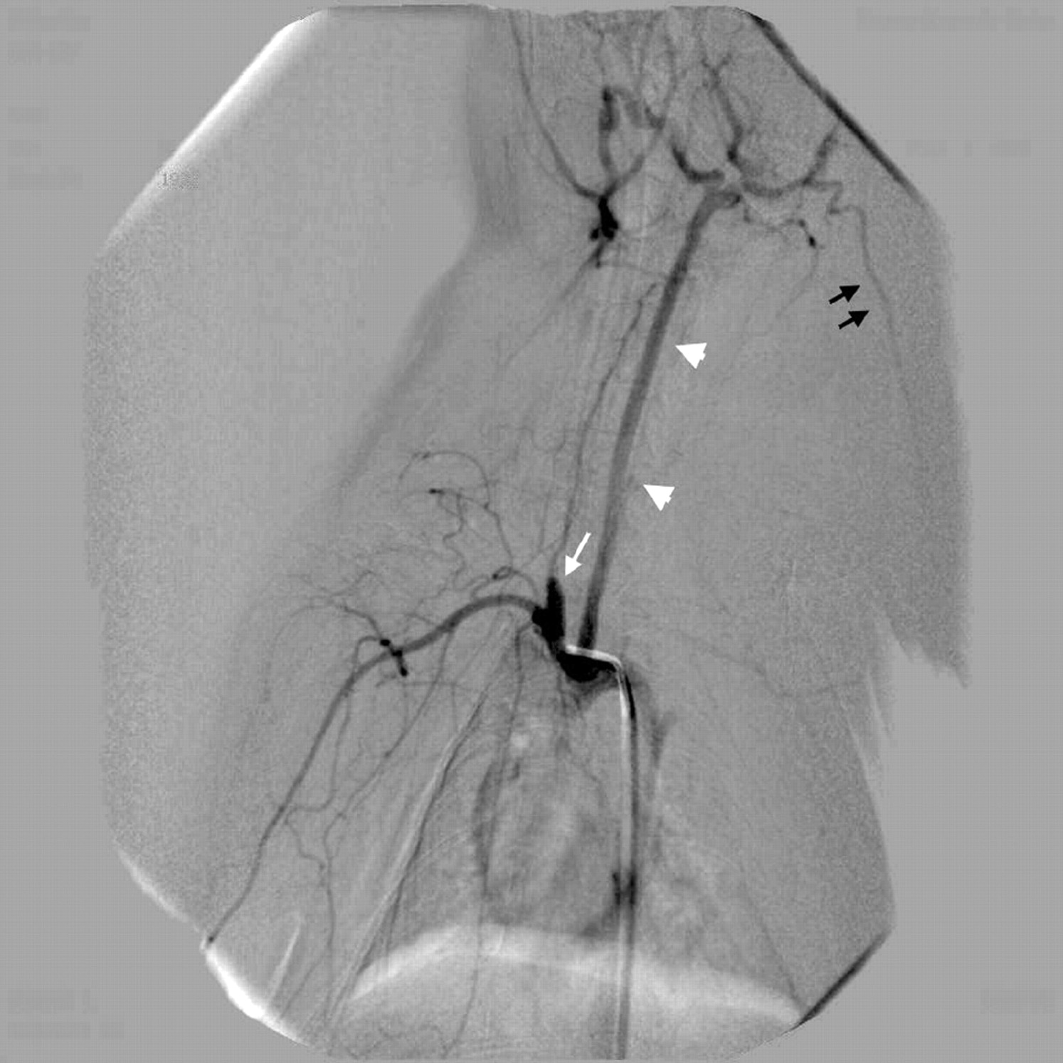
- Fig 1.
Vascular anatomy of the central artery of the ear, left antero-oblique view. Transcatheter contrast injection opacifies the BCA with the elastase-treated right CCA origin forming an aneurysm sac (white arrow) and the left CCA (white arrowheads) with its branches. The central artery of the ear (black arrows) originates from the external carotid artery territory as one of its major branches.
- Fig 2.
A, Left ear injection 3 months after stent implant and coiling of the elastase-induced aneurysm densely opacifies the left CCA (white arrowheads) and the BCA along with its branches. Notice the excellent opacification of the right VA (black arrow) and its derivative muscular branches. This angiogram was performed by hand injection of 5 mL of undiluted Visipaque via a 21-gauge plastic venous cannule into the left central ear artery.
B, Aortogram in the same animal at 6 months opacifies the same vessels, but to a lesser degree. Both the left CCA (white arrowheads) and the right VA (black arrow) are less opacified when compared with the ear injection technique. Note the nonvisualization of the muscular branches of the right VA. This angiogram was performed by hand injection of 5 mL of undiluted Visipaque via a 4F diagnostic catheter without sideholes.
- Fig 3.
This angiogram represents the unfavorable human-type left CCA origin directly from the aortic arch. Contrast injected via the left central ear artery densely opacifies the left CCA (white arrowheads) but flushes caudally by the flow in the aortic arch. Injection parameters were similar to those described in Fig 2B.


In this issue


Jump to section
Related Articles
Cited By...
- In situ decellularization of a large animal saccular aneurysm model: sustained inflammation and active aneurysm wall remodeling
- Phosphorylcholine surface modified flow diverter associated with reduced intimal hyperplasia
- Communicating malapposition of flow diverters assessed with optical coherence tomography correlates with delayed aneurysm occlusion
- Acute thrombus formation on phosphorilcholine surface modified flow diverters