
Article Figures & Data
Figures
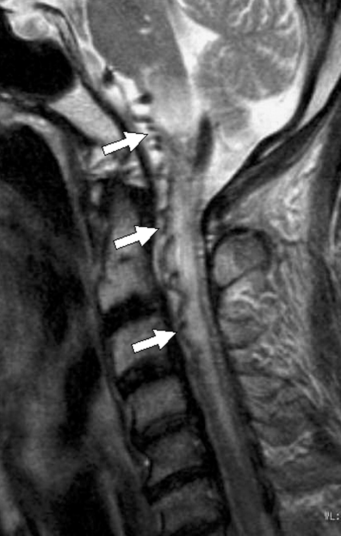
- Fig 1.
Preoperative T2-weighted MR image. The medulla oblongata and cervical spinal cord show diffuse high signal intensity. Note the tortuous signal intensity voids in the premedullary subarachnoid space (arrows).
- Fig 2.
Frontal (A) and lateral (B) views of preoperative external carotid arteriography. The dural arteriovenous fistula is fed by neuromeningeal branches of the ascending pharyngeal artery (white arrow) and a jugular branch of the occipital artery (black arrow) and drains into the pontomesencephalic vein (arrowheads).
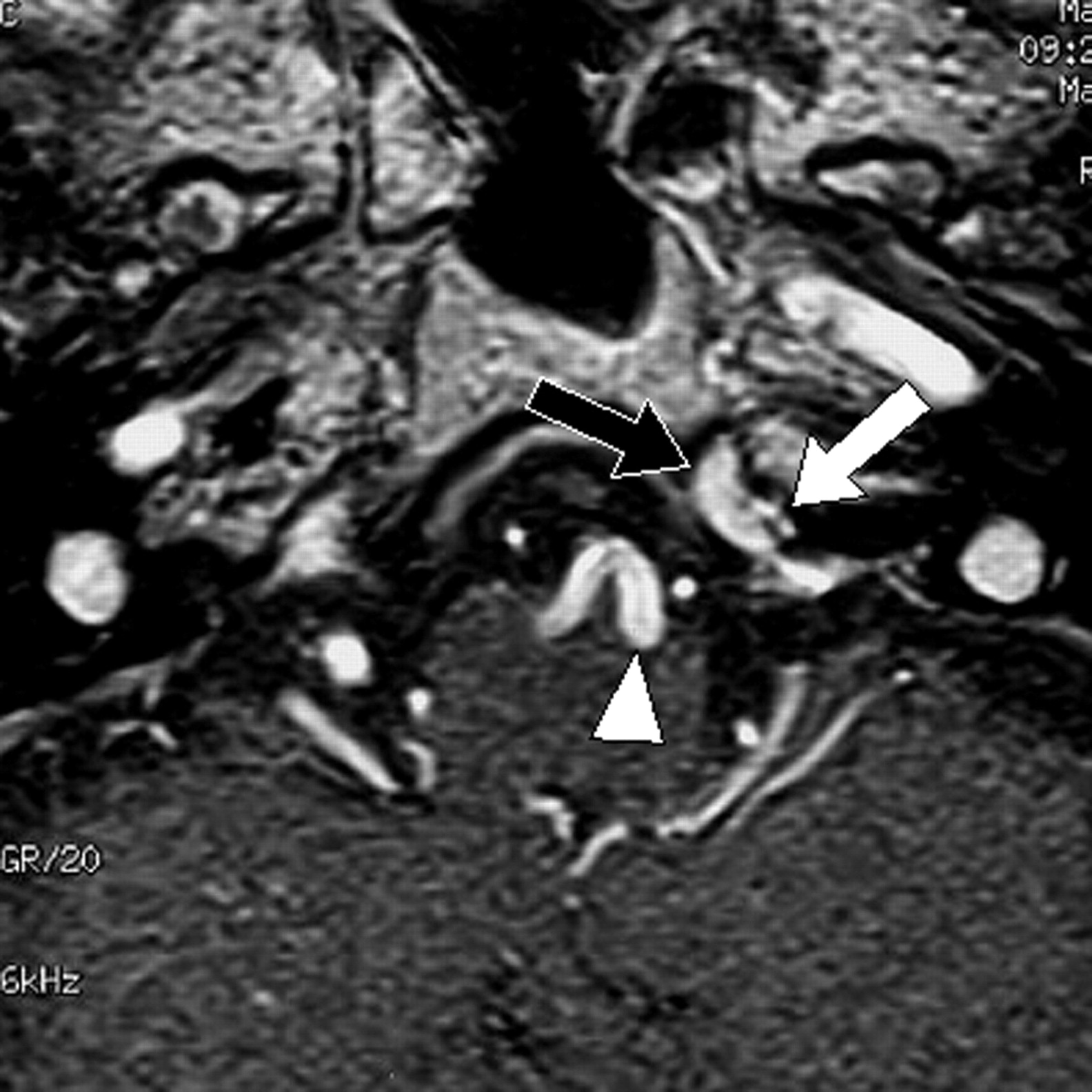
- Fig 3.
Source image of contrast-enhanced 3D MR angiography. The branches of the ascending pharyngeal artery (white arrow) flow into the anterior condylar vein (black arrow). The arrowhead indicates the dilated anterior pontomesencephalic vein draining from the anterior condylar vein.
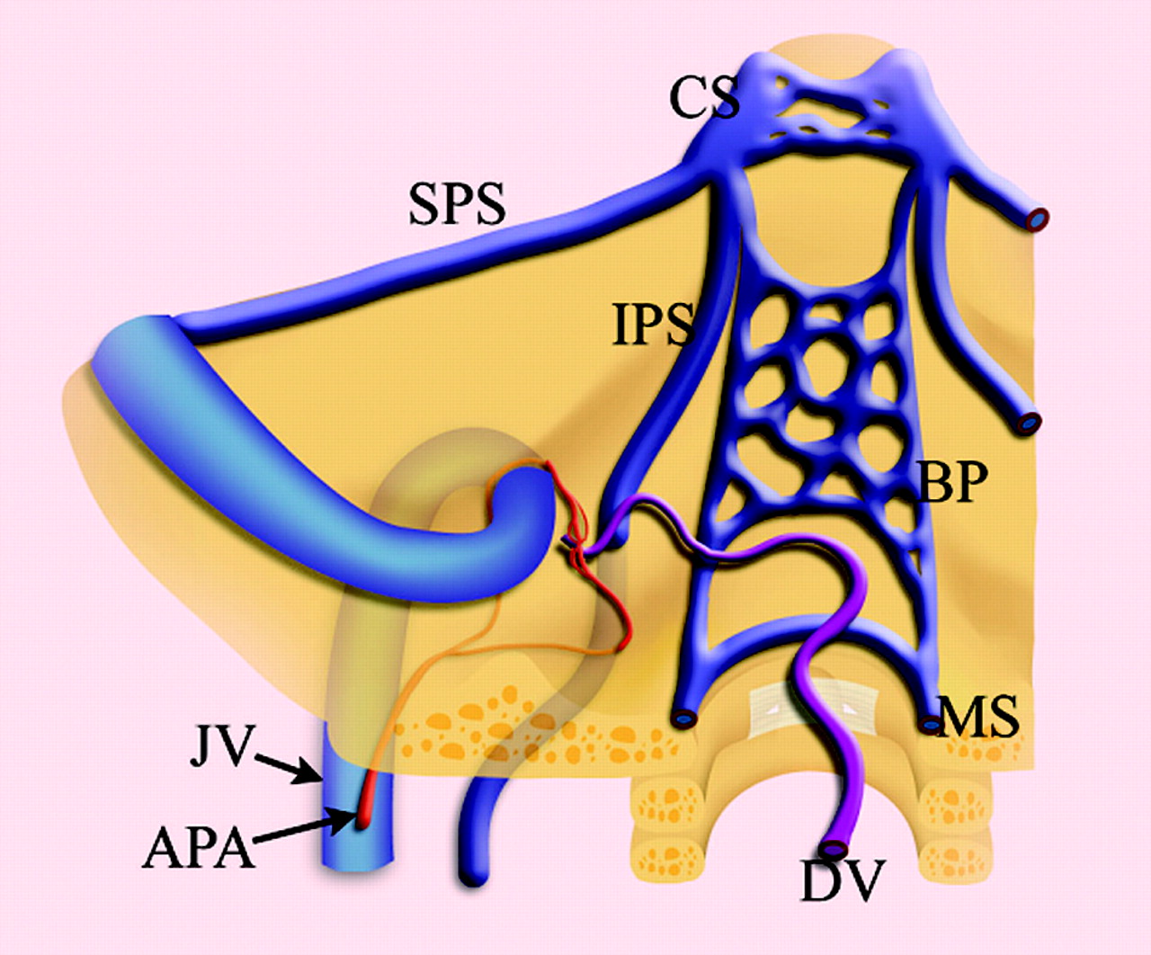
- Fig 4.
Schematic drawing of the dural arteriovenous fistula and surrounding venous structures in case 1. APA indicates the ascending pharyngeal artery; DV, drainage vein; CS, cavernous sinus; SPS, superior petrosal sinus; IPS, inferior petrosal sinus; BP, basilar plexus; MS, marginal sinus; JV, jugular vein.
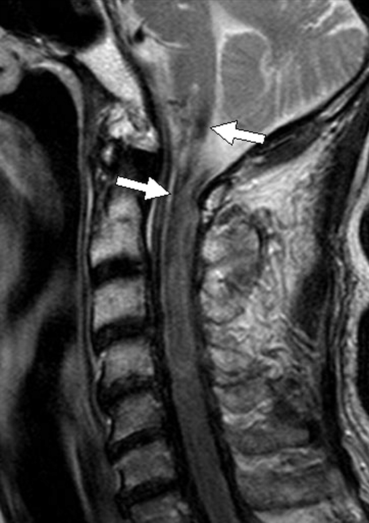
- Fig 5.
MR image obtained at 14 months after the procedure. The brain stem and spinal cord edema has completely disappeared. Low-signal-intensity areas (arrows) indicate deposition of hemosiderin due to previous venous congestion and infarction.
- Fig 6.
Preoperative T2-weighted MR image. A saccular signal intensity void is seen in the left hypoglossal canal (arrow).
- Fig 7.
Frontal (A) and lateral (B) views of preoperative external carotid arteriography. The dural arteriovenous fistula is fed by meningeal branches of the ascending pharyngeal artery and occipital artery. A dilated venous sac is seen at the fistulous point (arrow). Note the retrograde venous drainage via the inferior petrosal sinus and cavernous sinus (arrowheads).
- Fig 8.
Source image of time-of-flight MR angiography. The saccular high signal intensity indicates the venous sac of the anterior condylar dural arteriovenous fistula (arrow).
- Fig 9.
Frontal (A) and lateral (B) views of a postoperative angiogram. The dural arteriovenous fistula has completely disappeared. The arrows indicate the mass of metallic coils placed in the venous pouch.


In this issue


Jump to section
Related Articles
Cited By...
- Endovascular treatment strategy, technique, and outcomes for dural arteriovenous fistulas of the marginal sinus region
- Dural Arteriovenous Fistulas of the Foramen Magnum Region: Clinical Features and Angioarchitectural Phenotypes
- Onyx embolization of anterior condylar confluence dural arteriovenous fistula
- Onyx embolization of anterior condylar confluence dural arteriovenous fistula
- Intraosseous Cranial Dural Arteriovenous Fistula Treated with Transvenous Embolization