Advertisement

Main menu

  • Home
  • Content
    • Current Issue
    • Accepted Manuscripts
    • Article Preview
    • Past Issue Archive
    • Video Articles
    • AJNR Case Collection
    • Case of the Week Archive
    • Case of the Month Archive
    • Classic Case Archive
  • Special Collections
    • AJNR Awards
    • Low-Field MRI
    • Alzheimer Disease
    • ASNR Foundation Special Collection
    • Photon-Counting CT
    • View All
  • Multimedia
    • AJNR Podcasts
    • AJNR SCANtastic
    • Trainee Corner
    • MRI Safety Corner
    • Imaging Protocols
  • For Authors
    • Submit a Manuscript
    • Submit a Video Article
    • Submit an eLetter to the Editor/Response
    • Manuscript Submission Guidelines
    • Statistical Tips
    • Fast Publishing of Accepted Manuscripts
    • Graphical Abstract Preparation
    • Imaging Protocol Submission
    • Author Policies
  • About Us
    • About AJNR
    • Editorial Board
    • Editorial Board Alumni
  • More
    • Become a Reviewer/Academy of Reviewers
    • Subscribers
    • Permissions
    • Alerts
    • Feedback
    • Advertisers
    • ASNR Home

User menu

  • Alerts
  • Log in

Search

  • Advanced search
American Journal of Neuroradiology

American Journal of Neuroradiology

ASHNR American Society of Functional Neuroradiology ASHNR American Society of Pediatric Neuroradiology ASSR
  • Alerts
  • Log in

Advanced Search

  • Home
  • Content
    • Current Issue
    • Accepted Manuscripts
    • Article Preview
    • Past Issue Archive
    • Video Articles
    • AJNR Case Collection
    • Case of the Week Archive
    • Case of the Month Archive
    • Classic Case Archive
  • Special Collections
    • AJNR Awards
    • Low-Field MRI
    • Alzheimer Disease
    • ASNR Foundation Special Collection
    • Photon-Counting CT
    • View All
  • Multimedia
    • AJNR Podcasts
    • AJNR SCANtastic
    • Trainee Corner
    • MRI Safety Corner
    • Imaging Protocols
  • For Authors
    • Submit a Manuscript
    • Submit a Video Article
    • Submit an eLetter to the Editor/Response
    • Manuscript Submission Guidelines
    • Statistical Tips
    • Fast Publishing of Accepted Manuscripts
    • Graphical Abstract Preparation
    • Imaging Protocol Submission
    • Author Policies
  • About Us
    • About AJNR
    • Editorial Board
    • Editorial Board Alumni
  • More
    • Become a Reviewer/Academy of Reviewers
    • Subscribers
    • Permissions
    • Alerts
    • Feedback
    • Advertisers
    • ASNR Home
  • Follow AJNR on Twitter
  • Visit AJNR on Facebook
  • Follow AJNR on Instagram
  • Join AJNR on LinkedIn
  • RSS Feeds

AJNR Awards, New Junior Editors, and more. Read the latest AJNR updates

Research ArticleNeurointervention

Comparison of Technetium Tc 99m Hexamethylpropyleneamine Oxime Single-Photon Emission Tomograph with Stump Pressure During the Balloon Occlusion Test of the Internal Carotid Artery

Noriaki Tomura, Koichi Omachi, Satoshi Takahashi, Ikuo Sakuma, Takahiro Otani, Jiro Watarai, Kazuo Ishikawa, Hiroyuki Kinouchi and Kazuo Mizoi
American Journal of Neuroradiology September 2005, 26 (8) 1937-1942;
Noriaki Tomura
  • Find this author on Google Scholar
  • Find this author on PubMed
  • Search for this author on this site
Koichi Omachi
  • Find this author on Google Scholar
  • Find this author on PubMed
  • Search for this author on this site
Satoshi Takahashi
  • Find this author on Google Scholar
  • Find this author on PubMed
  • Search for this author on this site
Ikuo Sakuma
  • Find this author on Google Scholar
  • Find this author on PubMed
  • Search for this author on this site
Takahiro Otani
  • Find this author on Google Scholar
  • Find this author on PubMed
  • Search for this author on this site
Jiro Watarai
  • Find this author on Google Scholar
  • Find this author on PubMed
  • Search for this author on this site
Kazuo Ishikawa
  • Find this author on Google Scholar
  • Find this author on PubMed
  • Search for this author on this site
Hiroyuki Kinouchi
  • Find this author on Google Scholar
  • Find this author on PubMed
  • Search for this author on this site
Kazuo Mizoi
  • Find this author on Google Scholar
  • Find this author on PubMed
  • Search for this author on this site
  • Article
  • Figures & Data
  • Info & Metrics
  • Responses
  • References
  • PDF
Loading

Article Figures & Data

Figures

  • Fig 1.
    • Download figure
    • Open in new tab
    • Download powerpoint
    Fig 1.

    Graph shows the relationship between the minimum mean stump pressure and number of regions of interest (ROIs) with hypoperfusion. There was a significant correlation (P < .005 by Spearman rank correlation).

  • Fig 2.
    • Download figure
    • Open in new tab
    • Download powerpoint
    Fig 2.

    Graph shows that the number of regions of interest (ROIs) with hypoperfusion was significantly (P < .001 by Mann–Whitney U test) greater in patients with a minMSP <40 mm Hg (31.5 ± 13.7) than in patients with minMSP ≥40 mm Hg (5.1 ± 4.0).
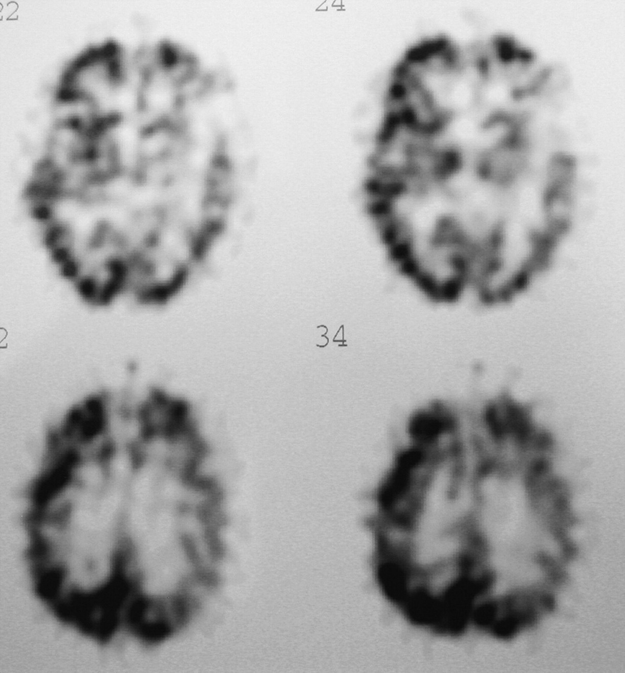
  • Fig 3.
    • Download figure
    • Open in new tab
    • Download powerpoint
    Fig 3.

    In a 73-year-old man with hypopharyngeal carcinoma on the left side, the minMSP was 30 mm Hg during balloon test occlusion of the left internal carotid artery. SPECT using technetium Tc 99m HMPAO showed hypoperfusion of the cerebral hemisphere on the left side.
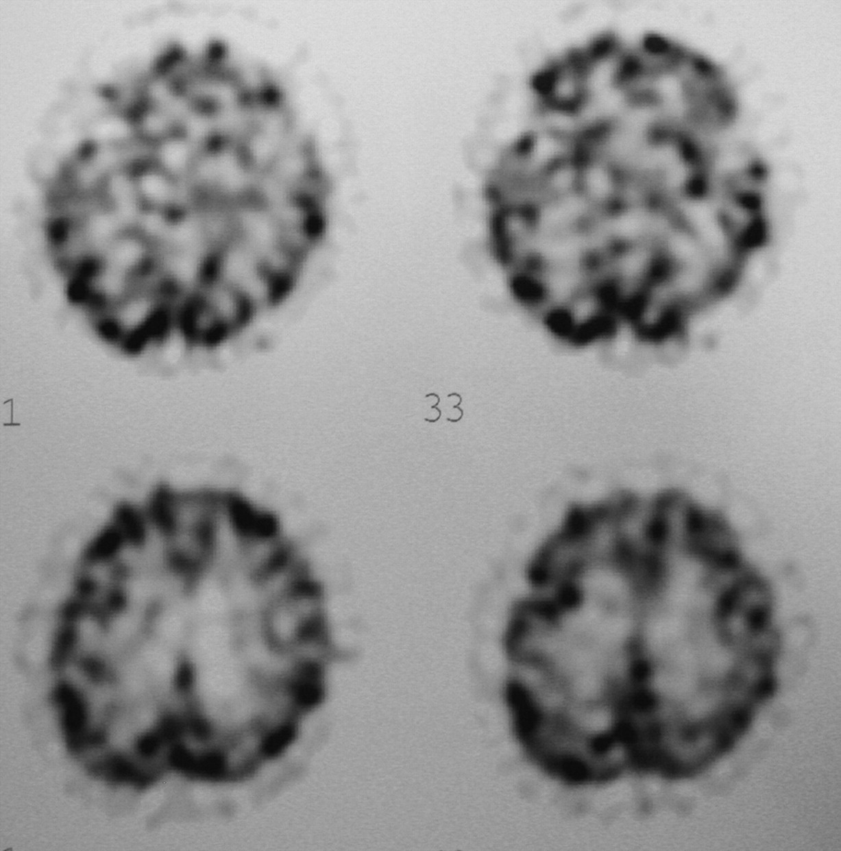
  • Fig 4.
    • Download figure
    • Open in new tab
    • Download powerpoint
    Fig 4.

    In a 64-year-old man with meningioma invading the left internal carotid artery, the MSP was 64 mm Hg during balloon test occlusion of the left internal carotid artery. SPECT using technetium Tc 99m HMPAO showed no hypoperfusion areas of the cerebral hemisphere on the left side.

  • Fig 5.
    • Download figure
    • Open in new tab
    • Download powerpoint
    Fig 5.

    Graph shows the relationship between the number of regions of interest (ROIs) with hypoperfusion and the pressure ratio of the minMSP to the minimum mean systemic pressure.

  • Fig 6.
    • Download figure
    • Open in new tab
    • Download powerpoint
    Fig 6.

    Graph shows that the number of regions of interest (ROIs) with hypoperfusion was significantly (P < .001 by Mann–Whitney U test) greater in patients with a pressure ratio <0.5 (26.7 ± 15.8) than in patients with a pressure ratio ≥0.5 (4.5 ± 3.5)

  • Fig 7.
    • Download figure
    • Open in new tab
    • Download powerpoint
    Fig 7.

    Graph shows the relationship between the minMSP and the mean L/n ratio (the radioactivity count of the occluded side / the radioactivity count of the contralateral normal side). There was a significant positive relationship (linear regression analysis; y = mean L/n ratio, x = minMSP; y = 0.71 + 0.005x; r = 0.497, P = .0084).

PreviousNext
Back to top

In this issue

American Journal of Neuroradiology: 26 (8)
American Journal of Neuroradiology
Vol. 26, Issue 8
1 Sep 2005
  • Table of Contents
  • Index by author
Advertisement
Print
Download PDF
Email Article

Thank you for your interest in spreading the word on American Journal of Neuroradiology.

NOTE: We only request your email address so that the person you are recommending the page to knows that you wanted them to see it, and that it is not junk mail. We do not capture any email address.

Enter multiple addresses on separate lines or separate them with commas.
Comparison of Technetium Tc 99m Hexamethylpropyleneamine Oxime Single-Photon Emission Tomograph with Stump Pressure During the Balloon Occlusion Test of the Internal Carotid Artery
(Your Name) has sent you a message from American Journal of Neuroradiology
(Your Name) thought you would like to see the American Journal of Neuroradiology web site.
CAPTCHA
This question is for testing whether or not you are a human visitor and to prevent automated spam submissions.
Cite this article
Noriaki Tomura, Koichi Omachi, Satoshi Takahashi, Ikuo Sakuma, Takahiro Otani, Jiro Watarai, Kazuo Ishikawa, Hiroyuki Kinouchi, Kazuo Mizoi
Comparison of Technetium Tc 99m Hexamethylpropyleneamine Oxime Single-Photon Emission Tomograph with Stump Pressure During the Balloon Occlusion Test of the Internal Carotid Artery
American Journal of Neuroradiology Sep 2005, 26 (8) 1937-1942;

Citation Manager Formats

  • BibTeX
  • Bookends
  • EasyBib
  • EndNote (tagged)
  • EndNote 8 (xml)
  • Medlars
  • Mendeley
  • Papers
  • RefWorks Tagged
  • Ref Manager
  • RIS
  • Zotero
0 Responses
Respond to this article
Share
Bookmark this article
Comparison of Technetium Tc 99m Hexamethylpropyleneamine Oxime Single-Photon Emission Tomograph with Stump Pressure During the Balloon Occlusion Test of the Internal Carotid Artery
Noriaki Tomura, Koichi Omachi, Satoshi Takahashi, Ikuo Sakuma, Takahiro Otani, Jiro Watarai, Kazuo Ishikawa, Hiroyuki Kinouchi, Kazuo Mizoi
American Journal of Neuroradiology Sep 2005, 26 (8) 1937-1942;
del.icio.us logo Twitter logo Facebook logo Mendeley logo
  • Tweet Widget
  • Facebook Like
  • Google Plus One

Jump to section

  • Article
    • Abstract
    • Methods
    • Results
    • Discussion
    • Conclusion
    • References
  • Figures & Data
  • Info & Metrics
  • Responses
  • References
  • PDF

Related Articles

  • No related articles found.
  • PubMed
  • Google Scholar

Cited By...

  • No citing articles found.
  • Crossref
  • Google Scholar

This article has not yet been cited by articles in journals that are participating in Crossref Cited-by Linking.

More in this TOC Section

  • Factors Associated with Major Re-Recanalization following Second Coiling for Recanalized Aneurysms: A Multicenter Experience over 20 Years during Long-Term Follow-up
  • A Key Factor Shapes LS-DAVFs EVT Outcome
  • Optimizing Voxel Size in 3D Rotational Angiography
Show more NEUROINTERVENTION

Similar Articles

Advertisement

Indexed Content

  • Current Issue
  • Accepted Manuscripts
  • Article Preview
  • Past Issues
  • Editorials
  • Editor's Choice
  • Fellows' Journal Club
  • Letters to the Editor
  • Video Articles

Cases

  • Case Collection
  • Archive - Case of the Week
  • Archive - Case of the Month
  • Archive - Classic Case

More from AJNR

  • Trainee Corner
  • Imaging Protocols
  • MRI Safety Corner

Multimedia

  • AJNR Podcasts
  • AJNR Scantastics

Resources

  • Turnaround Time
  • Submit a Manuscript
  • Submit a Video Article
  • Submit an eLetter to the Editor/Response
  • Manuscript Submission Guidelines
  • Statistical Tips
  • Fast Publishing of Accepted Manuscripts
  • Graphical Abstract Preparation
  • Imaging Protocol Submission
  • Evidence-Based Medicine Level Guide
  • Publishing Checklists
  • Author Policies
  • Become a Reviewer/Academy of Reviewers
  • News and Updates

About Us

  • About AJNR
  • Editorial Board
  • Editorial Board Alumni
  • Alerts
  • Permissions
  • Not an AJNR Subscriber? Join Now
  • Advertise with Us
  • Librarian Resources
  • Feedback
  • Terms and Conditions
  • AJNR Editorial Board Alumni

American Society of Neuroradiology

  • Not an ASNR Member? Join Now

© 2025 by the American Society of Neuroradiology All rights, including for text and data mining, AI training, and similar technologies, are reserved.
Print ISSN: 0195-6108 Online ISSN: 1936-959X

Powered by HighWire