
Article Figures & Data
Figures
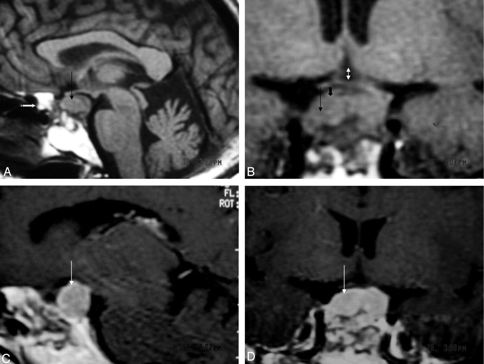
- Fig 1.
Case 1, a 32-year-old woman with chronic headaches and amenorrhea.
A, T1-weighted (TR, 760; TE, 15) sagittal MR demonstrating an intrasellar tumor with suprasellar extension (thin black arrow). There is evidence of previous surgery, as seen by fat packing in the sphenoid sinus (stippled white arrow).
B, T1 weighted (TR, 760; TE, 15) coronal MR showing the sellar mass (thin black arrow) with suprasellar extension (short black arrow) of tumor causing chiasmal (double-headed white arrow) compression.
C and D, T1-weighted contrast-enhanced sagittal and coronal MR showing dense heterogeneous enhancement of the tumor (thin white arrow).
- Fig 2.
Case 1.
A, Histology with H & E stain and 100× magnification reveals a tumor composed of spindle-shaped cells with elongated nuclei arranged in fascicles. Focal infiltrates of mature lympocytes are present.
B, Although Rosenthal fibers were not observed, hyaline bodies were focally seen (upper left).
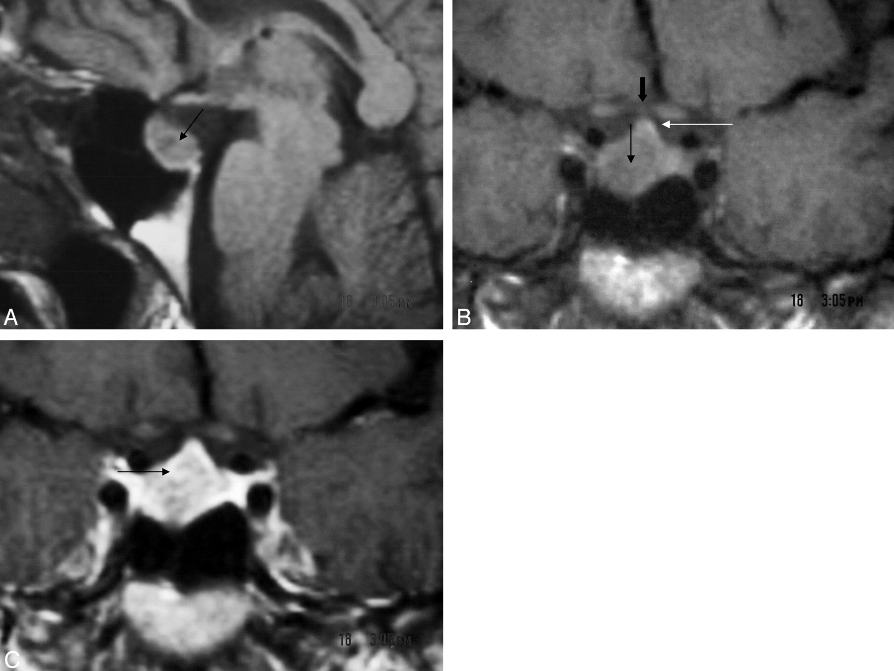
- Fig 3.
Case 2, a 45-year-old woman with uncontrollable chronic headaches.
A, T1-weighted coronal MR demonstrates suprasellar extension of the mass (thin black arrow), pituitary stalk (thin white arrow), and optic chiasm (bold black arrow).
B, T1-weighted (TR, 760; TE, 15) sagittal MR showing an intrasellar mass (thin black arrow).
C, T1-weighted contrast-enhanced coronal MR shows dense nonhomogenous enhancement of the tumor (thin black arrow).
- Fig 4.
Case 2.
A, H & E stain and 100× magnification shows compact areas of spindle cells with oval to fusiform nuclei.
B, GFAP immunohistochemical (100× magnification) highlights the fibrillary cellular processes.


