
Article Figures & Data
Figures
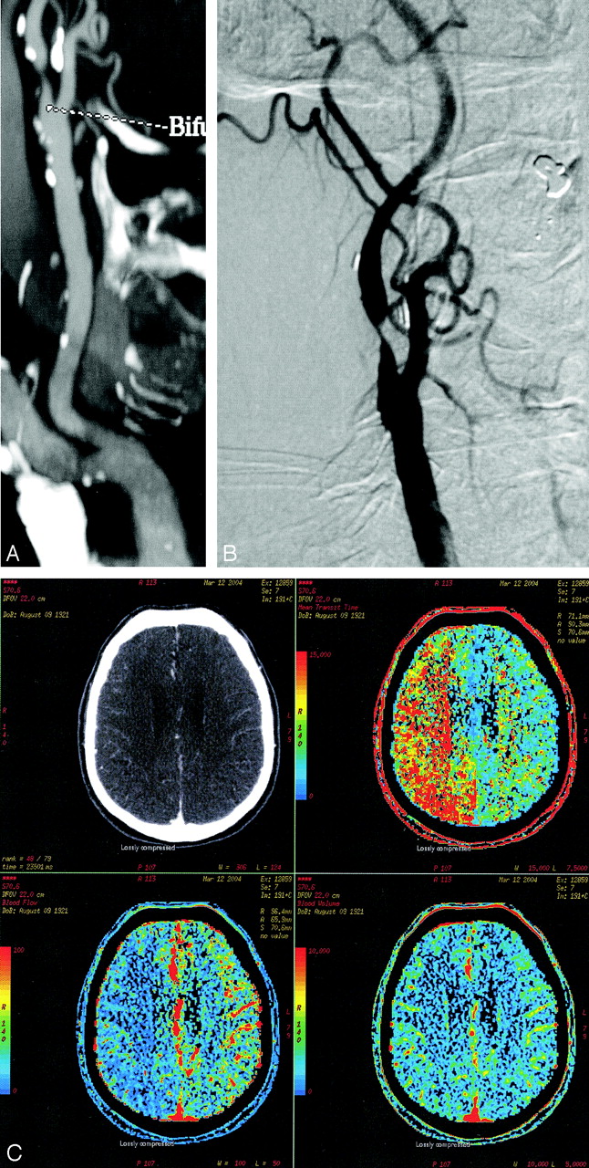
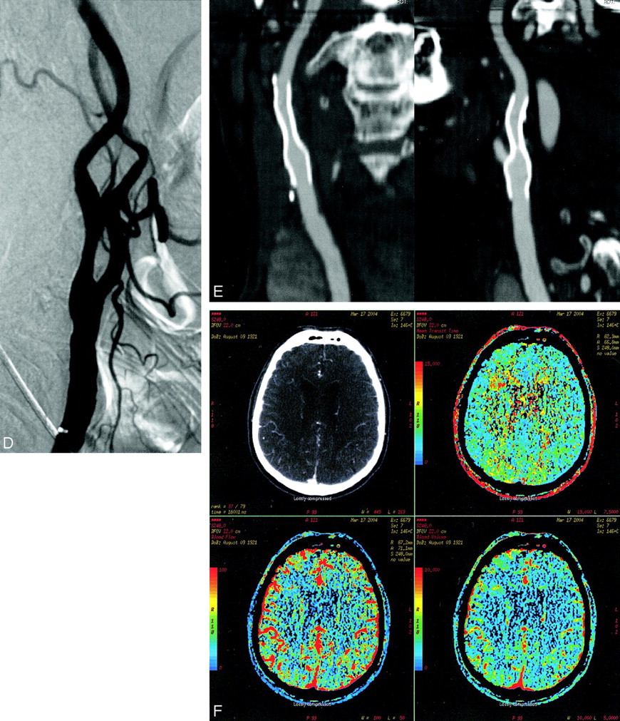
- Fig 1.
Sample patient with cervical internal carotid artery stenosis.
A, CTA 2D reformation showing the innominate, right common, and internal carotid arteries with a significant stenosis in the proximal ICA.
B, DSA showing right ICA stenosis.
C, Prestent CT perfusion demonstrating abnormal prolongation of mean transit time (MTT; upper right), decreased cerebral blood flow (CBF; lower left), and normal cerebral blood volume (CBV; bottom right).
D, DSA after stent placement.
E, CTA 2D multiplanar reformation with orthogonal projections through the stent.
F, CT perfusion after stent placement demonstrates normalization of MTT (upper right), CBF (bottom left), and CBV (bottom right).


Tables
Patient Number Age (years) Side of Stenosis Stenotic Vessel Urgent/Elective Etiology of Vessel Stenosis % Vessel Stenosis Prestent Perfusion Poststent Perfusion Cognitive Score Time of Imaging F/U (days) Time of Clinical F/U (month) Presence of Acom No. of Pcom(s) Functional COW 1 84 R ICA E A 65 Yes 2 7 70 14 − 0 N 2 66 L ICA E A 80 No ND 6 4 3 − 2 Y 3 83 R ICA E A >90 Yes 2 9 1 3 − 2 Y 4 88 R ICA U A >90 Yes 2 −8 0 5 − 1 N 5A 83 L ICA E A 70 No ND 6 51 5 − 0 N 5B 83 R ICA E A 70 No ND 6 47 5 − 0 N 6 63 R ICA E A 90 Yes 2 32 90 9 + 0 Y 7 63 R ICA E A 75 Yes 2 32 0 12 + 1 Y 8 62 R ECA E A >90 Yes 1 21 0 5 − 2 Y 9 31 L ICA U D >90 Yes 2 8 1 6 + 0 Y Note.—Cases of stents placed for extracranial carotid stenoses according to etiology of the stenoses (A indicates atherosclerosis; D, dissection); whether the stent was placed urgently (U) or electively (E); whether the case had prestent perfusion abnormality on imaging; whether the poststent perfusion abnormality normalized (2), improved (1), or did not change (0) (ND indicates patients who did not have prestent perfusion abnormality). The circle of Willis (COW) anatomy in each case is indicated by the presence or absence of anterior communicating artery (Acom) and the number of posterior communicating arteries (Pcom). The presence of a functional COW means that there is either a competent Acom or presence of a Pcom ipsilateral to the stenosis. Time of imaging follow-up is the number of days from the procedure that the follow-up perfusion imaging was performed. Time of clinical follow-up is the number of months from the procedure that the IQ-CODE was administered. L indicates left; R, right; ICA, internal cartoid artery; ECA, external carotid artery.
PatientNumber Age Side ofStenosis StenoticVessel Urgent/Elective Etiologyof VesselStenosis % Vessel Stenosis PrestentPerfusion PoststentPerfusion CognitiveScore Time of Imaging F/U (days) Time of Clinical F/U (month) Presenceof Acom No. ofPcom(s) FunctionalCOW 10 68 R ICA E A >85 Yes 2 9 9 4 − 1 N 11 69 L ICA E A >80 Yes 1 1 0 4 + 1 Y 12 58 R ICA E A 70 Yes 2 27 25 7 − 2 Y 13 66 L ICA E A 75 Yes 1 16 2 9 + 0 Y Note.—Cases of stents placed for intracranial carotid stenoses according to etiology of the stenoses (A indicates atherosclerosis; D, dissection); whether the stent was placed urgently (U) or electively (E); whether the case had prestent perfusion abnormality on imaging; whether the poststent perfusion abnormality normalized (2), improved (1), or did not change (0). The circle of Willis (COW) anatomy in each case is indicated by the presence or absence of anterior communicating artery (Acom) and number of posterior communicating arteries (Pcom). The presence of a functional COW means that there is either a competent Acom or presence of a Pcom ipsilateral to the stenosis. Time of imaging follow-up is the number of days from the procedure that the follow-up perfusion imaging was performed. Time of clinical follow-up is the number of months from the procedure that the IQ-CODE was administered. L indicates left; R, right; ICA, internal carotid artery.
PatientNumber Age Side ofStenosis StenoticVessel Urgent/Elective Etiologyof VesselStenosis % VesselStenosis PrestentPerfusion PoststentPerfusion CognitiveScore Time ofImagingF/U(days) Time ofClinicalF/U(month) Presenceof Acom No. ofPcom(s) FunctionalCOW 14 79 R V E A >85 Yes 1 21 0 3 − 0 N 15 66 L V E A 70 No ND 3 NA 6 + 1 Y 16 62 L V E A 70 No ND 0 30 5 − 1 Y 17 51 R VB U A 80 Yes 2 LFU 6 LFU − 0 N 18 69 R VB E A 80 Yes 0 3 4 10 + 0 N 19 56 M B U A 80 Yes 0 −3 1 3 − 2 Y 20 70 M B E A >75 No ND −1 21 4 − 2 Y Note.—Cases of stents placed for vertebrobasilar stenoses according to etiology of the stenoses (A indicates atherosclerosis; D, dissection); whether the stent was placed urgently (U) or electively (E); whether the case had prestent perfusion abnormality on imaging; whether the poststent perfusion abnormality normalized (2), improved (1), or did not change (0) (ND indicates patients who did not have prestent perfusion abnormality). The circle of Willis (COW) anatomy in each case is indicated by the presence or absence of anterior communicating artery (Acom) and the number of posterior communicating arteries (Pcom). The presence of a functional COW means that there is at least one Pcom. Time of imaging follow-up is the number of days from the procedure that the follow-up perfusion imaging was performed (NA indicates case number 15 did not have follow-up imaging). Time of clinical follow up is the number of months from the procedure that the IQ-CODE was administered. L indicates left; R, right; M, midline; V, vertebral; VB, vertebrobasilar junction; LFU, lost to follow-up.
In this issue


Jump to section
Related Articles
Cited By...
- Considering Psychological and Cognitive Factors in Interventional Neuroradiology: A Systematic Literature Review
- The association between vertebrobasilar insufficiency and the risk of dementia: a nationwide register-based retrospective cohort study in Taiwan
- Improvement of working memory after stenting for cervicocerebral artery stenosis
- Hemodynamic Alterations in Vertebrobasilar Large Artery Disease Assessed by Arterial Spin-Labeling MR Imaging
- The Role of Carotid Artery Stenting and Carotid Endarterectomy in Cognitive Performance: A Systematic Review
- New Brain Lesions After Carotid Stenting Versus Carotid Endarterectomy: A Systematic Review of the Literature