Advertisement

Main menu

  • Home
  • Content
    • Current Issue
    • Accepted Manuscripts
    • Article Preview
    • Past Issue Archive
    • Video Articles
    • AJNR Case Collection
    • Case of the Week Archive
    • Case of the Month Archive
    • Classic Case Archive
  • Special Collections
    • AJNR Awards
    • Low-Field MRI
    • Alzheimer Disease
    • ASNR Foundation Special Collection
    • Photon-Counting CT
    • View All
  • Multimedia
    • AJNR Podcasts
    • AJNR SCANtastic
    • Trainee Corner
    • MRI Safety Corner
    • Imaging Protocols
  • For Authors
    • Submit a Manuscript
    • Submit a Video Article
    • Submit an eLetter to the Editor/Response
    • Manuscript Submission Guidelines
    • Statistical Tips
    • Fast Publishing of Accepted Manuscripts
    • Graphical Abstract Preparation
    • Imaging Protocol Submission
    • Author Policies
  • About Us
    • About AJNR
    • Editorial Board
    • Editorial Board Alumni
  • More
    • Become a Reviewer/Academy of Reviewers
    • Subscribers
    • Permissions
    • Alerts
    • Feedback
    • Advertisers
    • ASNR Home

User menu

  • Alerts
  • Log in

Search

  • Advanced search
American Journal of Neuroradiology

American Journal of Neuroradiology

ASHNR American Society of Functional Neuroradiology ASHNR American Society of Pediatric Neuroradiology ASSR
  • Alerts
  • Log in

Advanced Search

  • Home
  • Content
    • Current Issue
    • Accepted Manuscripts
    • Article Preview
    • Past Issue Archive
    • Video Articles
    • AJNR Case Collection
    • Case of the Week Archive
    • Case of the Month Archive
    • Classic Case Archive
  • Special Collections
    • AJNR Awards
    • Low-Field MRI
    • Alzheimer Disease
    • ASNR Foundation Special Collection
    • Photon-Counting CT
    • View All
  • Multimedia
    • AJNR Podcasts
    • AJNR SCANtastic
    • Trainee Corner
    • MRI Safety Corner
    • Imaging Protocols
  • For Authors
    • Submit a Manuscript
    • Submit a Video Article
    • Submit an eLetter to the Editor/Response
    • Manuscript Submission Guidelines
    • Statistical Tips
    • Fast Publishing of Accepted Manuscripts
    • Graphical Abstract Preparation
    • Imaging Protocol Submission
    • Author Policies
  • About Us
    • About AJNR
    • Editorial Board
    • Editorial Board Alumni
  • More
    • Become a Reviewer/Academy of Reviewers
    • Subscribers
    • Permissions
    • Alerts
    • Feedback
    • Advertisers
    • ASNR Home
  • Follow AJNR on Twitter
  • Visit AJNR on Facebook
  • Follow AJNR on Instagram
  • Join AJNR on LinkedIn
  • RSS Feeds

AJNR Awards, New Junior Editors, and more. Read the latest AJNR updates

Research ArticleBRAIN

Trigeminal Ganglion and its Divisions: Detailed Anatomic MR Imaging with Contrast-Enhanced 3D Constructive Interference in the Steady State Sequences

Indra Yousry, Bernhard Moriggl, Urs D. Schmid, Thomas P. Naidich and Tarek A. Yousry
American Journal of Neuroradiology May 2005, 26 (5) 1128-1135;
Indra Yousry
  • Find this author on Google Scholar
  • Find this author on PubMed
  • Search for this author on this site
Bernhard Moriggl
  • Find this author on Google Scholar
  • Find this author on PubMed
  • Search for this author on this site
Urs D. Schmid
  • Find this author on Google Scholar
  • Find this author on PubMed
  • Search for this author on this site
Thomas P. Naidich
  • Find this author on Google Scholar
  • Find this author on PubMed
  • Search for this author on this site
Tarek A. Yousry
  • Find this author on Google Scholar
  • Find this author on PubMed
  • Search for this author on this site
  • Article
  • Figures & Data
  • Info & Metrics
  • Responses
  • References
  • PDF
Loading

Article Figures & Data

Figures

  • Tables
  • Fig 1.
    • Download figure
    • Open in new tab
    • Download powerpoint
    Fig 1.

    Sagittal cryomicrotomic section through the Meckel cave in the plane of the trigeminal nerves was obtained from the right medial aspect of a cadaveric specimen from a 74-year-old woman. Small portion of the sensory root (SeR) is seen at its entry into the cave. Trigeminal ganglion (thick white arrow) is at the anterior aspect of the cave, and anterior margin of the ganglion adheres to the dural wall (white arrowhead). Multiple sensory rootlets (long black arrows) arise from the concave medial surface of the ganglion (sinus ganglii) (short black arrow) to course in the trigeminal cistern (TC). Superior and inferior lips of the ganglion (thin white arrows) are particularly well displayed. Portion of the maxillary nerve (V2) shown in this plane is surrounded by venous channels (black arrowheads) along the inferior border of the cavernous sinus. Ant = anterior, CA = carotid artery, PC = prepontine cistern, Post = posterior, TE = cerebellar tentorium, TL = temporal lobe.

  • Fig 2.
    • Download figure
    • Open in new tab
    • Download powerpoint
    Fig 2.

    Sagittal nonenhanced 3D CISS image through the right side of the Meckel cave in a 58-year-old woman. Sensory root (SeR) is shown from its apparent origin at the pons (P) to the entrance of the cave. Ganglion (thick white arrow) at the anterior aspect of the cave is difficult to identify, and anterior margin of the trigeminal ganglion cannot be distinguished from the dural wall (arrowhead) of the cave. Only the superior lip of the ganglion (thin white arrow) is well defined. One smaller and one larger sensory rootlet (long black arrows) arise from the sinus ganglii (short black arrow) and course in the trigeminal cistern (TC). Maxillary nerve is not depicted at the inferior border of the cavernous sinus. CA = carotid artery, PC = prepontine cistern, TE = cerebellar tentorium, TL = temporal lobe.
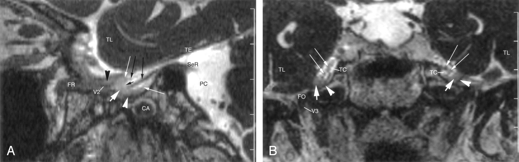
  • Fig 3.
    • Download figure
    • Open in new tab
    • Download powerpoint
    Fig 3.

    Enhanced 3D CISS images through the right side of the Meckel cave in a 62-year-old woman. TL = temporal lobe.

    A, Sagittal image shows a small portion of the sensory root (SeR) at its entry into the cave. Trigeminal ganglion (thick white arrow) and its lips (thin white arrows) are shown at the anterior end of the cave. Anterior margin of the ganglion is clearly differentiated from the dural wall (white arrowhead) of the cave. Two sensory rootlets (long black arrows) emerge from the sinus ganglii (short black arrow) and pass through the trigeminal cistern. Maxillary nerve (V2) courses along the inferior wall of the cavernous sinus, surrounded by enhancing venous channels (black arrowhead). CA = carotid artery, FR = foramen rotundum, PC = prepontine cistern, TE = cerebellar tentorium.

    B, Coronal image shows the cave bilaterally. Trigeminal ganglion (thick white arrows) lies along the anteroinferior border of the cave. Less-enhancing dura (arrowheads) is clearly differentiated from the ganglion. Multiple, small sensory rootlets (thin white arrows) are seen in the trigeminal cistern (TC). Mandibular nerve (V3) is partly shown on the right side in the foramen ovale (FO).

  • Fig 4.
    • Download figure
    • Open in new tab
    • Download powerpoint
    Fig 4.

    Sagittal enhanced 3D CISS image through the right side of the Meckel cave in a 54-year-old man. Sensory root (SeR) is displayed from its apparent origin at the pons (P) to the entrance of the cave. Trigeminal ganglion (thick white arrow) is at the anteroinferior border of the cave. Anterior margin of the enhancing ganglion is clearly distinguished from the less-enhancing dura (white arrowhead) of the cave. Lips of the ganglion (thin white arrows) define the sinus ganglii (short black arrow) from which small sensory rootlets (long black arrows) arise. Maxillary nerve (V2) is depicted along the inferior border of the cavernous sinus, surrounded by venous channels (black arrowhead) en route to the foramen rotundum (FR). CA = carotid artery, MoR = motor root of the trigeminal nerve at its point of exit, PC = prepontine cistern, TE = cerebellar tentorium, TL = temporal lobe.

  • Fig 5.
    • Download figure
    • Open in new tab
    • Download powerpoint
    Fig 5.

    Sagittal enhanced 3D TOF image through the right side of the Meckel cave in a 53-year-old-man. Enhancing ganglion (thick white arrow) is shown at the anteroinferior margin of the cave, in continuity with the dural wall (white arrowhead). Superior and inferior lips of the ganglion (thin white arrows) are well depicted. No sensory rootlets are seen in the trigeminal cistern (TC). Ophthalmic nerve (V1) and maxillary nerve (V2) are hypointense, linear structures surrounded by strongly enhancing venous channels (black arrowheads) in the lateral wall and along the inferior border, respectively, of the cavernous sinus. V2 enters the foramen rotundum (FR), while V1 passes to the superior orbital fissure (SOF). CA = carotid artery, PC = prepontine cistern, TE = cerebellar tentorium, TL = temporal lobe.

Tables

  • Figures
    • View popup
    TABLE 1:

    Identification of the trigeminal ganglion

    StudyPlane and Score*
    SagittalTransverseCoronal
    210210210
    Nonenhanced 3D CISS (44 sides)7 (15.9)27 (61.4)10 (22.7)2 (4.5)19 (43.2)33 (75)2 (4.5)16 (36.4)26 (59.1)
    Enhanced 3D CISS (44 sides)35 (79.5)9 (20.5)0 (0)32 (72.7)12 (27.3)0 (0)22 (50)19 (43.2)3 (6.8)
    Enhanced 3D TOF (42 sides)32 (76.2)7 (16.7)3 (7.1)16 (38.1)17 (40.5)9 (21.4)20 (47.6)14 (33.3)8 (19)
    • Note.—Data are numbers of ganglia (percentage).

    • * Scores: 2 = positive identification, 1 = highly probable identification, 0 = no identification.

    • View popup
    TABLE 2:

    Thickness of the trigeminal ganglion in the sagittal plane

    StudyThickness of Ganglion
    Not Determined1 mm2 mm3 mm
    Nonenhanced 3D CISS (44 sides)18 (40.9)11 (25)14 (31.8)1 (2.3)
    Enhanced 3D CISS (44 sides)1 (2.3)12 (27.3)31 (70.5)0 (0)
    Enhanced 3D TOF (42 sides)3 (7.1)12 (28.6)27 (64.3)0 (0)
    • Note.—Data are numbers of ganglia (percentage).

    • View popup
    TABLE 3:

    MR imaging identification of upper and lower lips of the ganglion and sinus ganglii in the sagittal plane

    StudyScore*
    210
    Nonenhanced 3D CISS (44 sides)19 (43.2)15 (34.1)10 (22.7)
    Enhanced 3D CISS (44 sides)42 (95.5)1 (2.5)1 (2.5)
    Enhanced 3D TOF (42 sides)4 (9.5)29 (69)8 (19)
    • Note.—Data are numbers of ganglia (percentage).

    • * Scores: 2 = positive identification, 1 = highly probable identification, 0 = no identification.

    • View popup
    TABLE 4:

    MR imaging identification of the mandibular, maxillary, and ophthalmic nerves

    Study and PlaneV3 *V2 †V1 ‡
    210210210
    Nonenhanced 3D CISS (44 sides)
        Transverse2 (4.5)0 (0)42 (95.5)0 (0)0 (0)44 (100)0 (0)1 (2.3)43 (97.7)
        Sagittal7 (15.9)11 (25)24 (54.5)0 (0)5 (11.4)39 (88.6)0 (0)18 (18.2)36 (81.1)
        Coronal6 (13.6)15 (34.1)23 (52.3)0 (0)0 (0)22 (100)0 (0)0 (0)44 (100)
    Enhanced 3D CISS (44 sides)
        Transverse26 (59.1)9 (20.5)9 (20.5)18 (41)22 (50)4 (9.1)35 (79.5)9 (20.5)0 (0)
        Sagittal44 (100)0 (0)0 (0)24 (54.5)17 (38.6)3 (6.8)39 (88.6)4 (9.1)1 (2.3)
        Coronal44 (100)0 (0)0 (0)13 (29.5)24 (54.5)7 (15.9)28 (63.6)12 (27.3)4 (9.1)
    Enhanced 3D TOF (42 sides)
        Transverse34 (81)18 (42.9)0 (0)33 (78.6)8 (19)0 (0)39 (92.9)3 (7.1)0 (0)
        Sagittal39 (92.9)3 (7.1)0 (0)31 (73.8)11 (26.2)0 (0)38 (90.5)4 (9.5)0 (0)
        Coronal42 (100)0 (0)0 (0)30 (71.4)11 (26.2)1 (2.4)30 (71.4)12 (28.6)0 (0)
    • Note.—Data are the numbers of nerves (percentage).

    • * Inside foramen ovale.

    • † At the inferior border of the cavernous sinus and inside the foramen rotundum.

    • ‡ Inside the lateral wall of the cavernous sinus and the superior orbital fissure.

PreviousNext
Back to top

In this issue

American Journal of Neuroradiology: 26 (5)
American Journal of Neuroradiology
Vol. 26, Issue 5
1 May 2005
  • Table of Contents
  • Index by author
Advertisement
Print
Download PDF
Email Article

Thank you for your interest in spreading the word on American Journal of Neuroradiology.

NOTE: We only request your email address so that the person you are recommending the page to knows that you wanted them to see it, and that it is not junk mail. We do not capture any email address.

Enter multiple addresses on separate lines or separate them with commas.
Trigeminal Ganglion and its Divisions: Detailed Anatomic MR Imaging with Contrast-Enhanced 3D Constructive Interference in the Steady State Sequences
(Your Name) has sent you a message from American Journal of Neuroradiology
(Your Name) thought you would like to see the American Journal of Neuroradiology web site.
CAPTCHA
This question is for testing whether or not you are a human visitor and to prevent automated spam submissions.
Cite this article
Indra Yousry, Bernhard Moriggl, Urs D. Schmid, Thomas P. Naidich, Tarek A. Yousry
Trigeminal Ganglion and its Divisions: Detailed Anatomic MR Imaging with Contrast-Enhanced 3D Constructive Interference in the Steady State Sequences
American Journal of Neuroradiology May 2005, 26 (5) 1128-1135;

Citation Manager Formats

  • BibTeX
  • Bookends
  • EasyBib
  • EndNote (tagged)
  • EndNote 8 (xml)
  • Medlars
  • Mendeley
  • Papers
  • RefWorks Tagged
  • Ref Manager
  • RIS
  • Zotero
0 Responses
Respond to this article
Share
Bookmark this article
Trigeminal Ganglion and its Divisions: Detailed Anatomic MR Imaging with Contrast-Enhanced 3D Constructive Interference in the Steady State Sequences
Indra Yousry, Bernhard Moriggl, Urs D. Schmid, Thomas P. Naidich, Tarek A. Yousry
American Journal of Neuroradiology May 2005, 26 (5) 1128-1135;
del.icio.us logo Twitter logo Facebook logo Mendeley logo
  • Tweet Widget
  • Facebook Like
  • Google Plus One

Jump to section

  • Article
    • Abstract
    • Methods
    • Results
    • Discussion
    • Conclusion
    • References
  • Figures & Data
  • Info & Metrics
  • Responses
  • References
  • PDF

Related Articles

  • No related articles found.
  • PubMed
  • Google Scholar

Cited By...

  • Contrast-Enhanced CISS Imaging for Evaluation of Neurovascular Compression in Trigeminal Neuralgia: Improved Correlation with Symptoms and Prediction of Surgical Outcomes
  • Detection of the Stellate and Thoracic Sympathetic Chain Ganglia with High-Resolution 3D-CISS MR Imaging
  • Visualization of the Trochlear Nerve in the Cistern with Use of High-Resolution Turbo Spin-Echo Multisection Motion-Sensitized Driven Equilibrium
  • 3D Double-Echo Steady-State with Water Excitation MR Imaging of the Intraparotid Facial Nerve at 1.5T: A Pilot Study
  • Is All "Communicating" Hydrocephalus Really Communicating? Prospective Study on the Value of 3D-Constructive Interference in Steady State Sequence at 3T
  • The Jugular Foramen: Imaging Strategy and Detailed Anatomy at 3T
  • Crossref
  • Google Scholar

This article has not yet been cited by articles in journals that are participating in Crossref Cited-by Linking.

More in this TOC Section

  • Usefulness of Quantitative Susceptibility Mapping for the Diagnosis of Parkinson Disease
  • White Matter Alterations in the Brains of Patients with Active, Remitted, and Cured Cushing Syndrome: A DTI Study
  • Qualitative and Quantitative Analysis of MR Imaging Findings in Patients with Middle Cerebral Artery Stroke Implanted with Mesenchymal Stem Cells
Show more BRAIN

Similar Articles

Advertisement

Indexed Content

  • Current Issue
  • Accepted Manuscripts
  • Article Preview
  • Past Issues
  • Editorials
  • Editor's Choice
  • Fellows' Journal Club
  • Letters to the Editor
  • Video Articles

Cases

  • Case Collection
  • Archive - Case of the Week
  • Archive - Case of the Month
  • Archive - Classic Case

More from AJNR

  • Trainee Corner
  • Imaging Protocols
  • MRI Safety Corner

Multimedia

  • AJNR Podcasts
  • AJNR Scantastics

Resources

  • Turnaround Time
  • Submit a Manuscript
  • Submit a Video Article
  • Submit an eLetter to the Editor/Response
  • Manuscript Submission Guidelines
  • Statistical Tips
  • Fast Publishing of Accepted Manuscripts
  • Graphical Abstract Preparation
  • Imaging Protocol Submission
  • Evidence-Based Medicine Level Guide
  • Publishing Checklists
  • Author Policies
  • Become a Reviewer/Academy of Reviewers
  • News and Updates

About Us

  • About AJNR
  • Editorial Board
  • Editorial Board Alumni
  • Alerts
  • Permissions
  • Not an AJNR Subscriber? Join Now
  • Advertise with Us
  • Librarian Resources
  • Feedback
  • Terms and Conditions
  • AJNR Editorial Board Alumni

American Society of Neuroradiology

  • Not an ASNR Member? Join Now

© 2025 by the American Society of Neuroradiology All rights, including for text and data mining, AI training, and similar technologies, are reserved.
Print ISSN: 0195-6108 Online ISSN: 1936-959X

Powered by HighWire