
Article Figures & Data
Figures
- Fig 1.
Example of a tissue sample and the matching area on postmortem MR images. WM lesions (WM), as well as type I lesions (I, mixed GM-WM), can be seen with relative ease on the different MR images. Intracortical lesions (IC) are difficult to detect and define, even in retrospect.
A, Photomicrograph (MBP immunohistochemical stain) reveals lesions (arrowheads) in the WM and cortical GM.
B, Short-echo T2-weighted SE image. Insert, a higher magnification of the intracortical lesion.
C, Long-echo T2-weighted SE image.
D, 3D FLAIR image.
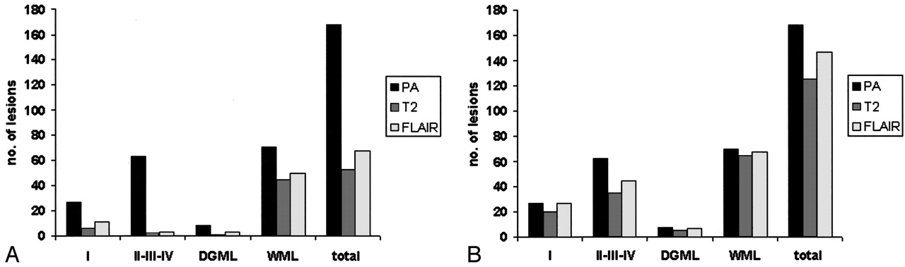
- Fig 2.
Lesion scores. I, type I cortical lesions; DGML indicates deep GM lesions; WML, WM lesions; total, total of all lesion categories; PA, lesions detected histopathologically; T2, lesions detected on postmortem T2SE imaging; and FLAIR, lesions detected on postmortem 3D FLAIR imaging.
A, Blinded.
B, After unblinding, numbers of detected lesions in all categories increased (retrospective scoring). However, a significant proportion of intracortical lesions remained undetectable.


Tables
Patient/Sex/Age (y) Postmortem Delay (h:min) Disease Duration (y) Type Cause of death 1/M/43 08:30 17 Secondary progressive Pneumonia 2/F/48 04:50 25 Secondary progressive Euthanasia 3/F/72 10:30 13 Unknown Pneumonia 4/M/77 04:15 32 Primary progressive Cerebral infarct 5/F/75 08:00 42 Secondary progressive Pneumonia 6/F/53 10:45 23 Secondary progressive Euthanasia 7/M/81 08:50 51 Primary progressive General deterioration 8/F/59 08:45 24 Secondary progressive Respiratory insufficiency 9/F/48 05:50 21 Unknown Congestive heart failure Note.—Mean age was 62 years, mean postmortem delay was 7:50, and mean disease duration was 28 y.
Type Lesions on Histopathology Prospective* Retrospective† T2SE FLAIR T2SE FLAIR I 27 6 (22) 11 (41) 20 (74) 27 (100) II 12 0 (0) 1 (8) 6 (50) 9 (75) III 41 1 (2) 2 (5) 22 (54) 26 (63) IV 10 1 (10) 0 (0) 7 (70) 10 (100) II-IV 63 2 (3) 3 (5) 35 (56) 45 (71) Deep GM 8 1 (13) 3 (38) 5 (63) 7 (88) WM 70 44 (63) 50 (71) 65 (93) 68 (97) Total 168 53 (32) 67 (40) 125 (74) 147 (88) Note.—Data in parentheses are the sensitivity (%).
* Lesions found without knowledge of histopathologic localization and type.
† Lesions found after the histopathologic localization of lesions was revealed.
In this issue


Jump to section
Related Articles
Cited By...
- Innate Immune Cell-Related Pathology in the Thalamus Signals a Risk for Disability Progression in Multiple Sclerosis
- T1/T2 Ratio Imaging Improves Cortical Lesion Contrast in Multiple Sclerosis on 3T MRI
- Imaging cortical multiple sclerosis lesions with ultra-high field MRI
- Pathologic correlates of the magnetization transfer ratio in multiple sclerosis
- Comparison of Multiple Sclerosis Cortical Lesion Types Detected by Multicontrast 3T and 7T MRI
- Detection of Leukocortical Lesions in Multiple Sclerosis and Their Association with Physical and Cognitive Impairment: A Comparison of Conventional and Synthetic Phase-Sensitive Inversion Recovery MRI
- Magnetic Resonance Imaging in Multiple Sclerosis
- Improved Visualization of Cortical Lesions in Multiple Sclerosis Using 7T MP2RAGE
- MRI evidence of acute inflammation in leukocortical lesions of patients with early multiple sclerosis
- Relationship of grey and white matter abnormalities with distance from the surface of the brain in multiple sclerosis
- Manual Segmentation of MS Cortical Lesions Using MRI: A Comparison of 3 MRI Reading Protocols
- A longitudinal study of cortical grey matter lesion subtypes in relapse-onset multiple sclerosis
- DIR-visible grey matter lesions and atrophy in multiple sclerosis: partners in crime?
- Ultra-High-Field MRI Visualization of Cortical Multiple Sclerosis Lesions with T2 and T2*: A Postmortem MRI and Histopathology Study
- Reduced grey matter perfusion without volume loss in early relapsing-remitting multiple sclerosis
- Multicontrast MR Imaging at 7T in Multiple Sclerosis: Highest Lesion Detection in Cortical Gray Matter with 3D-FLAIR
- Clinically feasible MTR is sensitive to cortical demyelination in MS
- Improved detection of cortical MS lesions with phase-sensitive inversion recovery MRI
- Exogenous Leukemia Inhibitory Factor Stimulates Oligodendrocyte Progenitor Cell Proliferation and Enhances Hippocampal Remyelination
- Postmortem verification of MS cortical lesion detection with 3D DIR
- Correlating Quantitative MR Imaging with Histopathology in X-Linked Adrenoleukodystrophy
- Identification and Clinical Impact of Multiple Sclerosis Cortical Lesions as Assessed by Routine 3T MR Imaging
- MS cortical lesion or not?: Double inversion recovery MRI reveals some answers and uncertainties
- Heterogeneity of small vessel disease: a systematic review of MRI and histopathology correlations
- Consensus recommendations for MS cortical lesion scoring using double inversion recovery MRI
- T2 lesion location really matters: a 10 year follow-up study in primary progressive multiple sclerosis
- Toward understanding cortical lesions in multiple sclerosis
- Reply:
- MRI criteria for MS in patients with clinically isolated syndromes
- What you see depends on how you look: Gray matter lesions in multiple sclerosis
- In vivo imaging of cortical pathology in multiple sclerosis using ultra-high field MRI
- First Clinical Study on Ultra-High-Field MR Imaging in Patients with Multiple Sclerosis: Comparison of 1.5T and 7T
- Magnetisation transfer ratio in the normal appearing white matter predicts progression of disability over 1 year in early primary progressive multiple sclerosis
- Thalamic atrophy and cognition in multiple sclerosis
- Gray matter involvement in multiple sclerosis