Advertisement

Main menu

  • Home
  • Content
    • Current Issue
    • Accepted Manuscripts
    • Article Preview
    • Past Issue Archive
    • Video Articles
    • AJNR Case Collection
    • Case of the Week Archive
    • Case of the Month Archive
    • Classic Case Archive
  • Special Collections
    • AJNR Awards
    • Low-Field MRI
    • Alzheimer Disease
    • ASNR Foundation Special Collection
    • Photon-Counting CT
    • View All
  • Multimedia
    • AJNR Podcasts
    • AJNR SCANtastic
    • Trainee Corner
    • MRI Safety Corner
    • Imaging Protocols
  • For Authors
    • Submit a Manuscript
    • Submit a Video Article
    • Submit an eLetter to the Editor/Response
    • Manuscript Submission Guidelines
    • Statistical Tips
    • Fast Publishing of Accepted Manuscripts
    • Graphical Abstract Preparation
    • Imaging Protocol Submission
    • Author Policies
  • About Us
    • About AJNR
    • Editorial Board
    • Editorial Board Alumni
  • More
    • Become a Reviewer/Academy of Reviewers
    • Subscribers
    • Permissions
    • Alerts
    • Feedback
    • Advertisers
    • ASNR Home

User menu

  • Alerts
  • Log in

Search

  • Advanced search
American Journal of Neuroradiology

American Journal of Neuroradiology

ASHNR American Society of Functional Neuroradiology ASHNR American Society of Pediatric Neuroradiology ASSR
  • Alerts
  • Log in

Advanced Search

  • Home
  • Content
    • Current Issue
    • Accepted Manuscripts
    • Article Preview
    • Past Issue Archive
    • Video Articles
    • AJNR Case Collection
    • Case of the Week Archive
    • Case of the Month Archive
    • Classic Case Archive
  • Special Collections
    • AJNR Awards
    • Low-Field MRI
    • Alzheimer Disease
    • ASNR Foundation Special Collection
    • Photon-Counting CT
    • View All
  • Multimedia
    • AJNR Podcasts
    • AJNR SCANtastic
    • Trainee Corner
    • MRI Safety Corner
    • Imaging Protocols
  • For Authors
    • Submit a Manuscript
    • Submit a Video Article
    • Submit an eLetter to the Editor/Response
    • Manuscript Submission Guidelines
    • Statistical Tips
    • Fast Publishing of Accepted Manuscripts
    • Graphical Abstract Preparation
    • Imaging Protocol Submission
    • Author Policies
  • About Us
    • About AJNR
    • Editorial Board
    • Editorial Board Alumni
  • More
    • Become a Reviewer/Academy of Reviewers
    • Subscribers
    • Permissions
    • Alerts
    • Feedback
    • Advertisers
    • ASNR Home
  • Follow AJNR on Twitter
  • Visit AJNR on Facebook
  • Follow AJNR on Instagram
  • Join AJNR on LinkedIn
  • RSS Feeds

AJNR Awards, New Junior Editors, and more. Read the latest AJNR updates

Research ArticleBRAIN

Cranial MR Findings in Chronic Toluene Abuse by Inhalation

Kubilay Aydin, Serra Sencer, Turkay Demir, Kultekin Ogel, Atadan Tunaci and Ozenc Minareci
American Journal of Neuroradiology August 2002, 23 (7) 1173-1179;
Kubilay Aydin
  • Find this author on Google Scholar
  • Find this author on PubMed
  • Search for this author on this site
Serra Sencer
  • Find this author on Google Scholar
  • Find this author on PubMed
  • Search for this author on this site
Turkay Demir
  • Find this author on Google Scholar
  • Find this author on PubMed
  • Search for this author on this site
Kultekin Ogel
  • Find this author on Google Scholar
  • Find this author on PubMed
  • Search for this author on this site
Atadan Tunaci
  • Find this author on Google Scholar
  • Find this author on PubMed
  • Search for this author on this site
Ozenc Minareci
  • Find this author on Google Scholar
  • Find this author on PubMed
  • Search for this author on this site
  • Article
  • Figures & Data
  • Info & Metrics
  • Responses
  • References
  • PDF
Loading

Article Figures & Data

Figures

  • Tables
  • Fig 1.
    • Download figure
    • Open in new tab
    • Download powerpoint
    Fig 1.

    Axial T2-weighted (2900/85/2 [TR/TE/NEX]) images in a 16-year-old patient who had inhaled toluene for 6 years.

    A, High signal intensity is seen in the centrum semiovale (arrows) on both sides. The peripheral cerebral white matter and gray matter-white matter differentiation are preserved.

    B, High-signal-intensity changes involve the frontal and parietal periventricular white matter (arrowheads). The pattern of white matter changes is compatible with that of the restricted type. Note that the lateral ventricles and cerebral sulci are enlarged.
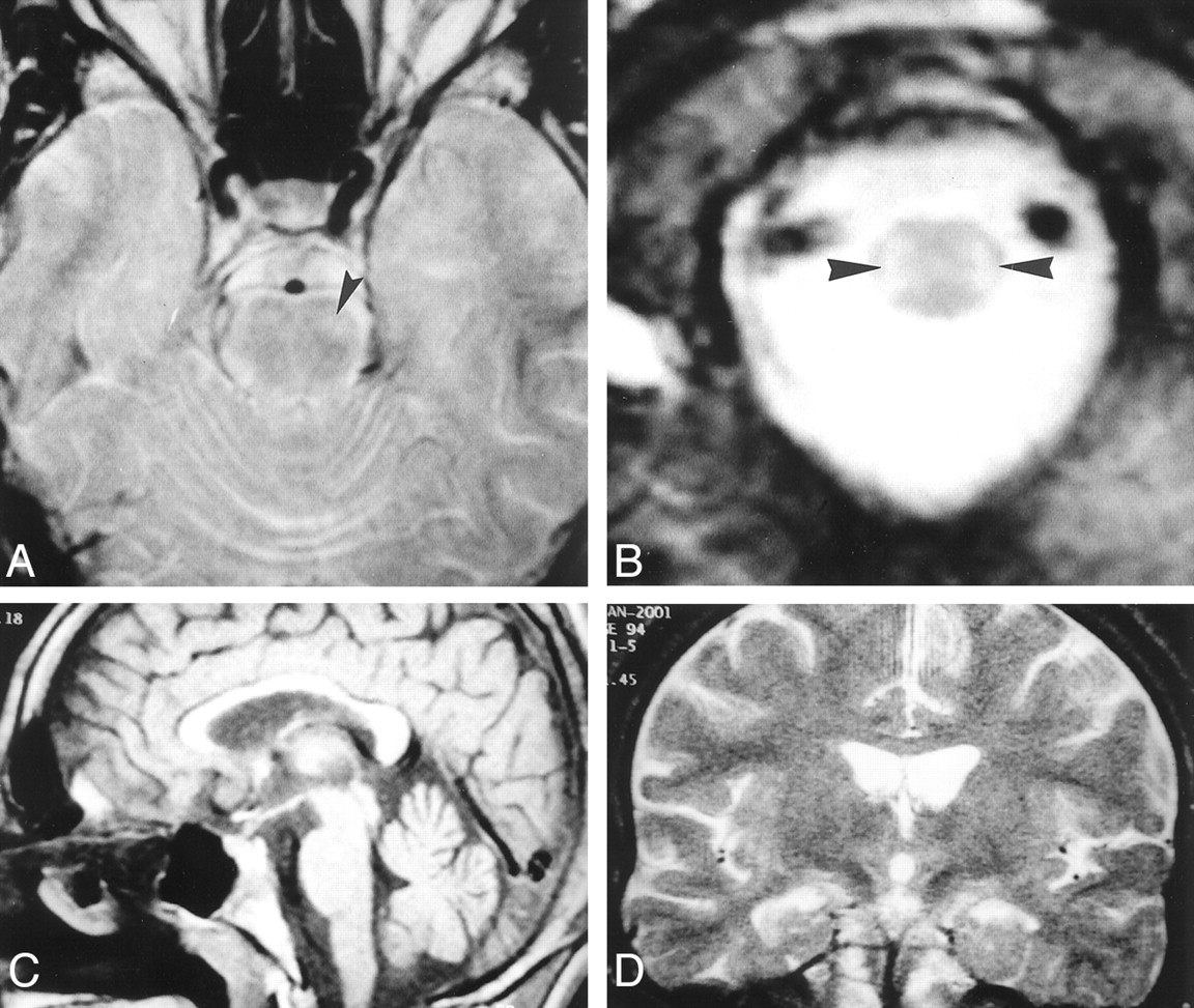
  • Fig 2.
    • Download figure
    • Open in new tab
    • Download powerpoint
    Fig 2.

    MR images in a 19-year-old patient who had abused toluene for 8 years.

    A, Axial T2-weighted (2620/85/2 [TR/TE/NEX]) image at the level of the brain stem shows a hyperintense lesion in the anterolateral part of pons (arrowhead). Note that gray matter-white matter differentiation is lost in the anterior part of the left temporal lobe.

    B, Axial T2-weighted (2620/85/2) image at the level of lower medulla oblongata shows symmetric hyperintensity along the spinocerebellar tracts (arrowheads).

    C, Sagittal T1-weighted (600/15/1) midline image shows thinning of the corpus callosum, which is more prominent in the body and genu. Note the enlarged cerebellar sulci.

    D, Coronal T2-weighted image (4800/100/2) reveals a lack of signal intensity abnormality in the corpus callosum.
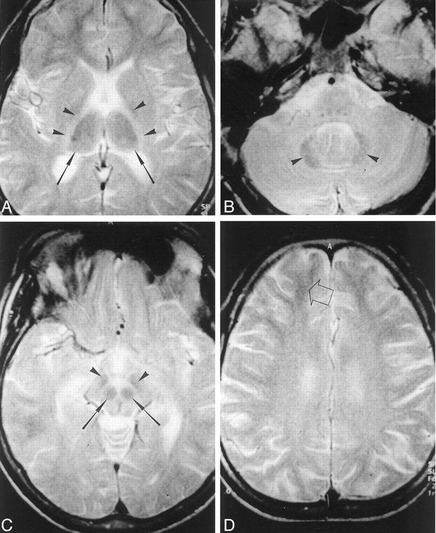
  • Fig 3.
    • Download figure
    • Open in new tab
    • Download powerpoint
    Fig 3.

    Axial T2-weighted MR images (3000/80/2 [TR/TE/NEX]) in a 22-year-old patient who had abused thinner for 11 years.

    A, Symmetric hypointensity is present in the thalami (arrows). Symmetric hyperintensity exists in the posterior limbs of the internal capsule (arrowheads).

    B, At the level of the posterior fossa, generalized increased signal intensity in the cerebellar white matter emphasizes the dentate nucleus (arrowheads).

    C, At the level of midbrain, red nuclei (arrows) and substantia nigra (arrowheads) are hypointense.

    D, Diffuse high-signal-intensity in the centrum semiovale causes loss of gray matter-white matter differentiation. Gray matter-white matter differentiation is preserved only in the subcortical-frontal white matter (arrow). The cerebral sulci are mildly dilated.

  • Fig 4.
    • Download figure
    • Open in new tab
    • Download powerpoint
    Fig 4.

    Axial T2-weighted (3000/80/2 [TR/TE/NEX]) images in a 16-year-old patient who had inhaled thinner for 3 years.

    A and B, Bilateral, symmetric hyperintensity is present in the parietal periventricular white matter (arrowheads). Note that gray matter-white matter differentiation is preserved. The widths of the cerebral sulci are normal. A suggests that the hyperintense findings may be caused by terminal myelination, which is a normal MR imaging finding in children. However, B shows that the lesions extend to the bilateral centrum semiovale, indicating that hyperintense findings were true lesions rather than terminal myelination.

Tables

  • Figures
  • Distribution of clinical and MR imaging findings with respect to the duration of toluene abuse

    Duration of Abuse (y)No. of PatientsSymptoms*Neurologic Signs*Cases with Positive MR Imaging Finding (n)
    White Matter ChangesThalamic HypointensityThin Corpus Callosum
    RestrictedDiffuse
    18IS (5), FG (4), ANS (5), TN (1)None0000
    24IS (4), FG (3), ANS (3), TN (1)None0000
    33IS (2), FG (3), ANS (2)ATX (2)1000
    45IS (5), FG (5), ANS (3)ATX (2), TM (2), SP (1)3000
    56IS (4), FG (5), ANS (3), TN (3)ATX (1), TM (1)1001
    64IS (3), FG (3), ANS (3), TN (3)ATX (3), TM (2)1211
    73IS (3), FG (3), ANS (3), TN (2)ATX (3), TM (3), SP (2), DA (1)1222
    84IS (4), FG (4), ANS (4), TN (2)ATX (4), TM (3), SP (3), NYS (1), DA (1)2222
    93IS (3), FG (3), ANS (3), TN (2)ATX (3), TM (3), SP (3), NYS (1), DA (1)1222
    111IS, FG, ANS, TNATX, TM, SP, NYS, DA0111
    • * ANS indicates anosmia; ATX, ataxia; DA, dysarthria; FG, forgetfulness; IS, insomnia; NYS, nystagmus; SP, spasticity; TM, tremors; and TN, tinnitus. Numbers in parentheses the number of cases.

PreviousNext
Back to top

In this issue

American Journal of Neuroradiology: 23 (7)
American Journal of Neuroradiology
Vol. 23, Issue 7
1 Aug 2002
  • Table of Contents
  • Index by author
Advertisement
Print
Download PDF
Email Article

Thank you for your interest in spreading the word on American Journal of Neuroradiology.

NOTE: We only request your email address so that the person you are recommending the page to knows that you wanted them to see it, and that it is not junk mail. We do not capture any email address.

Enter multiple addresses on separate lines or separate them with commas.
Cranial MR Findings in Chronic Toluene Abuse by Inhalation
(Your Name) has sent you a message from American Journal of Neuroradiology
(Your Name) thought you would like to see the American Journal of Neuroradiology web site.
CAPTCHA
This question is for testing whether or not you are a human visitor and to prevent automated spam submissions.
Cite this article
Kubilay Aydin, Serra Sencer, Turkay Demir, Kultekin Ogel, Atadan Tunaci, Ozenc Minareci
Cranial MR Findings in Chronic Toluene Abuse by Inhalation
American Journal of Neuroradiology Aug 2002, 23 (7) 1173-1179;

Citation Manager Formats

  • BibTeX
  • Bookends
  • EasyBib
  • EndNote (tagged)
  • EndNote 8 (xml)
  • Medlars
  • Mendeley
  • Papers
  • RefWorks Tagged
  • Ref Manager
  • RIS
  • Zotero
0 Responses
Respond to this article
Share
Bookmark this article
Cranial MR Findings in Chronic Toluene Abuse by Inhalation
Kubilay Aydin, Serra Sencer, Turkay Demir, Kultekin Ogel, Atadan Tunaci, Ozenc Minareci
American Journal of Neuroradiology Aug 2002, 23 (7) 1173-1179;
del.icio.us logo Twitter logo Facebook logo Mendeley logo
  • Tweet Widget
  • Facebook Like
  • Google Plus One

Jump to section

  • Article
    • Abstract
    • Methods
    • Results
    • Discussion
    • Conclusion
    • References
  • Figures & Data
  • Info & Metrics
  • Responses
  • References
  • PDF

Related Articles

  • No related articles found.
  • PubMed
  • Google Scholar

Cited By...

  • Addictive Illegal Drugs: Structural Neuroimaging
  • Smaller Gray Matter Volumes in Frontal and Parietal Cortices of Solvent Abusers Correlate with Cognitive Deficits
  • Crossref
  • Google Scholar

This article has not yet been cited by articles in journals that are participating in Crossref Cited-by Linking.

More in this TOC Section

  • Usefulness of Quantitative Susceptibility Mapping for the Diagnosis of Parkinson Disease
  • White Matter Alterations in the Brains of Patients with Active, Remitted, and Cured Cushing Syndrome: A DTI Study
  • Qualitative and Quantitative Analysis of MR Imaging Findings in Patients with Middle Cerebral Artery Stroke Implanted with Mesenchymal Stem Cells
Show more BRAIN

Similar Articles

Advertisement

Indexed Content

  • Current Issue
  • Accepted Manuscripts
  • Article Preview
  • Past Issues
  • Editorials
  • Editor's Choice
  • Fellows' Journal Club
  • Letters to the Editor
  • Video Articles

Cases

  • Case Collection
  • Archive - Case of the Week
  • Archive - Case of the Month
  • Archive - Classic Case

More from AJNR

  • Trainee Corner
  • Imaging Protocols
  • MRI Safety Corner
  • Book Reviews

Multimedia

  • AJNR Podcasts
  • AJNR Scantastics

Resources

  • Turnaround Time
  • Submit a Manuscript
  • Submit a Video Article
  • Submit an eLetter to the Editor/Response
  • Manuscript Submission Guidelines
  • Statistical Tips
  • Fast Publishing of Accepted Manuscripts
  • Graphical Abstract Preparation
  • Imaging Protocol Submission
  • Evidence-Based Medicine Level Guide
  • Publishing Checklists
  • Author Policies
  • Become a Reviewer/Academy of Reviewers
  • News and Updates

About Us

  • About AJNR
  • Editorial Board
  • Editorial Board Alumni
  • Alerts
  • Permissions
  • Not an AJNR Subscriber? Join Now
  • Advertise with Us
  • Librarian Resources
  • Feedback
  • Terms and Conditions
  • AJNR Editorial Board Alumni

American Society of Neuroradiology

  • Not an ASNR Member? Join Now

© 2025 by the American Society of Neuroradiology All rights, including for text and data mining, AI training, and similar technologies, are reserved.
Print ISSN: 0195-6108 Online ISSN: 1936-959X

Powered by HighWire