Advertisement
  • Main menu
  • User menu
  • Search
  • English ▼
    • English
    • Afrikaans
    • Albanian
    • Amharic
    • Arabic
    • Armenian
    • Azerbaijani
    • Basque
    • Belarusian
    • Bengali
    • Bosnian
    • Bulgarian
    • Catalan
    • Cebuano
    • Chichewa
    • Chinese (Simplified)
    • Chinese (Traditional)
    • Corsican
    • Croatian
    • Czech
    • Danish
    • Dutch
    • Esperanto
    • Estonian
    • Filipino
    • Finnish
    • French
    • Frisian
    • Galician
    • Georgian
    • German
    • Greek
    • Gujarati
    • Haitian Creole
    • Hausa
    • Hawaiian
    • Hebrew
    • Hindi
    • Hmong
    • Hungarian
    • Icelandic
    • Igbo
    • Indonesian
    • Irish
    • Italian
    • Japanese
    • Javanese
    • Kannada
    • Kazakh
    • Khmer
    • Korean
    • Kurdish (Kurmanji)
    • Kyrgyz
    • Lao
    • Latin
    • Latvian
    • Lithuanian
    • Luxembourgish
    • Macedonian
    • Malagasy
    • Malay
    • Malayalam
    • Maltese
    • Maori
    • Marathi
    • Mongolian
    • Myanmar (Burmese)
    • Nepali
    • Norwegian
    • Pashto
    • Persian
    • Polish
    • Portuguese
    • Punjabi
    • Romanian
    • Russian
    • Samoan
    • Scottish Gaelic
    • Serbian
    • Sesotho
    • Shona
    • Sindhi
    • Sinhala
    • Slovak
    • Slovenian
    • Somali
    • Spanish
    • Sudanese
    • Swahili
    • Swedish
    • Tajik
    • Tamil
    • Telugu
    • Thai
    • Turkish
    • Ukrainian
    • Urdu
    • Uzbek
    • Vietnamese
    • Welsh
    • Xhosa
    • Yiddish
    • Yoruba
    • Zulu

Main menu

  • Home
  • Content
    • Current Issue
    • Accepted Manuscripts
    • Article Preview
    • Past Issue Archive
    • Video Articles
    • AJNR Case Collection
    • Case of the Week Archive
    • Case of the Month Archive
    • Classic Case Archive
  • Special Collections
    • Advancing NeuroMRI with High-Relaxivity Contrast Agents
    • AJNR Awards
    • Low-Field MRI
    • Alzheimer Disease
    • ASNR Foundation Special Collection
    • Photon-Counting CT
    • View All
  • Multimedia
    • AJNR Podcasts
    • AJNR SCANtastic
    • Trainee Corner
    • MRI Safety Corner
    • Imaging Protocols
  • For Authors
    • Submit a Manuscript
    • Submit a Video Article
    • Submit an eLetter to the Editor/Response
    • Manuscript Submission Guidelines
    • Statistical Tips
    • Fast Publishing of Accepted Manuscripts
    • Graphical Abstract Preparation
    • Imaging Protocol Submission
    • Author Policies
  • About Us
    • About AJNR
    • Editorial Board
    • Editorial Board Alumni
  • More
    • Become a Reviewer/Academy of Reviewers
    • Subscribers
    • Permissions
    • Alerts
    • Feedback
    • Advertisers
    • ASNR Home

User menu

  • Alerts
  • Log in

Search

  • Advanced search
American Journal of Neuroradiology

American Journal of Neuroradiology

ASHNR American Society of Functional Neuroradiology ASHNR American Society of Pediatric Neuroradiology ASSR
  • Alerts
  • Log in

Advanced Search

English ▼
  • English
  • Afrikaans
  • Albanian
  • Amharic
  • Arabic
  • Armenian
  • Azerbaijani
  • Basque
  • Belarusian
  • Bengali
  • Bosnian
  • Bulgarian
  • Catalan
  • Cebuano
  • Chichewa
  • Chinese (Simplified)
  • Chinese (Traditional)
  • Corsican
  • Croatian
  • Czech
  • Danish
  • Dutch
  • Esperanto
  • Estonian
  • Filipino
  • Finnish
  • French
  • Frisian
  • Galician
  • Georgian
  • German
  • Greek
  • Gujarati
  • Haitian Creole
  • Hausa
  • Hawaiian
  • Hebrew
  • Hindi
  • Hmong
  • Hungarian
  • Icelandic
  • Igbo
  • Indonesian
  • Irish
  • Italian
  • Japanese
  • Javanese
  • Kannada
  • Kazakh
  • Khmer
  • Korean
  • Kurdish (Kurmanji)
  • Kyrgyz
  • Lao
  • Latin
  • Latvian
  • Lithuanian
  • Luxembourgish
  • Macedonian
  • Malagasy
  • Malay
  • Malayalam
  • Maltese
  • Maori
  • Marathi
  • Mongolian
  • Myanmar (Burmese)
  • Nepali
  • Norwegian
  • Pashto
  • Persian
  • Polish
  • Portuguese
  • Punjabi
  • Romanian
  • Russian
  • Samoan
  • Scottish Gaelic
  • Serbian
  • Sesotho
  • Shona
  • Sindhi
  • Sinhala
  • Slovak
  • Slovenian
  • Somali
  • Spanish
  • Sudanese
  • Swahili
  • Swedish
  • Tajik
  • Tamil
  • Telugu
  • Thai
  • Turkish
  • Ukrainian
  • Urdu
  • Uzbek
  • Vietnamese
  • Welsh
  • Xhosa
  • Yiddish
  • Yoruba
  • Zulu
  • Home
  • Content
    • Current Issue
    • Accepted Manuscripts
    • Article Preview
    • Past Issue Archive
    • Video Articles
    • AJNR Case Collection
    • Case of the Week Archive
    • Case of the Month Archive
    • Classic Case Archive
  • Special Collections
    • Advancing NeuroMRI with High-Relaxivity Contrast Agents
    • AJNR Awards
    • Low-Field MRI
    • Alzheimer Disease
    • ASNR Foundation Special Collection
    • Photon-Counting CT
    • View All
  • Multimedia
    • AJNR Podcasts
    • AJNR SCANtastic
    • Trainee Corner
    • MRI Safety Corner
    • Imaging Protocols
  • For Authors
    • Submit a Manuscript
    • Submit a Video Article
    • Submit an eLetter to the Editor/Response
    • Manuscript Submission Guidelines
    • Statistical Tips
    • Fast Publishing of Accepted Manuscripts
    • Graphical Abstract Preparation
    • Imaging Protocol Submission
    • Author Policies
  • About Us
    • About AJNR
    • Editorial Board
    • Editorial Board Alumni
  • More
    • Become a Reviewer/Academy of Reviewers
    • Subscribers
    • Permissions
    • Alerts
    • Feedback
    • Advertisers
    • ASNR Home
  • Follow AJNR on Twitter
  • Visit AJNR on Facebook
  • Follow AJNR on Instagram
  • Join AJNR on LinkedIn
  • RSS Feeds

AJNR Awards, New Junior Editors, and more. Read the latest AJNR updates


Improved Turnaround Times | Median time to first decision: 12 days

OtherNeurointervention

Histopathologic Findings in Human Cerebral Aneurysms Embolized with Platinum Coils: Report of Two Cases and Review of the Literature

Shoichiro Ishihara, Michel E. Mawad, Kentaro Ogata, Chihiro Suzuki, Nobusuke Tsuzuki, Hiroshi Katoh, Akira Ohnuki, Takahito Miyazawa, Hiroshi Nawashiro, Tatsumi Kaji and Katsuji Shima
American Journal of Neuroradiology June 2002, 23 (6) 970-974;
Shoichiro Ishihara
aDepartment of Neurosurgery, National Defense Medical College, Tokorozawa, Japan
  • Find this author on Google Scholar
  • Find this author on PubMed
  • Search for this author on this site
Michel E. Mawad
eDepartment of Neuroradiology, The Methodist Hospital, Baylor College of Medicine, Houston, TX
  • Find this author on Google Scholar
  • Find this author on PubMed
  • Search for this author on this site
Kentaro Ogata
cDepartment of Pathology, National Defense Medical College, Tokorozawa, Japan
  • Find this author on Google Scholar
  • Find this author on PubMed
  • Search for this author on this site
Chihiro Suzuki
dDepartment of Neurosurgery, Musashino General Hospital, Kawagoe, Japan
  • Find this author on Google Scholar
  • Find this author on PubMed
  • Search for this author on this site
Nobusuke Tsuzuki
aDepartment of Neurosurgery, National Defense Medical College, Tokorozawa, Japan
  • Find this author on Google Scholar
  • Find this author on PubMed
  • Search for this author on this site
Hiroshi Katoh
aDepartment of Neurosurgery, National Defense Medical College, Tokorozawa, Japan
  • Find this author on Google Scholar
  • Find this author on PubMed
  • Search for this author on this site
Akira Ohnuki
aDepartment of Neurosurgery, National Defense Medical College, Tokorozawa, Japan
  • Find this author on Google Scholar
  • Find this author on PubMed
  • Search for this author on this site
Takahito Miyazawa
aDepartment of Neurosurgery, National Defense Medical College, Tokorozawa, Japan
  • Find this author on Google Scholar
  • Find this author on PubMed
  • Search for this author on this site
Hiroshi Nawashiro
aDepartment of Neurosurgery, National Defense Medical College, Tokorozawa, Japan
  • Find this author on Google Scholar
  • Find this author on PubMed
  • Search for this author on this site
Tatsumi Kaji
bDepartment of Radiology, National Defense Medical College, Tokorozawa, Japan
  • Find this author on Google Scholar
  • Find this author on PubMed
  • Search for this author on this site
Katsuji Shima
aDepartment of Neurosurgery, National Defense Medical College, Tokorozawa, Japan
  • Find this author on Google Scholar
  • Find this author on PubMed
  • Search for this author on this site
  • Article
  • Figures & Data
  • Info & Metrics
  • Responses
  • References
  • PDF
Loading

Article Figures & Data

Figures

  • Fig 1.
    • Download figure
    • Open in new tab
    • Download powerpoint
    Fig 1.

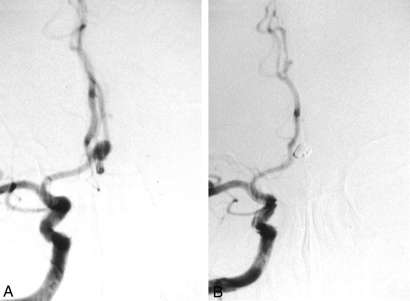
    Images from the case of a 65-year-old man with an unruptured aneurysm (case 1).

    A, Pretreatment cerebral angiogram shows the 5-mm anterior communicating artery aneurysm.

    B, Post-treatment cerebral angiogram shows embolization by interlocking detachable coils (4 mm × 8 cm and 3 mm × 6 cm).

  • Fig 2.
    • Download figure
    • Open in new tab
    • Download powerpoint
    Fig 2.

    Gross pathologic findings in case 1.

    A, Gross outlook appearance of the thin wall of the aneurysm through which embedded coils are seen.

    B, Intraluminal view shows a thin transparent layer of membrane covering the whole orifice of the aneurysm and bridged over the underlying coils.

    C, Scanning electron microscopy shows that the coils are covered by thick neointima at the orifice of the aneurysm. Part of the neointima was removed for transmission electron microscopic study (original magnification, ×50; bar = 100 μm).

    D, Superficial layer of neointima with a cobblestone appearance (original magnification, ×1000; bar = 10 μm).

  • Fig 3.
    • Download figure
    • Open in new tab
    • Download powerpoint
    Fig 3.

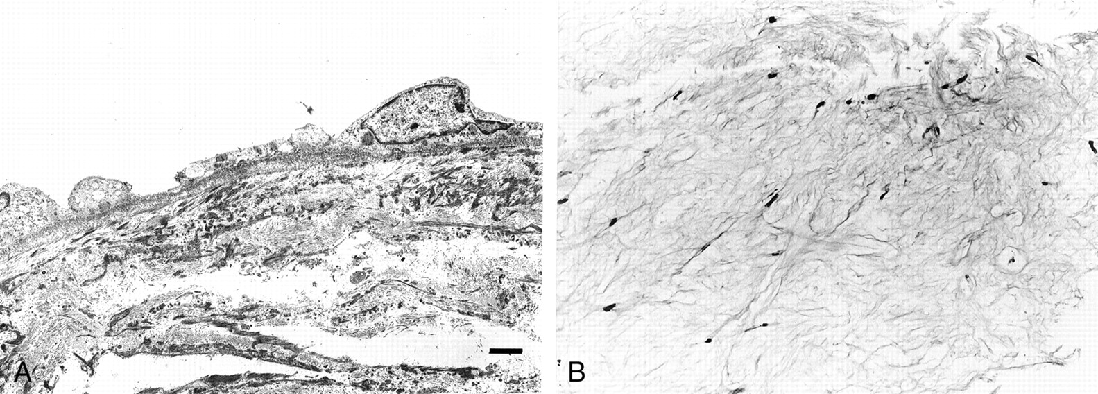
    Transmission electron microscopy and light microscopy (case 1).

    A, Basal lamina runs contiguously along the basal endothelial cytoplasmic membrane (original magnification, ×2000; bar = 2 μm).

    B, Light microscopy shows cotton-like white fibrous tissue in the dome of the aneurysm (hematoxylin and eosin stain; original magnification, ×74).

  • Fig 4.
    • Download figure
    • Open in new tab
    • Download powerpoint
    Fig 4.

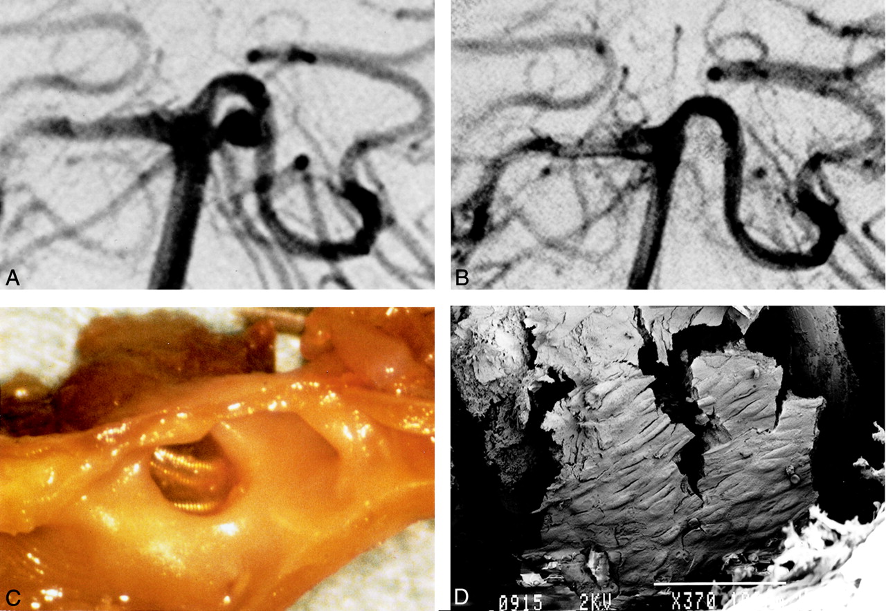
    Images from the case of a 62-year-old man with subarachnoid hemorrhage (case 2).

    A, Right pre-embolization vertebral angiography shows 4-mm left basilar artery-superior cerebellar artery aneurysm.

    B, After embolization by GDC (3 mm × 4 cm and 2 mm × 4 cm), almost total obliteration was achieved.

    C, Gross examination shows thin membrane on coils at orifice of aneurysm. The thin fragile covering bridges the whole space across the coils at the orifice.

    D, Scanning electron microscopy shows neointima partially covering the coils (GDC-10 soft). A cobblestone pattern can be seen (original magnification, ×370; bar = 100 μm).

PreviousNext
Back to top

In this issue

American Journal of Neuroradiology: 23 (6)
American Journal of Neuroradiology
Vol. 23, Issue 6
1 Jun 2002
  • Table of Contents
  • Index by author
Advertisement
Print
Download PDF
Email Article
Cite this article
0 Responses
Respond to this article
Share
Bookmark this article
Histopathologic Findings in Human Cerebral Aneurysms Embolized with Platinum Coils: Report of Two Cases and Review of the Literature
Shoichiro Ishihara, Michel E. Mawad, Kentaro Ogata, Chihiro Suzuki, Nobusuke Tsuzuki, Hiroshi Katoh, Akira Ohnuki, Takahito Miyazawa, Hiroshi Nawashiro, Tatsumi Kaji, Katsuji Shima
American Journal of Neuroradiology Jun 2002, 23 (6) 970-974;
del.icio.us logo Twitter logo Facebook logo Mendeley logo
  • Tweet Widget

Jump to section

  • Article
    • Abstract
    • Case Reports
    • Discussion
    • Conclusion
    • References
  • Figures & Data
  • Info & Metrics
  • Responses
  • References
  • PDF

Related Articles

  • No related articles found.
  • PubMed
  • Google Scholar

Cited By...

  • The Distribution and Role of M1 and M2 Macrophages in Aneurysm Healing after Platinum Coil Embolization
  • Assessment of endothelialization of aneurysm wall over time in a rabbit model through CD31 scoring
  • Combined endovascular coiling and intra-aneurysmal allogeneic mesenchymal stromal cell therapy for intracranial aneurysms in a rabbit model: a proof-of-concept study
  • Characterizing patterns of endothelialization following coil embolization: a whole-mount, dual immunostaining approach
  • Mechanisms of Healing in Coiled Intracranial Aneurysms: A Review of the Literature
  • New Generation of Flow Diverter (Surpass) for Unruptured Intracranial Aneurysms: A Prospective Single-Center Study in 37 Patients
  • Temporal Evolution of Susceptibility Artifacts from Coiled Aneurysms on MR Angiography: An In Vivo Canine Study
  • In Vivo Experimental Intracranial Aneurysm Models: A Systematic Review
  • Crossref
  • Google Scholar

This article has not yet been cited by articles in journals that are participating in Crossref Cited-by Linking.

More in this TOC Section

  • Prophylactic Endovascular Treatments for CBS
  • Smoking & Aneurysm Outcomes: A Meta-Analysis
  • “Buddy-wire anchoring” technique for TVE
Show more NEUROINTERVENTION

Similar Articles

  • Rabbit Elastase Aneurysm Model Mimics the Recurrence Rate of Human Intracranial Aneurysms following Platinum Coil Embolization
  • Histopathologic and Immunohistochemical Comparison of Human, Rabbit, and Swine Aneurysms Embolized with Platinum Coils
  • Bone Formation in Elastase-Induced Rabbit Aneurysms Embolized with Platinum Coils: Report of 2 Cases
  • Delayed Leukoencephalopathy: A Rare Complication after Coiling of Cerebral Aneurysms
  • Angiographic and Histologic Analysis of Experimental Aneurysms Embolized with Platinum Coils, Matrix, and HydroCoil
See more
Advertisement

Indexed Content

  • Current Issue
  • Accepted Manuscripts
  • Article Preview
  • Past Issues
  • Editorials
  • Editor's Choice
  • Fellows' Journal Club
  • Letters to the Editor
  • Video Articles

Cases

  • Case Collection
  • Archive - Case of the Week
  • Archive - Case of the Month
  • Archive - Classic Case

More from AJNR

  • Trainee Corner
  • Imaging Protocols
  • MRI Safety Corner
  • Book Reviews

Multimedia

  • AJNR Podcasts
  • AJNR Scantastics

Resources

  • Turnaround Time
  • Submit a Manuscript
  • Submit a Video Article
  • Submit an eLetter to the Editor/Response
  • Manuscript Submission Guidelines
  • Statistical Tips
  • Fast Publishing of Accepted Manuscripts
  • Graphical Abstract Preparation
  • Imaging Protocol Submission
  • Evidence-Based Medicine Level Guide
  • Publishing Checklists
  • Author Policies
  • Become a Reviewer/Academy of Reviewers
  • News and Updates

About Us

  • About AJNR
  • Editorial Board
  • Editorial Board Alumni
  • Alerts
  • Permissions
  • Not an AJNR Subscriber? Join Now
  • Advertise with Us
  • Librarian Resources
  • Feedback
  • Terms and Conditions
  • AJNR Editorial Board Alumni

American Society of Neuroradiology

  • Not an ASNR Member? Join Now

© 2025 by the American Society of Neuroradiology All rights, including for text and data mining, AI training, and similar technologies, are reserved.
Print ISSN: 0195-6108 Online ISSN: 1936-959X

Powered by HighWire
Email this Article

Thank you for your interest in spreading the word on American Journal of Neuroradiology.

NOTE: We only request your email address so that the person you are recommending the page to knows that you wanted them to see it, and that it is not junk mail. We do not capture any email address.

Enter multiple addresses on separate lines or separate them with commas.
Histopathologic Findings in Human Cerebral Aneurysms Embolized with Platinum Coils: Report of Two Cases and Review of the Literature
(Your Name) has sent you a message from American Journal of Neuroradiology
(Your Name) thought you would like to see the American Journal of Neuroradiology web site.
CAPTCHA
This question is for testing whether or not you are a human visitor and to prevent automated spam submissions.
Cite this article
Shoichiro Ishihara, Michel E. Mawad, Kentaro Ogata, Chihiro Suzuki, Nobusuke Tsuzuki, Hiroshi Katoh, Akira Ohnuki, Takahito Miyazawa, Hiroshi Nawashiro, Tatsumi Kaji, Katsuji Shima
Histopathologic Findings in Human Cerebral Aneurysms Embolized with Platinum Coils: Report of Two Cases and Review of the Literature
American Journal of Neuroradiology Jun 2002, 23 (6) 970-974;

Citation Manager Formats

  • BibTeX
  • Bookends
  • EasyBib
  • EndNote (tagged)
  • EndNote 8 (xml)
  • Medlars
  • Mendeley
  • Papers
  • RefWorks Tagged
  • Ref Manager
  • RIS
  • Zotero

We use cookies on this site to enhance your user experience. By clicking any link on this page you are giving your consent for us to set cookies.