Advertisement

Main menu

  • Home
  • Content
    • Current Issue
    • Accepted Manuscripts
    • Article Preview
    • Past Issue Archive
    • Video Articles
    • AJNR Case Collection
    • Case of the Week Archive
    • Case of the Month Archive
    • Classic Case Archive
  • Special Collections
    • AJNR Awards
    • Low-Field MRI
    • Alzheimer Disease
    • ASNR Foundation Special Collection
    • Photon-Counting CT
    • View All
  • Multimedia
    • AJNR Podcasts
    • AJNR SCANtastic
    • Trainee Corner
    • MRI Safety Corner
    • Imaging Protocols
  • For Authors
    • Submit a Manuscript
    • Submit a Video Article
    • Submit an eLetter to the Editor/Response
    • Manuscript Submission Guidelines
    • Statistical Tips
    • Fast Publishing of Accepted Manuscripts
    • Graphical Abstract Preparation
    • Imaging Protocol Submission
    • Author Policies
  • About Us
    • About AJNR
    • Editorial Board
    • Editorial Board Alumni
  • More
    • Become a Reviewer/Academy of Reviewers
    • Subscribers
    • Permissions
    • Alerts
    • Feedback
    • Advertisers
    • ASNR Home

User menu

  • Alerts
  • Log in

Search

  • Advanced search
American Journal of Neuroradiology

American Journal of Neuroradiology

ASHNR American Society of Functional Neuroradiology ASHNR American Society of Pediatric Neuroradiology ASSR
  • Alerts
  • Log in

Advanced Search

  • Home
  • Content
    • Current Issue
    • Accepted Manuscripts
    • Article Preview
    • Past Issue Archive
    • Video Articles
    • AJNR Case Collection
    • Case of the Week Archive
    • Case of the Month Archive
    • Classic Case Archive
  • Special Collections
    • AJNR Awards
    • Low-Field MRI
    • Alzheimer Disease
    • ASNR Foundation Special Collection
    • Photon-Counting CT
    • View All
  • Multimedia
    • AJNR Podcasts
    • AJNR SCANtastic
    • Trainee Corner
    • MRI Safety Corner
    • Imaging Protocols
  • For Authors
    • Submit a Manuscript
    • Submit a Video Article
    • Submit an eLetter to the Editor/Response
    • Manuscript Submission Guidelines
    • Statistical Tips
    • Fast Publishing of Accepted Manuscripts
    • Graphical Abstract Preparation
    • Imaging Protocol Submission
    • Author Policies
  • About Us
    • About AJNR
    • Editorial Board
    • Editorial Board Alumni
  • More
    • Become a Reviewer/Academy of Reviewers
    • Subscribers
    • Permissions
    • Alerts
    • Feedback
    • Advertisers
    • ASNR Home
  • Follow AJNR on Twitter
  • Visit AJNR on Facebook
  • Follow AJNR on Instagram
  • Join AJNR on LinkedIn
  • RSS Feeds

AJNR Awards, New Junior Editors, and more. Read the latest AJNR updates

Research ArticleNeurointervention

Endovascular Access to the Meningohypophyseal Trunk

Neil M. Borden and Kenneth M. Liebman
American Journal of Neuroradiology April 2001, 22 (4) 725-727;
Neil M. Borden
  • Find this author on Google Scholar
  • Find this author on PubMed
  • Search for this author on this site
Kenneth M. Liebman
  • Find this author on Google Scholar
  • Find this author on PubMed
  • Search for this author on this site
  • Article
  • Figures & Data
  • Info & Metrics
  • Responses
  • References
  • PDF
Loading

Abstract

Summary: We describe a novel technique to selectively catheterize the meningohypophyseal trunk (MHT) and its branches. We emphasize the difficulty in accessing the MHT via an ipsilateral approach because of the geometric orientation of this vessel to the parent internal carotid artery.

A variety of lesions can derive blood supply from branches of the meningohypophyseal trunk (MHT) (1–3). Endovascular embolization of these branches may be curative or a significant adjunct to definitive treatment (1, 2). Despite advancements in microcatheterization and microguidewire technology, selective catheterization of the MHT and its branches is often difficult or impossible owing to the small size of the arterial supply and the geometric orientation of these branches relative to the internal carotid artery (ICA) (1) (Fig 1).

fig 1.
  • Download figure
  • Open in new tab
  • Download powerpoint
fig 1.

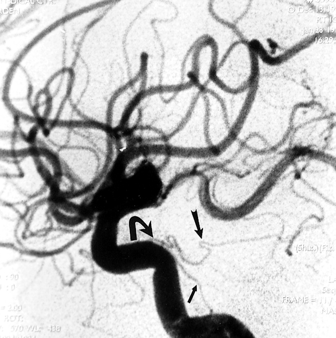
Lateral DSA, arterial phase, shows normal caliber dorsal clival (small arrow) and tentorial (large arrow) branches of the MHT in a different patient with no disease in this region. Note the difficult geometry of the MHT relative to the C4–C5 segments of the ICA if an anterograde approach to microcatheterization is attempted (curved arrow)

We report a patient in whom an iatrogenic dural carotid-cavernous fistula developed with arterial supply from a branch of the MHT and describe a new technique for selectively catheterizing a branch of the MHT.

Case Report

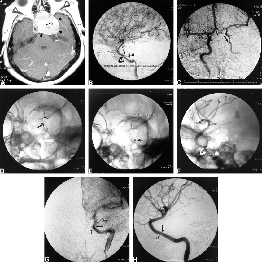
A 33-year-old right-handed woman presented with a recurrent pituitary macroadenoma approximately 20 months after her initial surgery. The most recent preoperative MR study showed a macroadenoma measuring approximately 4 cm in its largest transverse diameter invading both cavernous sinuses (Fig 2A). The patient underwent a second transphenoidal approach with a subtotal resection of the adenoma. Two hours after the surgery, the patient experienced brisk epistaxis of bright red blood. The bleeding was initially controlled by packing the nasal cavity. An emergency ICA angiogram obtained with digital subtraction angiography (DSA) revealed a slowly filling left carotid-cavernous fistula associated with an avulsed branch of the MHT bleeding into the cavernous sinus (Fig 2B). Transarterial embolization using a microcatheter and microguidewire from the left ICA was aborted after multiple unsuccessful attempts were made to selectively catheterize the MHT. Because the fistula was filling slowly, carotid cross compression therapy was instituted (4). This therapy required that the patient, with her right hand, compress the left carotid artery daily, gradually increasing the length of compression time. The patient was observed in the hospital, and 10 days later, a follow-up angiogram revealed patency of the fistula despite resolution of the epistaxis. An alternative, contralateral, approach for superselective catheterization of the MHT was attempted.

fig 2.
  • Download figure
  • Open in new tab
  • Download powerpoint
fig 2.

Case 1: 33-year-old right-handed woman with a recurrent pituitary macroadenoma approximately 20 months after initial surgery. A, Contrast-enhanced T1-weighted axial MR image shows a densely enhancing pituitary macroadenoma (arrowheads) invading the left cavernous sinus and encasing the intracavernous left ICA (arrow). B, Lateral DSA, late arterial phase, shows contrast material pooling in the tumor-invaded cavernous sinus (arrowhead). The avulsed branch of the MHT (small arrow) arises from the tumor-encased intracavernous ICA (large arrow). The tumor diffusely narrows and uncoils the intracavernous ICA. C, Anteroposterior DSA from right ICA injection with left carotid compression shows good cross filling through the ACoM (small arrow) with retrograde filling of the left carotid-cavernous fistula (curved arrow). D, Left anterior oblique (LAO) fluoroscopic image shows the microcatheter crossing from right to left through the ACoM (open arrow), then down the supraclinoid left ICA (small arrow) into the horizontal (C4) intracavernous left ICA (large arrow). E, LAO fluoroscopic image shows two 2-mm × 2-cm GDC-10 coils (arrow) after deployment into the venous side of the carotid-cavernous fistula. F, Steep LAO nonsubtracted DSA shows two 2-mm × 2-cm GDC-10 coils in the venous side of the carotid-cavernous fistula (arrow). G, Anteroposterior DSA from left ICA injection in the late arterial, early capillary phase shows complete obliteration of the carotid-cavernous fistula. The GDCs (small arrow) are faintly seen just medial to the C4–C5 junction of the left ICA (large arrow). H, Steep LAO DSA from the left ICA injection in the arterial phase shows complete obliteration of the carotid-cavernous fistula. The GDCs (small arrow) overlap the C4–C5 junction of the left ICA (large arrow) in this projection

The angiogram revealed a patent anterior communicating artery (ACoM) with brisk cross filling (Fig 2C). A guiding catheter was placed into the high cervical right ICA. A microcatheter and a microguidewire were placed coaxially through the guiding catheter and navigated under roadmap into the A1 segment of the right anterior cerebral artery. A diagnostic catheter was then placed into the cervical segment of the left ICA. Roadmapping through the left ICA was performed. The microcatheter was then navigated from right to left through the patent ACoM, down the left supraclinoid ICA to the horizontal (C4) intracavernous segment of the ICA (Fig 2D). The microcatheter/microguidewire combination was then passed from anterior to posterior through the horizontal intracavernous left ICA and then through the MHT into the fistula without difficulty. Two 2-mm × 2-cm Guglielmi detachable coils (GDC-10 soft coils) (Target Therapeutics, Fremont, CA) were then deployed into the cavernous sinus (Fig 2E and F). Postembolization angiography revealed marked slowing of the shunting. Because of the significant reduction in flow, the procedure was terminated and cross compression of the left ICA was continued. A 3-month follow-up angiography revealed complete obliteration of the arteriovenous fistula (Fig 2G and H).

Discussion

Our case illustrates an important and novel technique, which has not previously been described, that can transform an unsuccessful endovascular catheterization of the MHT into a relatively easy, fast, and successful procedure. Careful analysis of the difficult geometry in this region suggested an easier endovascular route. As illustrated in Figures 1 and 2B, the MHT arises from the ICA at the junction of the C4 and C5 segments (5–7). It projects posteriorly, nearly parallel to the horizontal ICA (C4 segment) and is easier to access in a retrograde fashion from anterior to posterior. In a contralateral, retrograde approach, the catheter does not need to make an acute turn to enter the orifice of the MHT. This is in contrast to an ipsilateral approach, which may result in failure to catheterize the MHT.

Conclusion

Many vascular lesions of the skull base derive blood supply from the MHT. Endovascular procedures are often difficult or impossible if attempted from an ipsilateral approach. In the technique described here, we used the contralateral ICA with retrograde catheterization of the carotid siphon. This provided better access to this difficult anatomy because of the more favorable anatomic geometry. This technique may be especially helpful when a transvenous route cannot be used.

References

  1. ↵
    Halbach VV, Higashida RT, Hieshima GB, Hardin CW. Embolization of branches arising from the cavernous portion of the internal carotid artery. AJNR Am J Neuroradiol 1989;10:143-150
    Abstract/FREE Full Text
  2. Lewis AI, Tomsick TA, Tew JM Jr. Management of tentorial dural arteriovenous malformations: transarterial embolization combined with stereotactic radiation or surgery. J Neurosurg 1994;81:851-859
    PubMed
  3. Pribram HFW, Boulter TR, McCormick WMF. The roentgenology of the meningohypophyseal trunk. AJNR Am J Neuroradiol 1966;98:583-594
  4. ↵
    Higashida RT, Hieshima GB, Halbach VV, Goto KG. Closure of carotid cavernous sinus fistula by external compression of cervical carotid artery and jugular vein. Acta Radiol (Suppl) 1986;369:580-583
  5. ↵
    Miyazaki Y, Yamamoto I, Shinozuka S, Sato O. Microsurgical anatomy of the cavernous sinus. Neurol Med Chir 1994;34:150-163
  6. Reisch R, Vutskits L, Patonay L, Fries G. The meningohypophyseal trunk and its blood supply to different intracranial structures: an anatomical study. Minim Invasive Neurosurg 1996;39:78-81
    CrossRefPubMed
  7. Robinson DH, Song JK, Eskridge JM. Embolization of meningohypophyseal and inferolateral branches of the cavernous internal carotid artery. AJNR Am J Neuroradiol 1999;20:1061-1067
    Abstract/FREE Full Text
  • Received July 31, 2000.
  • Copyright © American Society of Neuroradiology
View Abstract
PreviousNext
Back to top

In this issue

American Journal of Neuroradiology
Vol. 22, Issue 4
1 Apr 2001
  • Table of Contents
  • Index by author
Advertisement
Print
Download PDF
Email Article

Thank you for your interest in spreading the word on American Journal of Neuroradiology.

NOTE: We only request your email address so that the person you are recommending the page to knows that you wanted them to see it, and that it is not junk mail. We do not capture any email address.

Enter multiple addresses on separate lines or separate them with commas.
Endovascular Access to the Meningohypophyseal Trunk
(Your Name) has sent you a message from American Journal of Neuroradiology
(Your Name) thought you would like to see the American Journal of Neuroradiology web site.
CAPTCHA
This question is for testing whether or not you are a human visitor and to prevent automated spam submissions.
Cite this article
Neil M. Borden, Kenneth M. Liebman
Endovascular Access to the Meningohypophyseal Trunk
American Journal of Neuroradiology Apr 2001, 22 (4) 725-727;

Citation Manager Formats

  • BibTeX
  • Bookends
  • EasyBib
  • EndNote (tagged)
  • EndNote 8 (xml)
  • Medlars
  • Mendeley
  • Papers
  • RefWorks Tagged
  • Ref Manager
  • RIS
  • Zotero
0 Responses
Respond to this article
Share
Bookmark this article
Endovascular Access to the Meningohypophyseal Trunk
Neil M. Borden, Kenneth M. Liebman
American Journal of Neuroradiology Apr 2001, 22 (4) 725-727;
del.icio.us logo Twitter logo Facebook logo Mendeley logo
  • Tweet Widget
  • Facebook Like
  • Google Plus One

Jump to section

  • Article
    • Abstract
    • Case Report
    • Discussion
    • Conclusion
    • References
  • Figures & Data
  • Info & Metrics
  • Responses
  • References
  • PDF

Related Articles

  • No related articles found.
  • PubMed
  • Google Scholar

Cited By...

  • No citing articles found.
  • Crossref
  • Google Scholar

This article has not yet been cited by articles in journals that are participating in Crossref Cited-by Linking.

More in this TOC Section

  • Factors Associated with Major Re-Recanalization following Second Coiling for Recanalized Aneurysms: A Multicenter Experience over 20 Years during Long-Term Follow-up
  • A Key Factor Shapes LS-DAVFs EVT Outcome
  • Optimizing Voxel Size in 3D Rotational Angiography
Show more NEUROINTERVENTION

Similar Articles

Advertisement

Indexed Content

  • Current Issue
  • Accepted Manuscripts
  • Article Preview
  • Past Issues
  • Editorials
  • Editor's Choice
  • Fellows' Journal Club
  • Letters to the Editor
  • Video Articles

Cases

  • Case Collection
  • Archive - Case of the Week
  • Archive - Case of the Month
  • Archive - Classic Case

More from AJNR

  • Trainee Corner
  • Imaging Protocols
  • MRI Safety Corner

Multimedia

  • AJNR Podcasts
  • AJNR Scantastics

Resources

  • Turnaround Time
  • Submit a Manuscript
  • Submit a Video Article
  • Submit an eLetter to the Editor/Response
  • Manuscript Submission Guidelines
  • Statistical Tips
  • Fast Publishing of Accepted Manuscripts
  • Graphical Abstract Preparation
  • Imaging Protocol Submission
  • Evidence-Based Medicine Level Guide
  • Publishing Checklists
  • Author Policies
  • Become a Reviewer/Academy of Reviewers
  • News and Updates

About Us

  • About AJNR
  • Editorial Board
  • Editorial Board Alumni
  • Alerts
  • Permissions
  • Not an AJNR Subscriber? Join Now
  • Advertise with Us
  • Librarian Resources
  • Feedback
  • Terms and Conditions
  • AJNR Editorial Board Alumni

American Society of Neuroradiology

  • Not an ASNR Member? Join Now

© 2025 by the American Society of Neuroradiology All rights, including for text and data mining, AI training, and similar technologies, are reserved.
Print ISSN: 0195-6108 Online ISSN: 1936-959X

Powered by HighWire