
Article Figures & Data
Figures
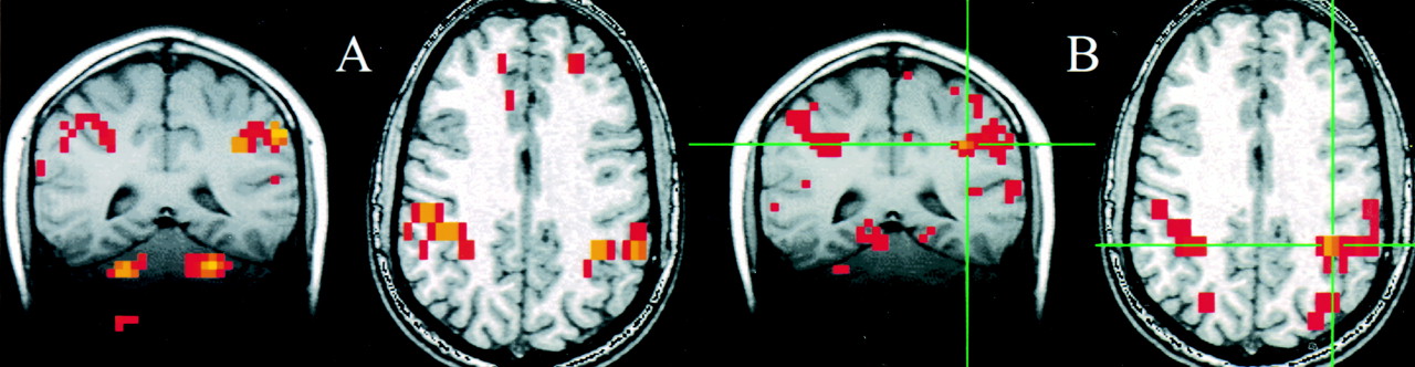
- fig 1.
A and B, Selected coronal and axial fMRI maps of voxels activated by the bilateral finger-tapping task (A) and of the voxels functionally connected to a seed voxel (crosshairs) in the sensorimotor cortex in a resting acquisition (B). The sensorimotor cortex is identified on both the fMRI and the fcMRI maps. Voxels within the dentate nucleus of the cerebellum were identified with fMRI but not with the functional connectivity study
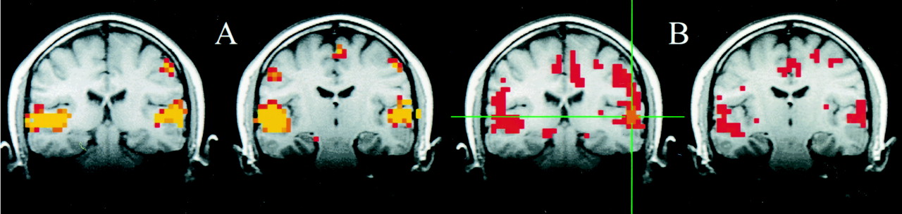
- fig 2.
A and B, Consecutive coronal fMRI maps of voxels activated by a text-listening task (A) and of the voxels functionally connected with a 2 × 2 seed voxel ROI (crosshairs) within the auditory cortex (B). Task activation was evident in the region of the primary and association auditory cortices in the superior temporal lobe. Many of the voxels functionally connected with the seed voxel in the left auditory cortex (crosshairs) have a similar distribution. A few other voxels with lower correlation coefficients (red) were identified outside the auditory cortices. For the connectivity study, data were collected without the patient performing a specific cognitive task
- fig 2.
fig 3. A and B, A coronal image showing task activation for the text-listening task (A) and connectivity for a seed voxel (crosshairs) in the left frontal lobe in a resting data set (B). The task produces activation in the superior temporal gyral regions bilaterally and in the left frontal lobe in or near Broca's region. Activation is also identified in the right frontal lobe. The voxels with connectivity to the seed voxel (crosshairs) appear to lie in similar locations. fig 4. A and B, Coronal images showing task activation for the word-generation task (A) and connectivity for the left frontal cortex in the region that showed task activation (B). The task produced activation in the left frontal lobe in or near Broca's area and in the superior temporal lobes bilaterally. The voxels with connectivity to the seed voxel (crosshairs) appear to lie in similar locations to the task activation produced by word generation and to the locations with connectivity in figure 3
- fig 5.
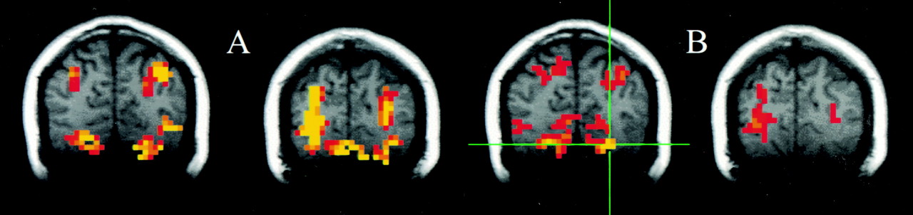
A and B, Consecutive coronal images showing task activation for the visual task (A) and connectivity for the visual cortex (B). The task produces activation in the striate cortex region and in the posterior parietal lobes bilaterally. The voxels with connectivity to the seed voxel (crosshairs) appear to lie in similar locations in the striate cortex region and in the posterior parietal lobe
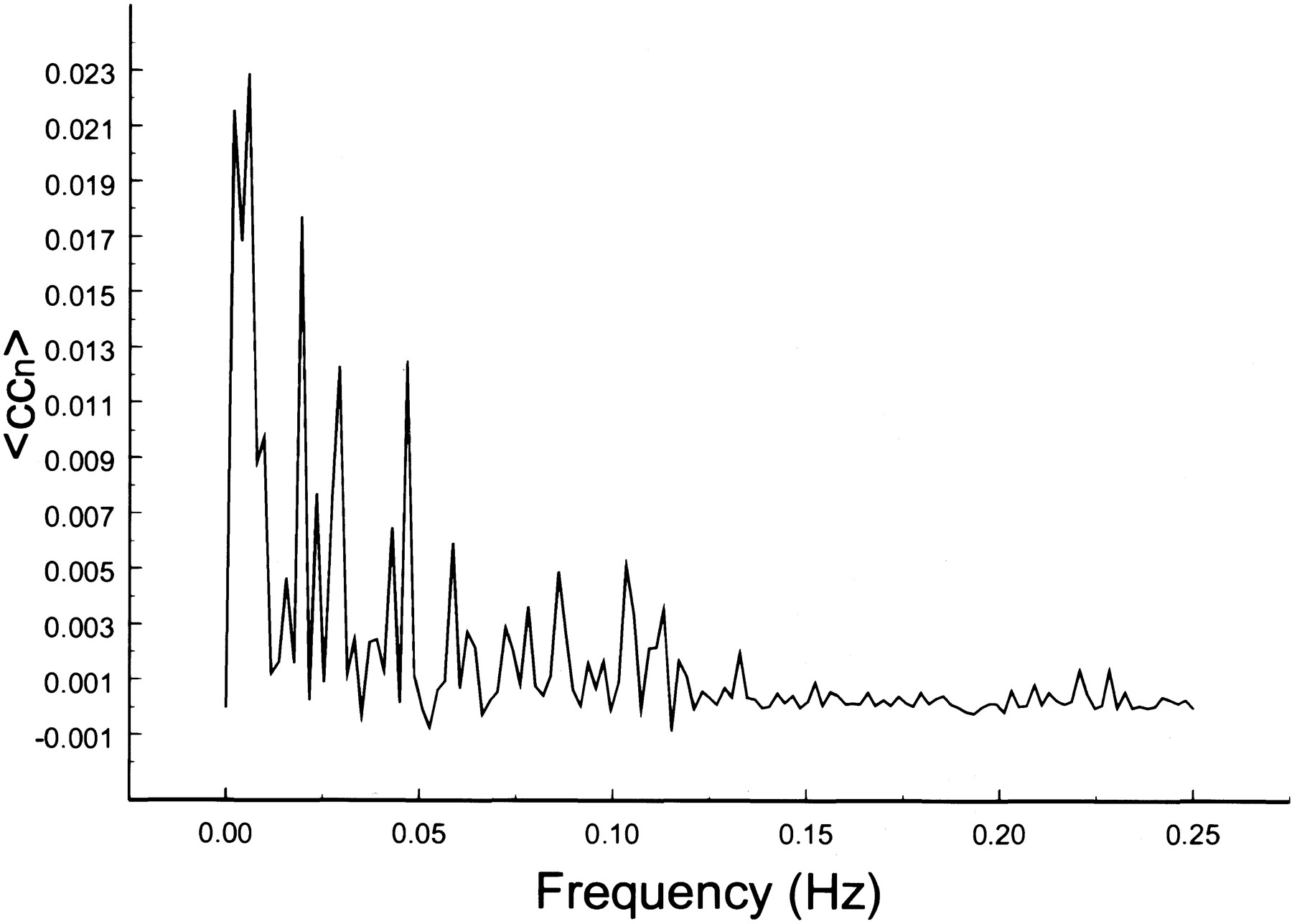
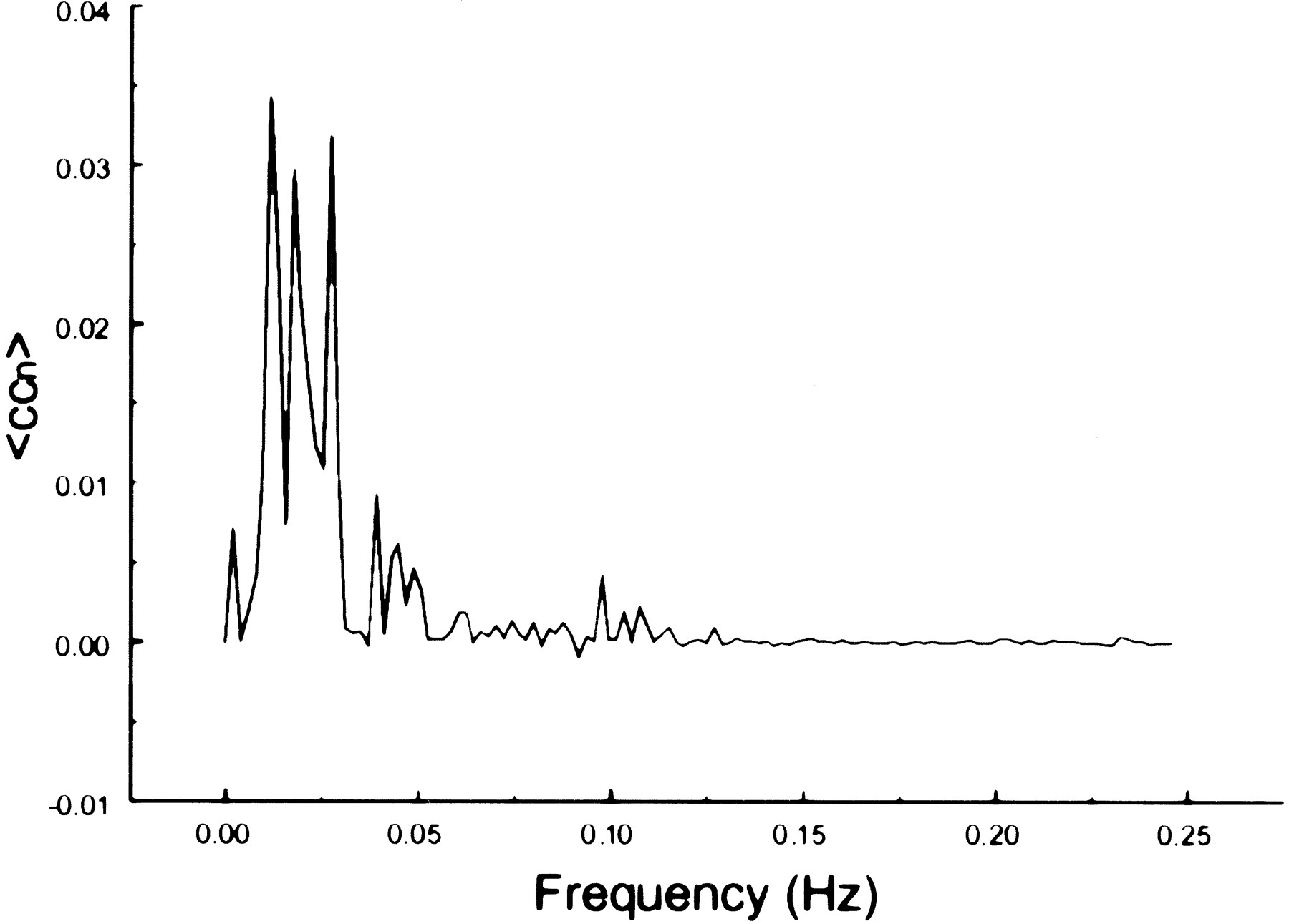
- fig 6.
Spectral decomposition of the average correlation coefficient for interregional connectivity in the motor cortex (same subject as in fig 1). Only frequency components between 0 and 0.05 Hz contribute significantly to the correlation coefficient
- fig 7.
Spectral decomposition of the average correlation coefficient for interregional connectivity in the auditory cortex (same subject as in fig 2). Only frequency components less than 0.05 Hz contribute significantly to the correlation coefficient
- fig 8.
Spectral decomposition of the average correlation coefficient for interregional connectivity in the prefrontal cortex (same subject as in fig 3). Only the same low frequency components contribute significantly to the correlation coefficient
- fig 9.
Spectral decomposition of the average correlation coefficient for interregional connectivity in the prefrontal cortex (same subject as in fig 4). Only the same low-frequency components contribute significantly to the correlation coefficient
- fig 10.
Spectral decomposition of the average correlation coefficient for interregional connectivity in the visual cortex (same subject as in fig 5). Only low-frequency components contribute significantly to the correlation coefficient
- fig 11.
Spectral decomposition of the average correlation coefficient from a seed voxel in the left jugular vein. Note the relative paucity of frequencies in the 0 to 0.05 Hz range, the spread over many frequencies, and the peak at 0.2 Hz
- fig 12.
Spectral decomposition of the average correlation coefficient using a seed voxel in the right middle cerebral artery. Low frequencies do not predominate. Peaks are present at multiple frequencies above 0.05 Hz
- fig 13.
Spectral decomposition of the average correlation coefficient using a seed voxel in the left ventricle. Frequencies from 0.025 to 0.075 predominate. Since no BOLD effect can be invoked in CSF, these frequencies represent likely aliased cardiac cycle effects


In this issue


Jump to section
Related Articles
Cited By...
- Dynamic fusion of structural and functional connectivity via joint connectivity matrix ICA
- Aging and the Spectral Properties of Brain Hemodynamics
- Prediction of dynamic balance state and recovery following stroke using fMRI graph analysis
- Unraveling the Neural Landscape of Mental Disorders using Double Functional Independent Primitives (dFIPs)
- Functional brain network dynamics of brooding in depression: insights from real-time fMRI neurofeedback
- Altered dynamic functional network connectivity in rheumatoid arthritis associated with peripheral inflammation and neuropsychiatric disorders
- Functional connectivity across the human subcortical auditory system using an autoregressive matrix-Gaussian copula graphical model approach with partial correlations
- Resting-state functional connectivity in children cooled for neonatal encephalopathy
- How can graph theory inform the dual-stream model of speech processing? a resting-state fMRI study of post-stroke aphasia
- Efficient estimation of time-dependent functional connectivity using Structural Connectivity constraints
- Single Ventricle Reconstruction III: Brain Connectome and Neurodevelopmental Outcomes: Design, Recruitment, and Technical Challenges of a Multicenter, Observational Neuroimaging Study
- Resting state networks of awake adolescent and adult squirrel monkeys using ultra-high field (9.4T) functional magnetic resonance imaging
- Efficient estimation of time-dependent functional connectivity using Structural Connectivity constraints
- Joint cmICA: auto-linking of structural and functional connectivity
- Acquisition and processing methods of whole-brain layer-fMRI VASO and BOLD: The Kenshu dataset
- A structure-function substrate of memory for spatial configurations in medial and lateral temporal cortices
- Resting-State Functional Networks of Different Topographic Representations in the Somatosensory Cortex of Macaque Monkeys and Humans
- A Paradigm for Measuring Resting State Functional Connectivity in Young Children Using fNIRS and Freeplay
- MOU-EC: model-based whole-brain effective connectivity to extract biomarkers for brain dynamics from fMRI data and study distributed cognition
- Trait-like variants in human functional brain networks
- Studying the precuneus reveals structure-function-affect correlation in long-term meditators
- The Relationship between BOLD and Neural Activity Arises from Temporally Sparse Events
- Large-scale intrinsic connectivity is consistent across varying task demands
- Extracting orthogonal subject- and behavior-specific signatures from fMRI data using whole-brain effective connectivity
- Analysis of fMRI data using noise-diffusion network models: a new covariance-coding perspective
- Stereotypical modulations in dynamic functional connectivity explained by changes in BOLD variance
- Effective connectivity inferred from fMRI transition dynamics during movie viewing points to a balanced reconfiguration of cortical interactions
- Resting State Networks in empirical and simulated dynamic functional connectivity
- Impact of gray matter signal regression in resting state and language task functional networks
- State-dependent modulation of functional connectivity in early blind individuals
- High spatial correspondence at a columnar level between activation and resting state fMRI signals and local field potentials
- Brain Age: A State-Of-Mind? On the Stability of Functional Connectivity across Behavioral States
- Functional connectivity arises from a slow rhythmic mechanism
- Imperceptible Somatosensory Stimulation Alters Sensorimotor Background Rhythm and Connectivity
- Noninvasive Functional and Anatomical Imaging of the Human Medial Temporal Lobe
- Intrinsically organized resting state networks in the human spinal cord
- Low-frequency calcium oscillations accompany deoxyhemoglobin oscillations in rat somatosensory cortex
- Time-varying functional network information extracted from brief instances of spontaneous brain activity
- Reorganization of Functional Connectivity of the Language Network in Patients with Brain Gliomas
- Unravelling the Intrinsic Functional Organization of the Human Lateral Frontal Cortex: A Parcellation Scheme Based on Resting State fMRI
- Functional Connectivity during Resting-State Functional MR Imaging: Study of the Correspondence between Independent Component Analysis and Region-of-Interest-Based Methods
- Prefrontal Transcranial Direct Current Stimulation Changes Connectivity of Resting-State Networks during fMRI
- Global Functional Connectivity Deficits in Schizophrenia Depend on Behavioral State
- Comparison of Hypothesis- and a Novel Hybrid Data/Hypothesis-Driven Method of Functional MR Imaging Analysis in Patients with Brain Gliomas
- Functional Connectivity MR Imaging of the Language Network in Patients with Drug-Resistant Epilepsy
- Growing Together and Growing Apart: Regional and Sex Differences in the Lifespan Developmental Trajectories of Functional Homotopy
- Neural basis of global resting-state fMRI activity
- Investigating the Functional Heterogeneity of the Default Mode Network Using Coordinate-Based Meta-Analytic Modeling
- Correspondence of the brain's functional architecture during activation and rest
- Decoupling of the brain's default mode network during deep sleep
- Distinct Cerebellar Contributions to Intrinsic Connectivity Networks
- The Blood Oxygen Level-Dependent Functional MR Imaging Signal Can Be Used to Identify Brain Tumors and Distinguish Them from Normal Tissue
- Regional Variation in Interhemispheric Coordination of Intrinsic Hemodynamic Fluctuations
- Resting-state networks in the infant brain
- Dissociable Intrinsic Connectivity Networks for Salience Processing and Executive Control
- Consistent resting-state networks across healthy subjects
- Spontaneous neuronal activity distinguishes human dorsal and ventral attention systems
- From The Cover: The human brain is intrinsically organized into dynamic, anticorrelated functional networks
- Frequencies Contributing to Functional Connectivity in the Cerebral Cortex in ""Resting-state"" Data