Advertisement

Main menu

  • Home
  • Content
    • Current Issue
    • Accepted Manuscripts
    • Article Preview
    • Past Issue Archive
    • Video Articles
    • AJNR Case Collection
    • Case of the Week Archive
    • Case of the Month Archive
    • Classic Case Archive
  • Special Collections
    • Advancing NeuroMRI with High-Relaxivity Contrast Agents
    • AJNR Awards
    • Low-Field MRI
    • Alzheimer Disease
    • ASNR Foundation Special Collection
    • Photon-Counting CT
    • View All
  • Multimedia
    • AJNR Podcasts
    • AJNR SCANtastic
    • Trainee Corner
    • MRI Safety Corner
    • Imaging Protocols
  • For Authors
    • Submit a Manuscript
    • Submit a Video Article
    • Submit an eLetter to the Editor/Response
    • Manuscript Submission Guidelines
    • Statistical Tips
    • Fast Publishing of Accepted Manuscripts
    • Graphical Abstract Preparation
    • Imaging Protocol Submission
    • Author Policies
  • About Us
    • About AJNR
    • Editorial Board
    • Editorial Board Alumni
  • More
    • Become a Reviewer/Academy of Reviewers
    • Subscribers
    • Permissions
    • Alerts
    • Feedback
    • Advertisers
    • ASNR Home

User menu

  • Alerts
  • Log in

Search

  • Advanced search
American Journal of Neuroradiology

American Journal of Neuroradiology

ASHNR American Society of Functional Neuroradiology ASHNR American Society of Pediatric Neuroradiology ASSR
  • Alerts
  • Log in

Advanced Search

  • Home
  • Content
    • Current Issue
    • Accepted Manuscripts
    • Article Preview
    • Past Issue Archive
    • Video Articles
    • AJNR Case Collection
    • Case of the Week Archive
    • Case of the Month Archive
    • Classic Case Archive
  • Special Collections
    • Advancing NeuroMRI with High-Relaxivity Contrast Agents
    • AJNR Awards
    • Low-Field MRI
    • Alzheimer Disease
    • ASNR Foundation Special Collection
    • Photon-Counting CT
    • View All
  • Multimedia
    • AJNR Podcasts
    • AJNR SCANtastic
    • Trainee Corner
    • MRI Safety Corner
    • Imaging Protocols
  • For Authors
    • Submit a Manuscript
    • Submit a Video Article
    • Submit an eLetter to the Editor/Response
    • Manuscript Submission Guidelines
    • Statistical Tips
    • Fast Publishing of Accepted Manuscripts
    • Graphical Abstract Preparation
    • Imaging Protocol Submission
    • Author Policies
  • About Us
    • About AJNR
    • Editorial Board
    • Editorial Board Alumni
  • More
    • Become a Reviewer/Academy of Reviewers
    • Subscribers
    • Permissions
    • Alerts
    • Feedback
    • Advertisers
    • ASNR Home
  • Follow AJNR on Twitter
  • Visit AJNR on Facebook
  • Follow AJNR on Instagram
  • Join AJNR on LinkedIn
  • RSS Feeds

AJNR Awards, New Junior Editors, and more. Read the latest AJNR updates


Improved Turnaround Times | Median time to first decision: 12 days

Research ArticleBrain

Diffusion-weighted Imaging of Patients with Subacute Cerebral Ischemia: Comparison with Conventional and Contrast-enhanced MR Imaging

Michael Augustin, Roland Bammer, Josef Simbrunner, Rudolf Stollberger, Hans-Peter Hartung and Franz Fazekas
American Journal of Neuroradiology October 2000, 21 (9) 1596-1602;
Michael Augustin
  • Find this author on Google Scholar
  • Find this author on PubMed
  • Search for this author on this site
Roland Bammer
  • Find this author on Google Scholar
  • Find this author on PubMed
  • Search for this author on this site
Josef Simbrunner
  • Find this author on Google Scholar
  • Find this author on PubMed
  • Search for this author on this site
Rudolf Stollberger
  • Find this author on Google Scholar
  • Find this author on PubMed
  • Search for this author on this site
Hans-Peter Hartung
  • Find this author on Google Scholar
  • Find this author on PubMed
  • Search for this author on this site
Franz Fazekas
  • Find this author on Google Scholar
  • Find this author on PubMed
  • Search for this author on this site
  • Article
  • Figures & Data
  • Info & Metrics
  • Responses
  • References
  • PDF
Loading

Article Figures & Data

Figures

  • Tables
  • fig 1.
    • Download figure
    • Open in new tab
    • Download powerpoint
    fig 1.

    Graph compares the probability of lesion detection for the subgroup of 41 patients with evidence of subacute ischemic damage on any of the MR sequences used, including contrast-enhanced T1-weighted images. The first and second columns indicate the percentage of patients with a subacute lesion on T2-weighted sequences without and with knowledge of the patients' symptoms. The third column shows the relative sensitivity of DWI without clinical information

  • fig 2.
    • Download figure
    • Open in new tab
    • Download powerpoint
    fig 2.

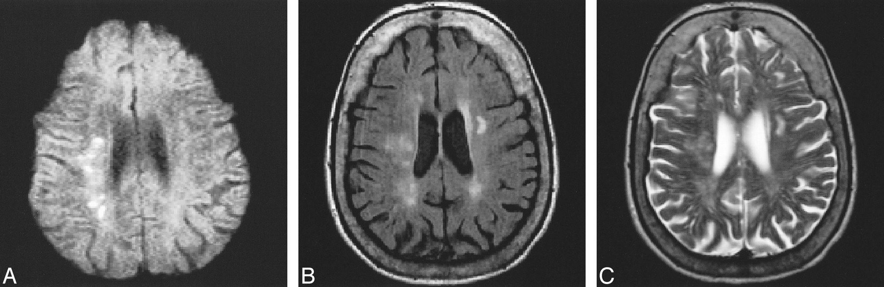
    80-year-old patient with left hemiparesis and ataxia.

    A–C, DWI (TR/TEdiff/TE = approximately 1500/115/18; 2 RR intervals) study (A) clearly shows multiple recent small ischemic lesions in the right centrum semiovale. These lesions are less visible on FLAIR image (6000/130/2, TI = 1900) (B) and T2-weighted-FSE image (2900/120/1) (C) and could not be identified as areas of subacute infarction on these sequences.

  • fig 3.
    • Download figure
    • Open in new tab
    • Download powerpoint
    fig 3.

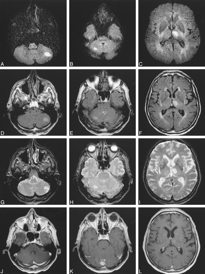
    63-year-old patient with right hemiparesis.

    A–L, Three days after stroke, DWI studies (approximately 1500/115/18; 2 RR intervals) (A–C) show two more clinically unexpected ischemic lesions in the cerebellum (A, B), suggestive of embolism. These two lesions would not have been definitively labeled as subacute on FLAIR (D–F) (6000/130/2, TI = 1900) or T2-weighted (2500/90/1) (G–I) sequences, especially in the presence of negative findings on contrast-enhanced images (556/14/2) (J–L).

  • fig 4.
    • Download figure
    • Open in new tab
    • Download powerpoint
    fig 4.

    A–D, Multiple tiny cortico-subcortical lesions in the right motor region on DWI (approximately 1500/115/18; 2 RR intervals) study (A) confirm the ischemic origin of mild hemiparesis 4 days after stroke. These changes might have gone undetected on T2-weighted FSE (2900/120/1) (B), FLAIR (6000/130/2, TI = 1900) (C), or contrast-enhanced (588/14/2) (D) images

  • fig 5.
    • Download figure
    • Open in new tab
    • Download powerpoint
    fig 5.

    Graph compares the percentage of patients with subacute ischemic lesions on DWI and contrast-enhanced T1-weighted sequences in the subgroup of 28 patients in whom contrast-enhanced studies were obtained

  • fig 6.
    • Download figure
    • Open in new tab
    • Download powerpoint
    fig 6.

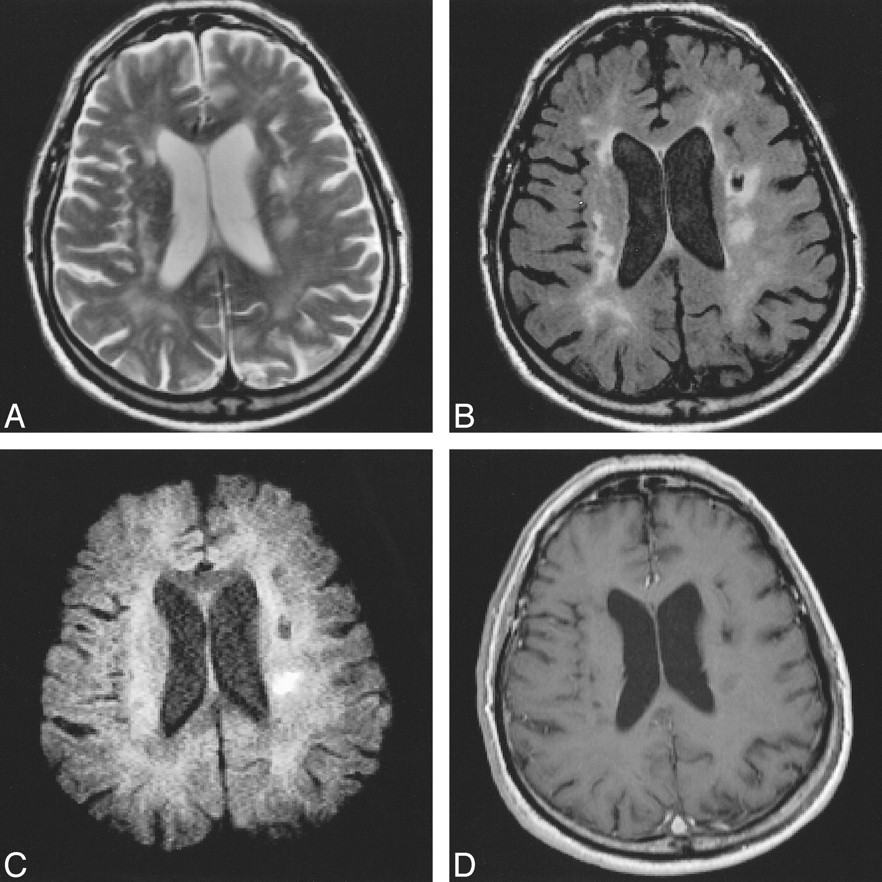
    63-year-old patient with preexisting ischemic lesions.

    A–D, Mild hemiparesis is well seen on FSE (2900/120/1) (A) and FLAIR (6000/130/2, TI = 1900) (B) sequences. DWI (approximately 1500/115/18; 2 RR intervals) sequence (C) depicts the area of recent ischemic damage 7 days after stroke. Contrast-enhanced MR image (588/14/2) (D) remains negative.

Tables

  • Figures
  • Table1

    Clinical and imaging findings in patients undergoing diffusion-weighted MR imaging

PreviousNext
Back to top

In this issue

American Journal of Neuroradiology
Vol. 21, Issue 9
1 Oct 2000
  • Table of Contents
  • Index by author
Advertisement
Print
Download PDF
Email Article

Thank you for your interest in spreading the word on American Journal of Neuroradiology.

NOTE: We only request your email address so that the person you are recommending the page to knows that you wanted them to see it, and that it is not junk mail. We do not capture any email address.

Enter multiple addresses on separate lines or separate them with commas.
Diffusion-weighted Imaging of Patients with Subacute Cerebral Ischemia: Comparison with Conventional and Contrast-enhanced MR Imaging
(Your Name) has sent you a message from American Journal of Neuroradiology
(Your Name) thought you would like to see the American Journal of Neuroradiology web site.
CAPTCHA
This question is for testing whether or not you are a human visitor and to prevent automated spam submissions.
Cite this article
Michael Augustin, Roland Bammer, Josef Simbrunner, Rudolf Stollberger, Hans-Peter Hartung, Franz Fazekas
Diffusion-weighted Imaging of Patients with Subacute Cerebral Ischemia: Comparison with Conventional and Contrast-enhanced MR Imaging
American Journal of Neuroradiology Oct 2000, 21 (9) 1596-1602;

Citation Manager Formats

  • BibTeX
  • Bookends
  • EasyBib
  • EndNote (tagged)
  • EndNote 8 (xml)
  • Medlars
  • Mendeley
  • Papers
  • RefWorks Tagged
  • Ref Manager
  • RIS
  • Zotero
0 Responses
Respond to this article
Share
Bookmark this article
Diffusion-weighted Imaging of Patients with Subacute Cerebral Ischemia: Comparison with Conventional and Contrast-enhanced MR Imaging
Michael Augustin, Roland Bammer, Josef Simbrunner, Rudolf Stollberger, Hans-Peter Hartung, Franz Fazekas
American Journal of Neuroradiology Oct 2000, 21 (9) 1596-1602;
del.icio.us logo Twitter logo Facebook logo Mendeley logo
  • Tweet Widget
  • Facebook Like
  • Google Plus One

Jump to section

  • Article
    • Abstract
    • Methods
    • Results
    • Discussion
    • Conclusion
    • Footnotes
    • References
  • Figures & Data
  • Info & Metrics
  • Responses
  • References
  • PDF

Related Articles

  • No related articles found.
  • PubMed
  • Google Scholar

Cited By...

  • Proposed Standardized Neurological Endpoints for Cardiovascular Clinical Trials: An Academic Research Consortium Initiative
  • Acute small subcortical infarctions on diffusion weighted MRI: clinical presentation and aetiology
  • DWI abnormalities and clinical characteristics in TIA patients
  • Abnormalities on diffusion weighted magnetic resonance imaging performed several weeks after a minor stroke or transient ischaemic attack
  • Neonatal Cerebral Infarction Diagnosed by Diffusion-Weighted MRI: Pseudonormalization Occurs Early
  • Crossref
  • Google Scholar

This article has not yet been cited by articles in journals that are participating in Crossref Cited-by Linking.

More in this TOC Section

  • Statin Therapy Does Not Affect the Radiographic and Clinical Profile of Patients with TIA and Minor Stroke
  • Usefulness of Quantitative Susceptibility Mapping for the Diagnosis of Parkinson Disease
  • White Matter Alterations in the Brains of Patients with Active, Remitted, and Cured Cushing Syndrome: A DTI Study
Show more Brain

Similar Articles

Advertisement

Indexed Content

  • Current Issue
  • Accepted Manuscripts
  • Article Preview
  • Past Issues
  • Editorials
  • Editor's Choice
  • Fellows' Journal Club
  • Letters to the Editor
  • Video Articles

Cases

  • Case Collection
  • Archive - Case of the Week
  • Archive - Case of the Month
  • Archive - Classic Case

More from AJNR

  • Trainee Corner
  • Imaging Protocols
  • MRI Safety Corner
  • Book Reviews

Multimedia

  • AJNR Podcasts
  • AJNR Scantastics

Resources

  • Turnaround Time
  • Submit a Manuscript
  • Submit a Video Article
  • Submit an eLetter to the Editor/Response
  • Manuscript Submission Guidelines
  • Statistical Tips
  • Fast Publishing of Accepted Manuscripts
  • Graphical Abstract Preparation
  • Imaging Protocol Submission
  • Evidence-Based Medicine Level Guide
  • Publishing Checklists
  • Author Policies
  • Become a Reviewer/Academy of Reviewers
  • News and Updates

About Us

  • About AJNR
  • Editorial Board
  • Editorial Board Alumni
  • Alerts
  • Permissions
  • Not an AJNR Subscriber? Join Now
  • Advertise with Us
  • Librarian Resources
  • Feedback
  • Terms and Conditions
  • AJNR Editorial Board Alumni

American Society of Neuroradiology

  • Not an ASNR Member? Join Now

© 2025 by the American Society of Neuroradiology All rights, including for text and data mining, AI training, and similar technologies, are reserved.
Print ISSN: 0195-6108 Online ISSN: 1936-959X

Powered by HighWire