Advertisement

Main menu

  • Home
  • Content
    • Current Issue
    • Accepted Manuscripts
    • Article Preview
    • Past Issue Archive
    • Video Articles
    • AJNR Case Collection
    • Case of the Week Archive
    • Case of the Month Archive
    • Classic Case Archive
  • Special Collections
    • AJNR Awards
    • Low-Field MRI
    • Alzheimer Disease
    • ASNR Foundation Special Collection
    • Photon-Counting CT
    • View All
  • Multimedia
    • AJNR Podcasts
    • AJNR SCANtastic
    • Trainee Corner
    • MRI Safety Corner
    • Imaging Protocols
  • For Authors
    • Submit a Manuscript
    • Submit a Video Article
    • Submit an eLetter to the Editor/Response
    • Manuscript Submission Guidelines
    • Statistical Tips
    • Fast Publishing of Accepted Manuscripts
    • Graphical Abstract Preparation
    • Imaging Protocol Submission
    • Author Policies
  • About Us
    • About AJNR
    • Editorial Board
    • Editorial Board Alumni
  • More
    • Become a Reviewer/Academy of Reviewers
    • Subscribers
    • Permissions
    • Alerts
    • Feedback
    • Advertisers
    • ASNR Home

User menu

  • Alerts
  • Log in

Search

  • Advanced search
American Journal of Neuroradiology

American Journal of Neuroradiology

ASHNR American Society of Functional Neuroradiology ASHNR American Society of Pediatric Neuroradiology ASSR
  • Alerts
  • Log in

Advanced Search

  • Home
  • Content
    • Current Issue
    • Accepted Manuscripts
    • Article Preview
    • Past Issue Archive
    • Video Articles
    • AJNR Case Collection
    • Case of the Week Archive
    • Case of the Month Archive
    • Classic Case Archive
  • Special Collections
    • AJNR Awards
    • Low-Field MRI
    • Alzheimer Disease
    • ASNR Foundation Special Collection
    • Photon-Counting CT
    • View All
  • Multimedia
    • AJNR Podcasts
    • AJNR SCANtastic
    • Trainee Corner
    • MRI Safety Corner
    • Imaging Protocols
  • For Authors
    • Submit a Manuscript
    • Submit a Video Article
    • Submit an eLetter to the Editor/Response
    • Manuscript Submission Guidelines
    • Statistical Tips
    • Fast Publishing of Accepted Manuscripts
    • Graphical Abstract Preparation
    • Imaging Protocol Submission
    • Author Policies
  • About Us
    • About AJNR
    • Editorial Board
    • Editorial Board Alumni
  • More
    • Become a Reviewer/Academy of Reviewers
    • Subscribers
    • Permissions
    • Alerts
    • Feedback
    • Advertisers
    • ASNR Home
  • Follow AJNR on Twitter
  • Visit AJNR on Facebook
  • Follow AJNR on Instagram
  • Join AJNR on LinkedIn
  • RSS Feeds

AJNR Awards, New Junior Editors, and more. Read the latest AJNR updates

Research ArticleBRAIN

Cerebral Activation during Multiplication: A Functional MR Imaging Study of Number Processing

Robert K. Fulbright, David L. Molfese, Alexander A. Stevens, Pawel Skudlarski, Cheryl M. Lacadie and John C. Gore
American Journal of Neuroradiology June 2000, 21 (6) 1048-1054;
Robert K. Fulbright
  • Find this author on Google Scholar
  • Find this author on PubMed
  • Search for this author on this site
David L. Molfese
  • Find this author on Google Scholar
  • Find this author on PubMed
  • Search for this author on this site
Alexander A. Stevens
  • Find this author on Google Scholar
  • Find this author on PubMed
  • Search for this author on this site
Pawel Skudlarski
  • Find this author on Google Scholar
  • Find this author on PubMed
  • Search for this author on this site
Cheryl M. Lacadie
  • Find this author on Google Scholar
  • Find this author on PubMed
  • Search for this author on this site
John C. Gore
  • Find this author on Google Scholar
  • Find this author on PubMed
  • Search for this author on this site
  • Article
  • Figures & Data
  • Info & Metrics
  • Responses
  • References
  • PDF
Loading

Article Figures & Data

Figures

  • Tables
  • fig 1.
    • Download figure
    • Open in new tab
    • Download powerpoint
    fig 1.

    Regional brain activation during matching and multiplication tasks.

    Top and Bottom, Composite activation maps comparing control trials with matching (Top) and multiplication trials (Bottom), respectively. Each column (1–11) represents composite images every 1.5 seconds. The first two columns were collected during stimulus presentation. Columns 3–10 represent the 12 seconds during which the subjects performed either the task or the control. The last column was collected during the subjects' response to target stimuli. Rows a–g indicate the slice position along the z -axis of the Talairach atlas system (50, 40, 32, 24, 12, 4, and−4, respectively). Numbers represent brain regions that were more active (P < .005, red-yellow color scale) during the task trials than during the control trials. Letters indicate brain regions that were more active (P< .005, blue-purple color scale) during the control trials than during the task trials. Both tasks produced a similar activation pattern in columns 2–6. The main differences in activation include a larger area of activation early during the matching task than during the multiplication task in the middle frontal gyrus bilaterally (A, rows c and d, columns 2–4), and during multiplication, activation in the posterior aspect of the left middle frontal gyrus (B, rows c and d, columns 3–9) was slightly delayed in onset and was seen for a longer period. Activation in the left intraparietal sulcus also lasted slightly longer during the multiplication task compared with the matching task (B, row b, columns 2–7). Areas of activation were: 1) superior frontal gyrus, 2) central sulcus region, 3) superior parietal lobule, 4) precuneus, 5) anterior cingulate gyrus and sulcus, 6) intraparietal sulcus (becomes superior occipital sulcus in row d), 7) superior frontal sulcus and adjacent gyri, 8) precentral sulcus and gyrus 9) inferior frontal gyrus and inferior frontal sulcus, 10) middle frontal gyrus 11) thalamus, 12) basal ganglia, 13) calcarine sulcus and cuneus, 14) anterior aspect of superior temporal gyrus, 15) lateral occipital gyrus, 16) lingual gyrus, 17) posterior aspect of middle temporal gyrus, 18) inferior occipital gyrus, 19) supramarginal gyrus, 20) head of caudate nucleus, (a) middle aspect of cingulate gyrus and sulcus, (b) posterior cingulate gyrus and sulcus, (c) postcentral gyrus (d) angular gyrus (e) posterior superior temporal sulcus and adjacent superior and middle temporal gyri and (f) lateral occipital gyri.

Tables

  • Figures
  • Table1

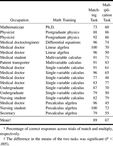
    Occupation, math education, and average percent accuracy* on the fMR imaging tasks for eighteen subjects

PreviousNext
Back to top

In this issue

American Journal of Neuroradiology
Vol. 21, Issue 6
1 Jun 2000
  • Table of Contents
  • Index by author
Advertisement
Print
Download PDF
Email Article

Thank you for your interest in spreading the word on American Journal of Neuroradiology.

NOTE: We only request your email address so that the person you are recommending the page to knows that you wanted them to see it, and that it is not junk mail. We do not capture any email address.

Enter multiple addresses on separate lines or separate them with commas.
Cerebral Activation during Multiplication: A Functional MR Imaging Study of Number Processing
(Your Name) has sent you a message from American Journal of Neuroradiology
(Your Name) thought you would like to see the American Journal of Neuroradiology web site.
CAPTCHA
This question is for testing whether or not you are a human visitor and to prevent automated spam submissions.
Cite this article
Robert K. Fulbright, David L. Molfese, Alexander A. Stevens, Pawel Skudlarski, Cheryl M. Lacadie, John C. Gore
Cerebral Activation during Multiplication: A Functional MR Imaging Study of Number Processing
American Journal of Neuroradiology Jun 2000, 21 (6) 1048-1054;

Citation Manager Formats

  • BibTeX
  • Bookends
  • EasyBib
  • EndNote (tagged)
  • EndNote 8 (xml)
  • Medlars
  • Mendeley
  • Papers
  • RefWorks Tagged
  • Ref Manager
  • RIS
  • Zotero
0 Responses
Respond to this article
Share
Bookmark this article
Cerebral Activation during Multiplication: A Functional MR Imaging Study of Number Processing
Robert K. Fulbright, David L. Molfese, Alexander A. Stevens, Pawel Skudlarski, Cheryl M. Lacadie, John C. Gore
American Journal of Neuroradiology Jun 2000, 21 (6) 1048-1054;
del.icio.us logo Twitter logo Facebook logo Mendeley logo
  • Tweet Widget
  • Facebook Like
  • Google Plus One

Jump to section

  • Article
    • Abstract
    • Methods
    • Results
    • Discussion
    • Conclusion
    • Footnotes
    • References
  • Figures & Data
  • Info & Metrics
  • Responses
  • References
  • PDF

Related Articles

  • No related articles found.
  • PubMed
  • Google Scholar

Cited By...

  • Investigating the Triple Code Model in Numerical Cognition Using Stereotactic Electroencephalography
  • Does deliberation really need more effort than intuition? A test using event-related brain potential
  • Intraoperative mapping of the cortical areas involved in multiplication and subtraction: an electrostimulation study in a patient with a left parietal glioma
  • Crossref
  • Google Scholar

This article has not yet been cited by articles in journals that are participating in Crossref Cited-by Linking.

More in this TOC Section

  • Multimodal CT Provides Improved Performance for Lacunar Infarct Detection
  • Optimal MRI Sequence for Identifying Occlusion Location in Acute Stroke: Which Value of Time-Resolved Contrast-Enhanced MRA?
  • Evaluating the Effects of White Matter Multiple Sclerosis Lesions on the Volume Estimation of 6 Brain Tissue Segmentation Methods
Show more BRAIN

Similar Articles

Advertisement

Indexed Content

  • Current Issue
  • Accepted Manuscripts
  • Article Preview
  • Past Issues
  • Editorials
  • Editor's Choice
  • Fellows' Journal Club
  • Letters to the Editor
  • Video Articles

Cases

  • Case Collection
  • Archive - Case of the Week
  • Archive - Case of the Month
  • Archive - Classic Case

More from AJNR

  • Trainee Corner
  • Imaging Protocols
  • MRI Safety Corner

Multimedia

  • AJNR Podcasts
  • AJNR Scantastics

Resources

  • Turnaround Time
  • Submit a Manuscript
  • Submit a Video Article
  • Submit an eLetter to the Editor/Response
  • Manuscript Submission Guidelines
  • Statistical Tips
  • Fast Publishing of Accepted Manuscripts
  • Graphical Abstract Preparation
  • Imaging Protocol Submission
  • Evidence-Based Medicine Level Guide
  • Publishing Checklists
  • Author Policies
  • Become a Reviewer/Academy of Reviewers
  • News and Updates

About Us

  • About AJNR
  • Editorial Board
  • Editorial Board Alumni
  • Alerts
  • Permissions
  • Not an AJNR Subscriber? Join Now
  • Advertise with Us
  • Librarian Resources
  • Feedback
  • Terms and Conditions
  • AJNR Editorial Board Alumni

American Society of Neuroradiology

  • Not an ASNR Member? Join Now

© 2025 by the American Society of Neuroradiology All rights, including for text and data mining, AI training, and similar technologies, are reserved.
Print ISSN: 0195-6108 Online ISSN: 1936-959X

Powered by HighWire