
Article Figures & Data
Figures
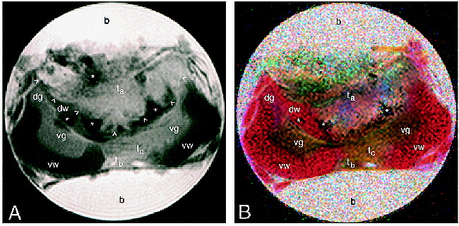
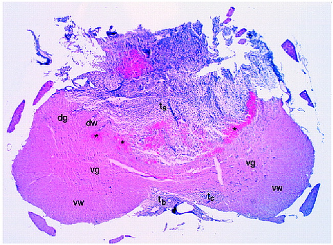
- fig 1.
MR images of a glioma engrafted within rat cervical spinal cord (b, buffer; dg, dorsal gray matter; dw, dorsal white matter; ta, main tumor mass; tb, tumor located in the meninges; tc, tumor infiltrating to the ventral side; vg, ventral gray matter; vw, ventral white matter; arrowheads, main tumor border; asterisks, blood cells).
A, Conventional T2-weighted (3000/44) spin-echo image. Fixation tends to reduce the T2 of rat spinal cord, typical values being in the range 25 to 35 milliseconds. T1 is typically 180 to 220 milliseconds for fixed rat cord at 14.1 T.
B, Colored trace image from the same image plane as in A, formed from the ADT image. Diffusion directions are assigned as follows: blue, x (ventral-dorsal); green, y (left-right); red, z (perpendicular to plane).


