Advertisement

Main menu

  • Home
  • Content
    • Current Issue
    • Accepted Manuscripts
    • Article Preview
    • Past Issue Archive
    • Video Articles
    • AJNR Case Collection
    • Case of the Week Archive
    • Case of the Month Archive
    • Classic Case Archive
  • Special Collections
    • AJNR Awards
    • Low-Field MRI
    • Alzheimer Disease
    • ASNR Foundation Special Collection
    • Photon-Counting CT
    • View All
  • Multimedia
    • AJNR Podcasts
    • AJNR SCANtastic
    • Trainee Corner
    • MRI Safety Corner
    • Imaging Protocols
  • For Authors
    • Submit a Manuscript
    • Submit a Video Article
    • Submit an eLetter to the Editor/Response
    • Manuscript Submission Guidelines
    • Statistical Tips
    • Fast Publishing of Accepted Manuscripts
    • Graphical Abstract Preparation
    • Imaging Protocol Submission
    • Author Policies
  • About Us
    • About AJNR
    • Editorial Board
    • Editorial Board Alumni
  • More
    • Become a Reviewer/Academy of Reviewers
    • Subscribers
    • Permissions
    • Alerts
    • Feedback
    • Advertisers
    • ASNR Home

User menu

  • Alerts
  • Log in

Search

  • Advanced search
American Journal of Neuroradiology

American Journal of Neuroradiology

ASHNR American Society of Functional Neuroradiology ASHNR American Society of Pediatric Neuroradiology ASSR
  • Alerts
  • Log in

Advanced Search

  • Home
  • Content
    • Current Issue
    • Accepted Manuscripts
    • Article Preview
    • Past Issue Archive
    • Video Articles
    • AJNR Case Collection
    • Case of the Week Archive
    • Case of the Month Archive
    • Classic Case Archive
  • Special Collections
    • AJNR Awards
    • Low-Field MRI
    • Alzheimer Disease
    • ASNR Foundation Special Collection
    • Photon-Counting CT
    • View All
  • Multimedia
    • AJNR Podcasts
    • AJNR SCANtastic
    • Trainee Corner
    • MRI Safety Corner
    • Imaging Protocols
  • For Authors
    • Submit a Manuscript
    • Submit a Video Article
    • Submit an eLetter to the Editor/Response
    • Manuscript Submission Guidelines
    • Statistical Tips
    • Fast Publishing of Accepted Manuscripts
    • Graphical Abstract Preparation
    • Imaging Protocol Submission
    • Author Policies
  • About Us
    • About AJNR
    • Editorial Board
    • Editorial Board Alumni
  • More
    • Become a Reviewer/Academy of Reviewers
    • Subscribers
    • Permissions
    • Alerts
    • Feedback
    • Advertisers
    • ASNR Home
  • Follow AJNR on Twitter
  • Visit AJNR on Facebook
  • Follow AJNR on Instagram
  • Join AJNR on LinkedIn
  • RSS Feeds

AJNR Awards, New Junior Editors, and more. Read the latest AJNR updates

Research ArticleNeurointervention

Development of a Biologically Active Guglielmi Detachable Coil for the Treatment of Cerebral Aneurysms. Part I: In Vitro Study

Yuichi Murayama, Yoshiaki Suzuki, Fernando Viñuela, Makoto Kaibara, Kimi Kurotobi, Masaya Iwaki and Toshiaki Abe
American Journal of Neuroradiology November 1999, 20 (10) 1986-1991;
Yuichi Murayama
  • Find this author on Google Scholar
  • Find this author on PubMed
  • Search for this author on this site
Yoshiaki Suzuki
  • Find this author on Google Scholar
  • Find this author on PubMed
  • Search for this author on this site
Fernando Viñuela
  • Find this author on Google Scholar
  • Find this author on PubMed
  • Search for this author on this site
Makoto Kaibara
  • Find this author on Google Scholar
  • Find this author on PubMed
  • Search for this author on this site
Kimi Kurotobi
  • Find this author on Google Scholar
  • Find this author on PubMed
  • Search for this author on this site
Masaya Iwaki
  • Find this author on Google Scholar
  • Find this author on PubMed
  • Search for this author on this site
Toshiaki Abe
  • Find this author on Google Scholar
  • Find this author on PubMed
  • Search for this author on this site
  • Article
  • Figures & Data
  • Info & Metrics
  • Responses
  • References
  • PDF
Loading

Article Figures & Data

Figures

  • fig 1.
    • Download figure
    • Open in new tab
    • Download powerpoint
    fig 1.

    Schematic diagram of surface modification by ion implantation to protein-coated material surface. Surface of PS dishes and platinum plates was coated with type I collagen. Ne+ implantation was performed on an area that was regulated by a 100 μm circular mask.

  • fig 2.
    • Download figure
    • Open in new tab
    • Download powerpoint
    fig 2.

    Schematic diagram of parallel plate-type flow chamber used for applying flow shear stress to endothelial cells cultured on PS surface. PS-coated slide glass on which endothelial cells (BAECs) were confluently cultured was mounted together with a silicone rubber gasket to a lower PMMA plate. Flow shear stress was imposed on BAECs by circulating 1% PBS maintained at 37°C in reservoir by using pump.
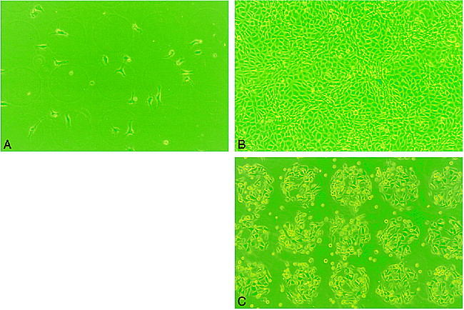
  • fig 3.
    • Download figure
    • Open in new tab
    • Download powerpoint
    fig 3.

    Phase-contrast micrograph of BAECs on PS surface patterned by ion implantation (original magnification, ×100; diameter of a circle, 100 μm).

    A, Day 1, no endothelial proliferation was observed on either protein-coated only (area A) or ion-implanted protein-coated (area B) surface.

    B, Day 5, areas A and B demonstrated uniform proliferation of the BAECs.

    C, Day 5, Post-trypsin treatment at 2 hours. Note most of BAECs were strongly attached on the ion-implanted circular area. Almost no BAECs were observed on surface coated with collagen only.

  • fig 4.
    • Download figure
    • Open in new tab
    • Download powerpoint
    fig 4.

    Scanning electron micrograph of BAECs on platinum plate patterned by ion implantation. (Note morphologic deformity of cells owing to effect of trypsin and glutaraldehyde.)

    A, Day 5, areas A and B demonstrated uniform proliferation of BAECs.

    B and C, Note strong BAEC adhesion only on circular ion-implanted protein-coated areas (original magnification, ×100 [for B]; ×400 [for C]).

  • fig 5.
    • Download figure
    • Open in new tab
    • Download powerpoint
    fig 5.

    Sequential changes in number of endothelial cells exposed to flow shear stress: 30 min (P < .001), 45 min (P < .001), 60 min (P < .05), 90 min (P < .01), 150 min (P < .05), 180 min (P = .1488).

  • fig 6.
    • Download figure
    • Open in new tab
    • Download powerpoint
    fig 6.

    Transmission electron micrograph of BAECs on polystyrene dish. Both simple collagen-coated surface and collagen-coated ion-implanted surface demonstrate BAECs attachment.

    A, Note thickness of coated collagen on simple coating surface (large arrow). Detachment of cell-collagen complex may appear to occur from interface between coated collagen and PS surface (small arrow).

    B, On the other hand, direct cell adhesion was observed on ion-implanted collagen-coated PS surface.

PreviousNext
Back to top

In this issue

American Journal of Neuroradiology
Vol. 20, Issue 10
1 Nov 1999
  • Table of Contents
  • Index by author
Advertisement
Print
Download PDF
Email Article

Thank you for your interest in spreading the word on American Journal of Neuroradiology.

NOTE: We only request your email address so that the person you are recommending the page to knows that you wanted them to see it, and that it is not junk mail. We do not capture any email address.

Enter multiple addresses on separate lines or separate them with commas.
Development of a Biologically Active Guglielmi Detachable Coil for the Treatment of Cerebral Aneurysms. Part I: In Vitro Study
(Your Name) has sent you a message from American Journal of Neuroradiology
(Your Name) thought you would like to see the American Journal of Neuroradiology web site.
CAPTCHA
This question is for testing whether or not you are a human visitor and to prevent automated spam submissions.
Cite this article
Yuichi Murayama, Yoshiaki Suzuki, Fernando Viñuela, Makoto Kaibara, Kimi Kurotobi, Masaya Iwaki, Toshiaki Abe
Development of a Biologically Active Guglielmi Detachable Coil for the Treatment of Cerebral Aneurysms. Part I: In Vitro Study
American Journal of Neuroradiology Nov 1999, 20 (10) 1986-1991;

Citation Manager Formats

  • BibTeX
  • Bookends
  • EasyBib
  • EndNote (tagged)
  • EndNote 8 (xml)
  • Medlars
  • Mendeley
  • Papers
  • RefWorks Tagged
  • Ref Manager
  • RIS
  • Zotero
0 Responses
Respond to this article
Share
Bookmark this article
Development of a Biologically Active Guglielmi Detachable Coil for the Treatment of Cerebral Aneurysms. Part I: In Vitro Study
Yuichi Murayama, Yoshiaki Suzuki, Fernando Viñuela, Makoto Kaibara, Kimi Kurotobi, Masaya Iwaki, Toshiaki Abe
American Journal of Neuroradiology Nov 1999, 20 (10) 1986-1991;
del.icio.us logo Twitter logo Facebook logo Mendeley logo
  • Tweet Widget
  • Facebook Like
  • Google Plus One

Jump to section

  • Article
    • Abstract
    • Methods
    • Results
    • Discussion
    • Conclusion
    • Footnotes
    • References
  • Figures & Data
  • Info & Metrics
  • Responses
  • References
  • PDF

Related Articles

  • No related articles found.
  • PubMed
  • Google Scholar

Cited By...

  • Bioactivity and bioinactivity: two sides of the same coin
  • Endovascular Histologic Effects of Ultrathin Gold- or Vitronectin-Coated Platinum Aneurysm Coils in a Rodent Arterial Occlusion Model: A Preliminary Investigation
  • Polyglycolide/Polylactide-Coated Platinum Coils for Patients With Ruptured and Unruptured Cerebral Aneurysms: A Single-Center Experience
  • Matrix and Bioabsorbable Polymeric Coils Accelerate Healing of Intracranial Aneurysms: Long-Term Experimental Study
  • A Collagen-Based Coil for Embolization of Saccular Aneurysms in a New Zealand White Rabbit Model
  • Cellular Responses of Bioabsorbable Polymeric Material and Guglielmi Detachable Coil in Experimental Aneurysms
  • Role of the Endothelial Lining in Persistence of Residual Lesions and Growth of Recurrences After Endovascular Treatment of Experimental Aneurysms
  • Crossref
  • Google Scholar

This article has not yet been cited by articles in journals that are participating in Crossref Cited-by Linking.

More in this TOC Section

  • Optimizing Voxel Size in 3D Rotational Angiography
  • Neuroform Atlas Stent for Intracranial Aneurysms
  • Neurologic Complication in Transradial Angiography
Show more Neurointervention

Similar Articles

Advertisement

Indexed Content

  • Current Issue
  • Accepted Manuscripts
  • Article Preview
  • Past Issues
  • Editorials
  • Editor's Choice
  • Fellows' Journal Club
  • Letters to the Editor
  • Video Articles

Cases

  • Case Collection
  • Archive - Case of the Week
  • Archive - Case of the Month
  • Archive - Classic Case

More from AJNR

  • Trainee Corner
  • Imaging Protocols
  • MRI Safety Corner

Multimedia

  • AJNR Podcasts
  • AJNR Scantastics

Resources

  • Turnaround Time
  • Submit a Manuscript
  • Submit a Video Article
  • Submit an eLetter to the Editor/Response
  • Manuscript Submission Guidelines
  • Statistical Tips
  • Fast Publishing of Accepted Manuscripts
  • Graphical Abstract Preparation
  • Imaging Protocol Submission
  • Evidence-Based Medicine Level Guide
  • Publishing Checklists
  • Author Policies
  • Become a Reviewer/Academy of Reviewers
  • News and Updates

About Us

  • About AJNR
  • Editorial Board
  • Editorial Board Alumni
  • Alerts
  • Permissions
  • Not an AJNR Subscriber? Join Now
  • Advertise with Us
  • Librarian Resources
  • Feedback
  • Terms and Conditions
  • AJNR Editorial Board Alumni

American Society of Neuroradiology

  • Not an ASNR Member? Join Now

© 2025 by the American Society of Neuroradiology All rights, including for text and data mining, AI training, and similar technologies, are reserved.
Print ISSN: 0195-6108 Online ISSN: 1936-959X

Powered by HighWire