Advertisement

Main menu

  • Home
  • Content
    • Current Issue
    • Accepted Manuscripts
    • Article Preview
    • Past Issue Archive
    • Video Articles
    • AJNR Case Collection
    • Case of the Week Archive
    • Case of the Month Archive
    • Classic Case Archive
  • Special Collections
    • AJNR Awards
    • Low-Field MRI
    • Alzheimer Disease
    • ASNR Foundation Special Collection
    • Photon-Counting CT
    • View All
  • Multimedia
    • AJNR Podcasts
    • AJNR SCANtastic
    • Trainee Corner
    • MRI Safety Corner
    • Imaging Protocols
  • For Authors
    • Submit a Manuscript
    • Submit a Video Article
    • Submit an eLetter to the Editor/Response
    • Manuscript Submission Guidelines
    • Statistical Tips
    • Fast Publishing of Accepted Manuscripts
    • Graphical Abstract Preparation
    • Imaging Protocol Submission
    • Author Policies
  • About Us
    • About AJNR
    • Editorial Board
    • Editorial Board Alumni
  • More
    • Become a Reviewer/Academy of Reviewers
    • Subscribers
    • Permissions
    • Alerts
    • Feedback
    • Advertisers
    • ASNR Home

User menu

  • Alerts
  • Log in

Search

  • Advanced search
American Journal of Neuroradiology

American Journal of Neuroradiology

ASHNR American Society of Functional Neuroradiology ASHNR American Society of Pediatric Neuroradiology ASSR
  • Alerts
  • Log in

Advanced Search

  • Home
  • Content
    • Current Issue
    • Accepted Manuscripts
    • Article Preview
    • Past Issue Archive
    • Video Articles
    • AJNR Case Collection
    • Case of the Week Archive
    • Case of the Month Archive
    • Classic Case Archive
  • Special Collections
    • AJNR Awards
    • Low-Field MRI
    • Alzheimer Disease
    • ASNR Foundation Special Collection
    • Photon-Counting CT
    • View All
  • Multimedia
    • AJNR Podcasts
    • AJNR SCANtastic
    • Trainee Corner
    • MRI Safety Corner
    • Imaging Protocols
  • For Authors
    • Submit a Manuscript
    • Submit a Video Article
    • Submit an eLetter to the Editor/Response
    • Manuscript Submission Guidelines
    • Statistical Tips
    • Fast Publishing of Accepted Manuscripts
    • Graphical Abstract Preparation
    • Imaging Protocol Submission
    • Author Policies
  • About Us
    • About AJNR
    • Editorial Board
    • Editorial Board Alumni
  • More
    • Become a Reviewer/Academy of Reviewers
    • Subscribers
    • Permissions
    • Alerts
    • Feedback
    • Advertisers
    • ASNR Home
  • Follow AJNR on Twitter
  • Visit AJNR on Facebook
  • Follow AJNR on Instagram
  • Join AJNR on LinkedIn
  • RSS Feeds

AJNR Awards, New Junior Editors, and more. Read the latest AJNR updates

Table of Contents

July 01, 2016; Volume 37,Issue 7

Perspectives

  • You have access
    Perspectives
    Ajeet Gordhan
    American Journal of Neuroradiology July 2016, 37 (7) 1191; DOI: https://doi.org/10.3174/ajnr.P0020

Review Article

  • PATIENT SAFETY
    Open Access
    Gadolinium-Based Contrast Agent Accumulation and Toxicity: An Update
    J. Ramalho, R.C. Semelka, M. Ramalho, R.H. Nunes, M. AlObaidy and M. Castillo
    American Journal of Neuroradiology July 2016, 37 (7) 1192-1198; DOI: https://doi.org/10.3174/ajnr.A4615

Patient Safety

  • FELLOWS' JOURNAL CLUBPediatric Neuroimaging
    Open Access
    Full Dose-Reduction Potential of Statistical Iterative Reconstruction for Head CT Protocols in a Predominantly Pediatric Population
    A.E. Mirro, S.L. Brady and R.A. Kaufman
    American Journal of Neuroradiology July 2016, 37 (7) 1199-1205; DOI: https://doi.org/10.3174/ajnr.A4754

    The authors set out to determine the maximum level of statistical iterative reconstruction that can be used to establish dose-reduced head CT protocols in a primarily pediatric population while maintaining similar appearance and level of image noise in the reconstructed image. Dose-reduced head protocols using an adaptive statistical iterative reconstruction were compared for image quality with the original filtered back-projection reconstructed protocols in a phantom and CT dose index and image noise magnitude were assessed in 737 pre- and post-dose-reduced examinations. Implementation of 40% and 60% adaptive statistical iterative reconstruction led to an average reduction in the volume CT dose index of 43% for brain, 41% for orbit, 30% for maxilla, 43% for sinus, and 42% for temporal bone protocols for patients between 1 month and 26 years of age, while improving the contrast-to-noise ratio of low-contrast soft-tissue targets.

Academic Perspectives

  • FELLOWS' JOURNAL CLUB
    You have access
    Non-Relative Value Unit–Generating Activities Represent One-Fifth of Academic Neuroradiologist Productivity
    M. Wintermark, M. Zeineh, G. Zaharchuk, A. Srivastava and N. Fischbein
    American Journal of Neuroradiology July 2016, 37 (7) 1206-1208; DOI: https://doi.org/10.3174/ajnr.A4701

    Four full-time neuroradiologists, working an average of 50% clinical and 50% academic activity, systematically recorded all the non-relative value unit– generating consults and conferences in which they were involved during 3 months. The 4 neuroradiologists working an average of 50% clinical activity interpreted 4241 RVU-generating imaging studies, representing 8152 work RVUs. During the same period, they recorded 792 non-RVU-generating study reviews as part of consults. The authors propose a simple Web-based smartphone app to record and quantify non-RVU-generating activities including consults, clinical conferences, and tumor boards.

General Contents

  • ADULT BRAIN
    Open Access
    Quantitative Susceptibility Mapping in Cerebral Cavernous Malformations: Clinical Correlations
    H. Tan, L. Zhang, A.G. Mikati, R. Girard, O. Khanna, M.D. Fam, T. Liu, Y. Wang, R.R. Edelman, G. Christoforidis and I.A. Awad
    American Journal of Neuroradiology July 2016, 37 (7) 1209-1215; DOI: https://doi.org/10.3174/ajnr.A4724
  • EDITOR'S CHOICEADULT BRAIN
    Open Access
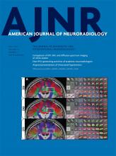
    Mapping the Orientation of White Matter Fiber Bundles: A Comparative Study of Diffusion Tensor Imaging, Diffusional Kurtosis Imaging, and Diffusion Spectrum Imaging
    G.R. Glenn, L.-W. Kuo, Y.-P. Chao, C.-Y. Lee, J.A. Helpern and J.H. Jensen
    American Journal of Neuroradiology July 2016, 37 (7) 1216-1222; DOI: https://doi.org/10.3174/ajnr.A4714

    The authors evaluated fiber bundle orientations from DTI and diffusional kurtosis compared with diffusion spectrum imaging as a criterion standard to assess the performance of each technique. DTI, diffusional kurtosis imaging, and diffusion spectrum imaging datasets were acquired during 2 independent sessions in 3 volunteers. While orientation estimates from all 3 techniques had comparable angular reproducibility, diffusional kurtosis imaging decreased angular error throughout the white matter compared with DTI. Diffusion spectrum imaging and diffusional kurtosis imaging enabled the detection of crossing-fiber bundles. They conclude that fiber bundle orientation estimates from diffusional kurtosis imaging have less systematic error than those from DTI.

  • EDITOR'S CHOICEADULT BRAIN
    Open Access
    Iron and Non-Iron-Related Characteristics of Multiple Sclerosis and Neuromyelitis Optica Lesions at 7T MRI
    S. Chawla, I. Kister, J. Wuerfel, J.-C. Brisset, S. Liu, T. Sinnecker, P. Dusek, E.M. Haacke, F. Paul and Y. Ge
    American Journal of Neuroradiology July 2016, 37 (7) 1223-1230; DOI: https://doi.org/10.3174/ajnr.A4729

    Twenty-one patients with MS and 21 patients with neuromyelitis optica underwent 7T high-resolution 2D-gradient-echo-T2* and 3D-susceptibility-weighted imaging. An in-house-developed algorithm was used to reconstruct quantitative susceptibility mapping from SWI. Of the patients with MS, 19 (90.5%) demonstrated at least 1 quantitative susceptibility mapping–hyperintense lesion, and 11/21 (52.4%) had iron-laden lesions. No quantitative susceptibility mapping–hyperintense or iron-laden lesions were observed in any patients with neuromyelitis optica. The authors conclude that ultra-high-field MR imaging may be useful in distinguishing MS from neuromyelitis optica.

  • ADULT BRAIN
    Open Access
    Assessment of Collateral Status by Dynamic CT Angiography in Acute MCA Stroke: Timing of Acquisition and Relationship with Final Infarct Volume
    I.R. van den Wijngaard, G. Holswilder, M.J.H. Wermer, J. Boiten, A. Algra, D.W.J. Dippel, J.W. Dankbaar, B.K. Velthuis, A.M.M. Boers, C.B.L.M. Majoie and M.A.A. van Walderveen
    American Journal of Neuroradiology July 2016, 37 (7) 1231-1236; DOI: https://doi.org/10.3174/ajnr.A4746
  • ADULT BRAIN
    Open Access
    Improving Perfusion Measurement in DSC–MR Imaging with Multiecho Information for Arterial Input Function Determination
    A.T. Newton, S. Pruthi, A.M. Stokes, J.T. Skinner and C.C. Quarles
    American Journal of Neuroradiology July 2016, 37 (7) 1237-1243; DOI: https://doi.org/10.3174/ajnr.A4700
  • ADULT BRAIN
    You have access
    Time-of-Flight MR Angiography for Detection of Cerebral Hyperperfusion Syndrome after Superficial Temporal Artery–Middle Cerebral Artery Anastomosis in Moyamoya Disease
    K. Sato, M. Yamada, H. Kuroda, D. Yamamoto, Y. Asano, Y. Inoue, K. Fujii and T. Kumabe
    American Journal of Neuroradiology July 2016, 37 (7) 1244-1248; DOI: https://doi.org/10.3174/ajnr.A4715
  • ADULT BRAIN
    You have access
    Comparison of CSF Distribution between Idiopathic Normal Pressure Hydrocephalus and Alzheimer Disease
    S. Yamada, M. Ishikawa and K. Yamamoto
    American Journal of Neuroradiology July 2016, 37 (7) 1249-1255; DOI: https://doi.org/10.3174/ajnr.A4695
  • FELLOWS' JOURNAL CLUBADULT BRAIN
    You have access
    Atypical Presentations of Intracranial Hypotension: Comparison with Classic Spontaneous Intracranial Hypotension
    A.A. Capizzano, L. Lai, J. Kim, M. Rizzo, L. Gray, M.K. Smoot and T. Moritani
    American Journal of Neuroradiology July 2016, 37 (7) 1256-1261; DOI: https://doi.org/10.3174/ajnr.A4706

    The authors evaluated the clinical records and neuroimaging of patients with spontaneous intracranial hypotension from September 2005 to August 2014. Patients with classic spontaneous intracranial hypotension (n = 33) were compared with those with intracranial hypotension with atypical clinical presentation (n = 8). There was no significant difference in dural enhancement, subdural hematomas, or cerebellar tonsil herniation. Patients with atypical spontaneous intracranial hypotension had significantly more elongated anteroposterior midbrain diameter compared with those with classic spontaneous intracranial hypotension, and shortened pontomammillary distance. In this population, patients with atypical spontaneous intracranial hypotension showed a more chronic syndrome compared with classic spontaneous intracranial hypotension, more severe brain sagging, lower rates of clinical response, and frequent relapses.

  • ADULT BRAIN
    You have access
    Quantitative Assessment of Circumferential Enhancement along the Wall of Cerebral Aneurysms Using MR Imaging
    S. Omodaka, H. Endo, K. Niizuma, M. Fujimura, T. Inoue, K. Sato, S.-i. Sugiyama and T. Tominaga
    American Journal of Neuroradiology July 2016, 37 (7) 1262-1266; DOI: https://doi.org/10.3174/ajnr.A4722
  • ADULT BRAIN
    Open Access
    Evaluating Permeability Surface-Area Product as a Measure of Blood-Brain Barrier Permeability in a Murine Model
    E.K. Weidman, C.P. Foley, O. Kallas, J.P. Dyke, A. Gupta, A.E. Giambrone, J. Ivanidze, H. Baradaran, D.J. Ballon and P.C. Sanelli
    American Journal of Neuroradiology July 2016, 37 (7) 1267-1274; DOI: https://doi.org/10.3174/ajnr.A4712
  • Neurointervention
    Open Access
    Multicenter Prospective Trial of Stent Placement in Patients with Symptomatic High-Grade Intracranial Stenosis
    P. Gao, D. Wang, Z. Zhao, Y. Cai, T. Li, H. Shi, W. Wu, W. He, L. Yin, S. Huang, F. Zhu, L. Jiao, X. Ji, A.I. Qureshi and F. Ling
    American Journal of Neuroradiology July 2016, 37 (7) 1275-1280; DOI: https://doi.org/10.3174/ajnr.A4698
  • EDITOR'S CHOICENeurointervention
    You have access
    Endovascular Management of Tandem Occlusion Stroke Related to Internal Carotid Artery Dissection Using a Distal to Proximal Approach: Insight from the RECOST Study
    G. Marnat, I. Mourand, O. Eker, P. Machi, C. Arquizan, C. Riquelme, X. Ayrignac, A. Bonafé and V. Costalat
    American Journal of Neuroradiology July 2016, 37 (7) 1281-1288; DOI: https://doi.org/10.3174/ajnr.A4752

    The authors analyzed all carotid artery dissection tandem occlusion strokes and isolated anterior circulation occlusions from their ongoing prospective stroke data base. For carotid artery dissection, the revascularization procedure consisted of initial distal recanalization by a stent retriever in the intracranial vessel. Following assessment of the circle of Willis, ICA stent placement was only performed in case of insufficiency. Two hundred fifty-eight patients with an anterior circulation stroke were analyzed, including 20 with carotid artery dissection–related occlusion. Only 5 carotid artery dissections (25%) necessitated cervical stent placement. No early ipsilateral stroke recurrence was recorded, despite the absence of stent placement in 15 patients (75%) with carotid artery dissection. Mechanical endovascular treatment of carotid artery dissection tandem occlusions is safe and effective compared with isolated anterior circulation occlusion stroke therapy. The authors favor a complete evaluation of the circle of Willis in these patients, which requires a contralateral femoral puncture, allowing selective contralateral common carotid and vertebrobasilar catheterizations.

  • Neurointervention
    You have access
    Hemodynamic and Anatomic Variations Require an Adaptable Approach during Intra-Arterial Chemotherapy for Intraocular Retinoblastoma: Alternative Routes, Strategies, and Follow-Up
    E. Bertelli, S. Leonini, D. Galimberti, S. Moretti, R. Tinturini, T. Hadjistilianou, S. De Francesco, D.G. Romano, I.M. Vallone, S. Cioni, P. Gennari, P. Galluzzi, I. Grazzini, S. Rossi and S. Bracco
    American Journal of Neuroradiology July 2016, 37 (7) 1289-1295; DOI: https://doi.org/10.3174/ajnr.A4741
  • Neurointervention
    You have access
    Prediction of Stent-Retriever Thrombectomy Outcomes by Dynamic Multidetector CT Angiography in Patients with Acute Carotid T or MCA Occlusions
    K.M. Thierfelder, W.H. Sommer, B. Ertl-Wagner, S.E. Beyer, F.G. Meinel, W.G. Kunz, G. Buchholz, M.F. Reiser and H. Janssen
    American Journal of Neuroradiology July 2016, 37 (7) 1296-1302; DOI: https://doi.org/10.3174/ajnr.A4694
  • Neurointervention
    Open Access
    C-Arm Conebeam CT Perfusion Imaging in the Angiographic Suite: A Comparison with Multidetector CT Perfusion Imaging
    K. Niu, P. Yang, Y. Wu, T. Struffert, A. Doerfler, S. Schafer, K. Royalty, C. Strother and G.-H. Chen
    American Journal of Neuroradiology July 2016, 37 (7) 1303-1309; DOI: https://doi.org/10.3174/ajnr.A4691
  • Neurointervention
    Open Access
    Thinner Regions of Intracranial Aneurysm Wall Correlate with Regions of Higher Wall Shear Stress: A 7T MRI Study
    R. Blankena, R. Kleinloog, B.H. Verweij, P. van Ooij, B. ten Haken, P.R. Luijten, G.J.E. Rinkel and J.J.M. Zwanenburg
    American Journal of Neuroradiology July 2016, 37 (7) 1310-1317; DOI: https://doi.org/10.3174/ajnr.A4734
  • Neurointervention
    You have access
    Inflow Jet Patterns of Unruptured Cerebral Aneurysms Based on the Flow Velocity in the Parent Artery: Evaluation Using 4D Flow MRI
    K. Futami, T. Kitabayashi, H. Sano, K. Misaki, N. Uchiyama, F. Ueda and M. Nakada
    American Journal of Neuroradiology July 2016, 37 (7) 1318-1323; DOI: https://doi.org/10.3174/ajnr.A4704
  • Head and Neck Imaging
    You have access
    The Impact of Middle Turbinate Concha Bullosa on the Severity of Inferior Turbinate Hypertrophy in Patients with a Deviated Nasal Septum
    C.M. Tomblinson, M.-R. Cheng, D. Lal and J.M. Hoxworth
    American Journal of Neuroradiology July 2016, 37 (7) 1324-1330; DOI: https://doi.org/10.3174/ajnr.A4705
  • Head and Neck Imaging
    Open Access
    Vestibular Aqueduct Measurements in the 45° Oblique (Pöschl) Plane
    A.F. Juliano, E.Y. Ting, V. Mingkwansook, L.M. Hamberg and H.D. Curtin
    American Journal of Neuroradiology July 2016, 37 (7) 1331-1337; DOI: https://doi.org/10.3174/ajnr.A4735
  • Pediatric Neuroimaging
    Open Access
    Brain Injury in Neonates with Complex Congenital Heart Disease: What Is the Predictive Value of MRI in the Fetal Period?
    M. Brossard-Racine, A. du Plessis, G. Vezina, R. Robertson, M. Donofrio, W. Tworetzky and C. Limperopoulos
    American Journal of Neuroradiology July 2016, 37 (7) 1338-1346; DOI: https://doi.org/10.3174/ajnr.A4716
  • Pediatric Neuroimaging
    Open Access
    Whole-Brain DTI Assessment of White Matter Damage in Children with Bilateral Cerebral Palsy: Evidence of Involvement beyond the Primary Target of the Anoxic Insult
    F. Arrigoni, D. Peruzzo, C. Gagliardi, C. Maghini, P. Colombo, F. Servodio Iammarrone, C. Pierpaoli, F. Triulzi and A.C. Turconi
    American Journal of Neuroradiology July 2016, 37 (7) 1347-1353; DOI: https://doi.org/10.3174/ajnr.A4717
  • Pediatric Neuroimaging
    You have access
    Decreased Superior Sagittal Sinus Diameter and Jugular Bulb Narrowing Are Associated with Poor Clinical Outcome in Vein of Galen Arteriovenous Malformation
    G. Saliou, P. Dirks, R.H. Sacho, L. Chen, K. terBrugge and T. Krings
    American Journal of Neuroradiology July 2016, 37 (7) 1354-1358; DOI: https://doi.org/10.3174/ajnr.A4697
  • Pediatric Neuroimaging
    You have access
    Development of the Fetal Vermis: New Biometry Reference Data and Comparison of 3 Diagnostic Modalities–3D Ultrasound, 2D Ultrasound, and MR Imaging
    E. Katorza, E. Bertucci, S. Perlman, S. Taschini, R. Ber, Y. Gilboa, V. Mazza and R. Achiron
    American Journal of Neuroradiology July 2016, 37 (7) 1359-1366; DOI: https://doi.org/10.3174/ajnr.A4725
  • Pediatric Neuroimaging
    You have access
    A Novel Methodology for Applying Multivoxel MR Spectroscopy to Evaluate Convection-Enhanced Drug Delivery in Diffuse Intrinsic Pontine Gliomas
    D.I. Guisado, R. Singh, S. Minkowitz, Z. Zhou, S. Haque, K.K. Peck, R.J. Young, A.J. Tsiouris, M.M. Souweidane and S.B. Thakur
    American Journal of Neuroradiology July 2016, 37 (7) 1367-1373; DOI: https://doi.org/10.3174/ajnr.A4713
  • Spine Imaging and Spine Image-Guided Interventions
    You have access
    Imaging Signs in Spontaneous Intracranial Hypotension: Prevalence and Relationship to CSF Pressure
    P.G. Kranz, T.P. Tanpitukpongse, K.R. Choudhury, T.J. Amrhein and L. Gray
    American Journal of Neuroradiology July 2016, 37 (7) 1374-1378; DOI: https://doi.org/10.3174/ajnr.A4689
  • Spine Imaging and Spine Image-Guided Interventions
    You have access
    The “Hyperdense Paraspinal Vein” Sign: A Marker of CSF-Venous Fistula
    P.G. Kranz, T.J. Amrhein, W.I. Schievink, I.O. Karikari and L. Gray
    American Journal of Neuroradiology July 2016, 37 (7) 1379-1381; DOI: https://doi.org/10.3174/ajnr.A4682

35 Years Ago in AJNR

  • You have access
    Celebrating 35 Years of the AJNR
    American Journal of Neuroradiology July 2016, 37 (7) 1382; DOI: https://doi.org/10.3174/ajnr.P0022
Back to top
PreviousNext

In this issue

American Journal of Neuroradiology: 37 (7)
American Journal of Neuroradiology
Vol. 37, Issue 7
1 Jul 2016
  • Table of Contents
  • Index by author
  • Complete Issue (PDF)
Sign up for alerts
Advertisement

Jump to

  • Perspectives
  • Review Article
  • Patient Safety
  • Academic Perspectives
  • General Contents
  • 35 Years Ago in AJNR
  • Letters
  • Most Read
  • Most Cited
Loading
Advertisement

Indexed Content

  • Current Issue
  • Accepted Manuscripts
  • Article Preview
  • Past Issues
  • Editorials
  • Editor's Choice
  • Fellows' Journal Club
  • Letters to the Editor
  • Video Articles

Cases

  • Case Collection
  • Archive - Case of the Week
  • Archive - Case of the Month
  • Archive - Classic Case

More from AJNR

  • Trainee Corner
  • Imaging Protocols
  • MRI Safety Corner
  • Book Reviews

Multimedia

  • AJNR Podcasts
  • AJNR Scantastics

Resources

  • Turnaround Time
  • Submit a Manuscript
  • Submit a Video Article
  • Submit an eLetter to the Editor/Response
  • Manuscript Submission Guidelines
  • Statistical Tips
  • Fast Publishing of Accepted Manuscripts
  • Graphical Abstract Preparation
  • Imaging Protocol Submission
  • Evidence-Based Medicine Level Guide
  • Publishing Checklists
  • Author Policies
  • Become a Reviewer/Academy of Reviewers
  • News and Updates

About Us

  • About AJNR
  • Editorial Board
  • Editorial Board Alumni
  • Alerts
  • Permissions
  • Not an AJNR Subscriber? Join Now
  • Advertise with Us
  • Librarian Resources
  • Feedback
  • Terms and Conditions
  • AJNR Editorial Board Alumni

American Society of Neuroradiology

  • Not an ASNR Member? Join Now

© 2025 by the American Society of Neuroradiology All rights, including for text and data mining, AI training, and similar technologies, are reserved.
Print ISSN: 0195-6108 Online ISSN: 1936-959X

Powered by HighWire