Advertisement
  • Main menu
  • User menu
  • Search
  • English ▼
    • English
    • Afrikaans
    • Albanian
    • Amharic
    • Arabic
    • Armenian
    • Azerbaijani
    • Basque
    • Belarusian
    • Bengali
    • Bosnian
    • Bulgarian
    • Catalan
    • Cebuano
    • Chichewa
    • Chinese (Simplified)
    • Chinese (Traditional)
    • Corsican
    • Croatian
    • Czech
    • Danish
    • Dutch
    • Esperanto
    • Estonian
    • Filipino
    • Finnish
    • French
    • Frisian
    • Galician
    • Georgian
    • German
    • Greek
    • Gujarati
    • Haitian Creole
    • Hausa
    • Hawaiian
    • Hebrew
    • Hindi
    • Hmong
    • Hungarian
    • Icelandic
    • Igbo
    • Indonesian
    • Irish
    • Italian
    • Japanese
    • Javanese
    • Kannada
    • Kazakh
    • Khmer
    • Korean
    • Kurdish (Kurmanji)
    • Kyrgyz
    • Lao
    • Latin
    • Latvian
    • Lithuanian
    • Luxembourgish
    • Macedonian
    • Malagasy
    • Malay
    • Malayalam
    • Maltese
    • Maori
    • Marathi
    • Mongolian
    • Myanmar (Burmese)
    • Nepali
    • Norwegian
    • Pashto
    • Persian
    • Polish
    • Portuguese
    • Punjabi
    • Romanian
    • Russian
    • Samoan
    • Scottish Gaelic
    • Serbian
    • Sesotho
    • Shona
    • Sindhi
    • Sinhala
    • Slovak
    • Slovenian
    • Somali
    • Spanish
    • Sudanese
    • Swahili
    • Swedish
    • Tajik
    • Tamil
    • Telugu
    • Thai
    • Turkish
    • Ukrainian
    • Urdu
    • Uzbek
    • Vietnamese
    • Welsh
    • Xhosa
    • Yiddish
    • Yoruba
    • Zulu

Main menu

  • Home
  • Content
    • Current Issue
    • Accepted Manuscripts
    • Article Preview
    • Past Issue Archive
    • Video Articles
    • AJNR Case Collection
    • Case of the Week Archive
    • Case of the Month Archive
    • Classic Case Archive
  • Special Collections
    • AJNR Awards
    • Low-Field MRI
    • Alzheimer Disease
    • ASNR Foundation Special Collection
    • Photon-Counting CT
    • View All
  • Multimedia
    • AJNR Podcasts
    • AJNR SCANtastic
    • Trainee Corner
    • MRI Safety Corner
    • Imaging Protocols
  • For Authors
    • Submit a Manuscript
    • Submit a Video Article
    • Submit an eLetter to the Editor/Response
    • Manuscript Submission Guidelines
    • Statistical Tips
    • Fast Publishing of Accepted Manuscripts
    • Graphical Abstract Preparation
    • Imaging Protocol Submission
    • Author Policies
  • About Us
    • About AJNR
    • Editorial Board
    • Editorial Board Alumni
  • More
    • Become a Reviewer/Academy of Reviewers
    • Subscribers
    • Permissions
    • Alerts
    • Feedback
    • Advertisers
    • ASNR Home

User menu

  • Alerts
  • Log in

Search

  • Advanced search
American Journal of Neuroradiology

American Journal of Neuroradiology

ASHNR American Society of Functional Neuroradiology ASHNR American Society of Pediatric Neuroradiology ASSR
  • Alerts
  • Log in

Advanced Search

English ▼
  • English
  • Afrikaans
  • Albanian
  • Amharic
  • Arabic
  • Armenian
  • Azerbaijani
  • Basque
  • Belarusian
  • Bengali
  • Bosnian
  • Bulgarian
  • Catalan
  • Cebuano
  • Chichewa
  • Chinese (Simplified)
  • Chinese (Traditional)
  • Corsican
  • Croatian
  • Czech
  • Danish
  • Dutch
  • Esperanto
  • Estonian
  • Filipino
  • Finnish
  • French
  • Frisian
  • Galician
  • Georgian
  • German
  • Greek
  • Gujarati
  • Haitian Creole
  • Hausa
  • Hawaiian
  • Hebrew
  • Hindi
  • Hmong
  • Hungarian
  • Icelandic
  • Igbo
  • Indonesian
  • Irish
  • Italian
  • Japanese
  • Javanese
  • Kannada
  • Kazakh
  • Khmer
  • Korean
  • Kurdish (Kurmanji)
  • Kyrgyz
  • Lao
  • Latin
  • Latvian
  • Lithuanian
  • Luxembourgish
  • Macedonian
  • Malagasy
  • Malay
  • Malayalam
  • Maltese
  • Maori
  • Marathi
  • Mongolian
  • Myanmar (Burmese)
  • Nepali
  • Norwegian
  • Pashto
  • Persian
  • Polish
  • Portuguese
  • Punjabi
  • Romanian
  • Russian
  • Samoan
  • Scottish Gaelic
  • Serbian
  • Sesotho
  • Shona
  • Sindhi
  • Sinhala
  • Slovak
  • Slovenian
  • Somali
  • Spanish
  • Sudanese
  • Swahili
  • Swedish
  • Tajik
  • Tamil
  • Telugu
  • Thai
  • Turkish
  • Ukrainian
  • Urdu
  • Uzbek
  • Vietnamese
  • Welsh
  • Xhosa
  • Yiddish
  • Yoruba
  • Zulu
  • Home
  • Content
    • Current Issue
    • Accepted Manuscripts
    • Article Preview
    • Past Issue Archive
    • Video Articles
    • AJNR Case Collection
    • Case of the Week Archive
    • Case of the Month Archive
    • Classic Case Archive
  • Special Collections
    • AJNR Awards
    • Low-Field MRI
    • Alzheimer Disease
    • ASNR Foundation Special Collection
    • Photon-Counting CT
    • View All
  • Multimedia
    • AJNR Podcasts
    • AJNR SCANtastic
    • Trainee Corner
    • MRI Safety Corner
    • Imaging Protocols
  • For Authors
    • Submit a Manuscript
    • Submit a Video Article
    • Submit an eLetter to the Editor/Response
    • Manuscript Submission Guidelines
    • Statistical Tips
    • Fast Publishing of Accepted Manuscripts
    • Graphical Abstract Preparation
    • Imaging Protocol Submission
    • Author Policies
  • About Us
    • About AJNR
    • Editorial Board
    • Editorial Board Alumni
  • More
    • Become a Reviewer/Academy of Reviewers
    • Subscribers
    • Permissions
    • Alerts
    • Feedback
    • Advertisers
    • ASNR Home
  • Follow AJNR on Twitter
  • Visit AJNR on Facebook
  • Follow AJNR on Instagram
  • Join AJNR on LinkedIn
  • RSS Feeds

AJNR Awards, New Junior Editors, and more. Read the latest AJNR updates

Research ArticleBrain

Relationships between Angiographic Findings and National Institutes of Health Stroke Scale Score in Cases of Hyperacute Carotid Ischemic Stroke

Makoto Nakajima, Kazumi Kimura, Toshiyasu Ogata, Tatsuro Takada, Makoto Uchino and Kazuo Minematsu
American Journal of Neuroradiology February 2004, 25 (2) 238-241;
Makoto Nakajima
  • Find this author on Google Scholar
  • Find this author on PubMed
  • Search for this author on this site
Kazumi Kimura
  • Find this author on Google Scholar
  • Find this author on PubMed
  • Search for this author on this site
Toshiyasu Ogata
  • Find this author on Google Scholar
  • Find this author on PubMed
  • Search for this author on this site
Tatsuro Takada
  • Find this author on Google Scholar
  • Find this author on PubMed
  • Search for this author on this site
Makoto Uchino
  • Find this author on Google Scholar
  • Find this author on PubMed
  • Search for this author on this site
Kazuo Minematsu
  • Find this author on Google Scholar
  • Find this author on PubMed
  • Search for this author on this site
  • Article
  • Figures & Data
  • Info & Metrics
  • Responses
  • References
  • PDF
Loading

Abstract

BACKGROUND AND PURPOSE: Stroke severity in cases of hyperacute carotid ischemic stroke may be related to site of arterial occlusion. We evaluated the relationships between National Institutes of Health Stroke Scale (NIHSS) scores and findings on intra-arterial digital subtraction angiograms (IA-DSA) of patients with ischemic stroke within 6 hr of stroke onset.

METHODS: A total of 43 consecutive patients (38 men and five women; mean age, 69.4 ± 8.7 years) with ischemic stroke in the carotid territory underwent IA-DSA within 6 hr of stroke onset. Baseline NIHSS score was assessed immediately before IA-DSA. Patients were divided into four groups according to site of arterial occlusion: 1) the internal carotid artery (ICA group, n = 10); 2) stem of the middle cerebral artery or stem of the anterior cerebral artery (Stem group, n = 14); 3) branches of middle cerebral artery or anterior cerebral artery (Branch group, n = 11); and 4) no arterial occlusion (Normal group, n = 8).

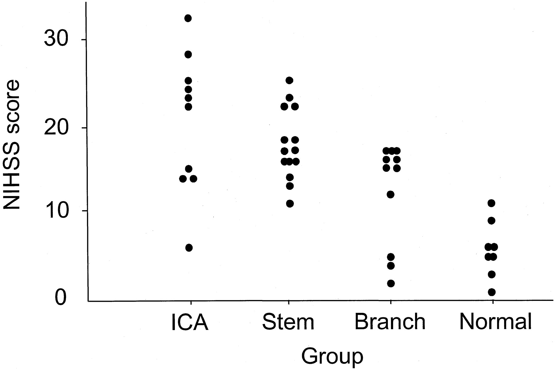
RESULTS: Mean (±SD) NIHSS score was 14.7 ± 7.4. The interval from stroke onset to IA-DSA study was 205 ± 76 min. NIHSS score was higher in the ICA group (median, 23; range, 6–32) than in the Branch (median, 17; range, 11–25; P = .02) or Normal (median, 15; range, 2–17; P < .001) groups but was not higher than in the Stem group (median, 6; range, 1–11; P = .73). Sensitivity-specificity curve analysis suggested an NIHSS score ≥10 as indicative of arterial occlusion of the carotid system. A total of 96.9% of patients with NIHSS scores ≥10 displayed arterial occlusion, and 63.6% of patients with NIHSS scores <10 displayed no arterial occlusion.

CONCLUSION: NIHSS score is related to site of arterial occlusion in cases of hyperacute carotid ischemic stroke. An NIHSS score of 10 seems to represent the cut-off for discriminating between patients with arterial occlusion and patients without.

The National Institutes of Health Stroke Scale (NIHSS) is a widely used and well-validated neurologic impairment scale, measuring speech and language, cognition, visual field deficits, motor and sensory impairments, and ataxia (1). NIHSS score assessed during the hyperacute phase of stroke strongly predicts the likelihood of patient recovery after stroke and has been used to include or exclude patients from trials of acute stroke therapy, including thrombolysis (2–7). The NIHSS therefore represents a standard part of clinical assessment for patients with acute stroke in many stroke centers.

Fink et al (8) reported a significant correlation between diffusion-weighted MR imaging lesion volume and NIHSS score. Several studies examined relationships between initial NIHSS score and vascular imaging techniques such as ultrasonography (9, 10), CT angiography (11), and MR angiography (12), reporting that a higher NIHSS score was associated with more severe vascular lesions in patients with acute stroke. However, vascular imaging methods have limitations in clearly displaying occlusion or stenosis of the main stem and branches of the middle cerebral artery (MCA) and anterior cerebral artery (ACA). Relationships between NIHSS score and site of arterial occlusion during the hyperacute phase of stroke, therefore, have yet to be accurately determined.

Intra-arterial digital subtraction angiography (IA-DSA) is superior to other methods for detecting the site of arterial occlusion and is considered to represent the gold standard for vascular imaging. During the hyperacute phase of stroke, detailed knowledge of arterial occlusion can be clinically important, particularly regarding thromobolysis (13, 14). The aim of the present study was to evaluate relationships between NIHSS score and IA-DSA findings in patients with ischemic stroke within 6 hr of stroke onset.

Methods

Patients and Techniques

Of the 112 patients admitted to our division within 6 hr of ischemic stroke onset between April 1999 and June 2002, IA-DSA was performed in 43 patients with carotid acute ischemic stroke (38 men and five women; mean age, 69.4 ± 8.7 years). We excluded the patients with posterior circulation strokes because anterior circulation and posterior circulation strokes were thought to be separate entities, with different underlying pathogenesis and natural histories. These 43 patients were enrolled into this study. All patients were assessed by using the NIHSS immediately before IA-DSA. If the patient was aware of symptoms on waking from sleep, time of onset was defined as the last time they were free from symptoms. A modified Rankin Scale (15) score ≥2 before stroke onset was used as an exclusion criterion.

Sex, age, history of stroke or transient ischemic attack, and modified Rankin Scale score were examined, along with vascular risk factors including hypertension, diabetes mellitus, hyperlipidemia, smoking, and potential embolic sources of emboli (atrial fibrillation, patent foramen ovale, left ventricular aneurysm, prosthetic heart valves, infective endocarditis, sick sinus syndrome, dilated cardiomyopathy, and complicated lesions in aortic arch).

Vascular risk factors were identified as follows: 1) use of antihypertensive agents for hypertension, with systolic blood pressure ≥160 mmHg or diastolic blood pressure ≥95 mmHg at admission for hypertension; 2) use of oral hypoglycemic agents, insulin, or glycosylated hemoglobin >6.4% for diabetes mellitus; 3) use of antihyperlipidemic agents or serum cholesterol level >220 mg/dL for hypercholesterolemia; or 4) history of smoking. To detect potential embolic sources of emboli, all patients underwent 12-lead ECG, 24-hr ECG monitoring, and transthoracic or transesophageal echocardiography.

Informed consent for performance of IA-DSA was obtained from patients and/or their family members. Selective IA-DSA was performed by using a biplane, high resolution angiography system (Angio Rex Super-G and DFP-2000A, Toshiba) with a matrix of 1024 × 1024 pixels. A catheter was inserted into the right brachial artery or femoral artery in accordance with the Seldinger method and then guided to the cerebral arteries for diagnostic four-vessel angiography.

Patients were divided into four groups according to the site of arterial occlusion: 1) occlusion of the internal carotid artery (ICA group); 2) occlusion of the main stem of the MCA or A1 segment of the ACA (Stem group); 3) occlusion of the MCA or ACA branch, including occlusion of M2 or A2 or more distal sites (Branch group); or 4) no arterial occlusion (Normal group). If a patient displayed two or more occluded arteries, the patient was placed in the largest artery group (eg, if occlusions of both the right main trunk of the MCA and the ipsilateral A2 portion were present, the patient was placed in the Stem group).

CT of the brain was performed immediately at admission and 3 days after stroke onset to evaluate ischemic lesions. Within 7 days of stroke onset, MR imaging was performed by using a 1.5-T system (Magnetom Vision, Siemens) equipped with single shot echo-planar imaging to obtain rapid diffusion images. MR imaging studies included axial T1-weighted, axial T2-weighted, and diffusion-weighted sequences (approximately 30 min of imaging time). Imaging parameters were as follows: 4000/103 (TR/TE); matrix, 128 × 128; field of view, 230 mm; section thickness, 4 mm; section gap, 2 mm. Two b values were used (0 and 1000 s/mm2). Diffusion gradients were applied in successive images in each of the x, y, and z directions, and diffusion-weighted images were formed from the mean of these values. Criteria for diagnosis of acute infarcts on diffusion-weighted images included focal hyperintensity judged not to be due to normal anisotropic diffusion or magnetic susceptibility artifact. These lesions were also categorized as cortical, subcortical, or lacunar infarcts according to location.

Statistical analysis was performed by using StatView 5.0 for Windows (SAS Institute, 1998). The χ2 test or Kruskal-Wallis U test was used to compare baseline characteristics among the four groups. Relationships between baseline NIHSS score and site of arterial occlusion were tested by using the Kruskal-Wallis U test, and the differences between NIHSS scores for each group were tested by post hoc analysis under Scheffe’s method. To obtain the NIHSS score as the cut-off point for discriminating between patients with arterial occlusion and those without, a sensitivity and specificity curve was drawn. The study protocol followed all principles outlined in the Declaration of Helsinki.

Results

Of the 43 patients enrolled in this study, 20 underwent IA-DSA within 3 hr of stroke onset. Intervals from stroke onset to arrival at hospital and to IA-DSA study were 88 ± 58 min and 205 ± 76 min, respectively.

The Stem group was the largest (n = 14), with the other groups in descending order being the Branch (n = 11), ICA (n = 10), and Normal (n = 8) groups. Demographic data and clinical features of each group are shown in Table 1. Atrial fibrillation was observed most frequently in the Branch group (P < .014). No other significant differences in baseline characteristics were observed.

View this table:
  • View inline
  • View popup
TABLE 1:

Clinical characteristics of all patients

In the ICA group, seven patients displayed ICA occlusion. For one, occlusion of both the ICA and ipsilateral ACA A2 portion was show, and for the remaining two, both ICA occlusion and ipsilateral MCA stem occlusion was shown, despite good collateral flow from the contralateral ACA via the anterior communicating artery. In the Stem group, 11 patients displayed MCA stem occlusion, one had bilateral MCA stem occlusion, one had both MCA stem occlusion and ipsilateral A2 occlusion, and the remaining had both MCA stem occlusion and occlusion of the distal site of ipsilateral ACA. In the Branch group, five patients had MCA M2 branch occlusion, two had M3 branch occlusion, two had both M2 and A2 portion occlusion, one had M2 and A4 portion occlusions, and the other one displayed M4 and A3 portion occlusions.

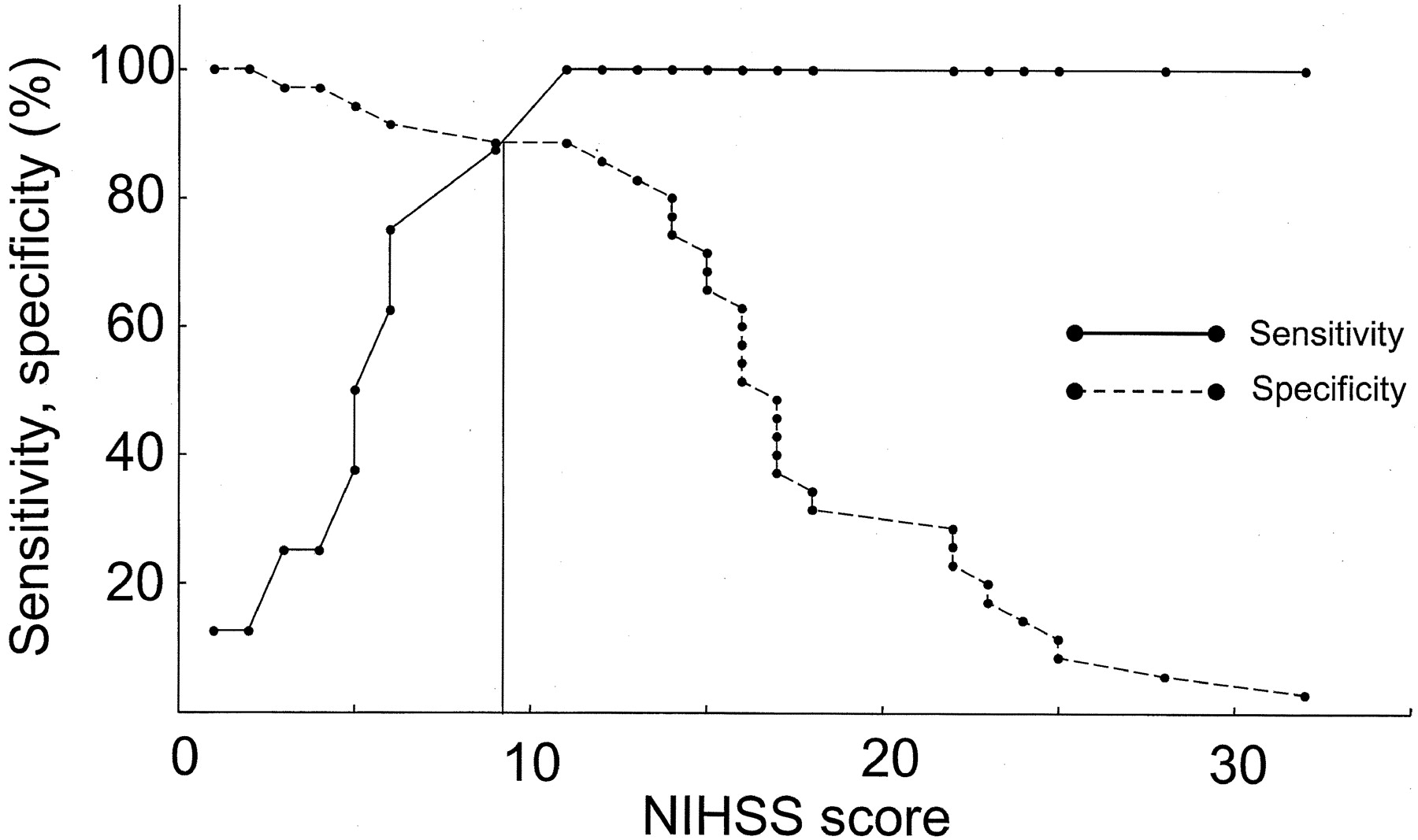
The median NIHSS score was 16 (range, 1–32). No significant differences were observed between NIHSS scores of patients with left- and right-sided stroke (P = .874, Mann-Whitney U test). NIHSS score was higher in the ICA group (median, 23; range, 6–32) than in the Branch (median, 15; range, 2–17; P = .02) or Normal (median, 6; range, 1–11; P < .001) groups but not higher than in the Stem group (median, 17; range, 11–25; P = .73). Patients in the Stem group displayed higher NIHSS scores than those in the Normal group (P < .001). In sensitivity-specificity curve analysis for predicting arterial occlusion, the optimal threshold value of the NIHSS score was 10 (Figs 1 and 2). Using an NIHSS score of 10 as the cut-off, sensitivity, specificity, positive predictive value, and negative predictive value for any arterial occlusion were 88.6%, 87.5%, 63.6%, and 96.9%, respectively.

Fig 1.
  • Download figure
  • Open in new tab
  • Download powerpoint
Fig 1.

Distribution of the NIHSS score of four groups.

Fig 2.
  • Download figure
  • Open in new tab
  • Download powerpoint
Fig 2.

Sensitivity-specificity curve analysis for predicting arterial occlusion. The optimal threshold value of the NIHSS score was 10.

MR imaging revealed 33 cortical infarcts, six subcortical infarcts, one lacunar infarct, and no ischemic lesions in three patients. Cortical and subcortical infarcts were present in seven and three of the 10 ICA group patients, respectively, 14 and 0 of the 14 MCA group patients, respectively, 10 and 0 of the 11 Branch group patients, respectively, and two and three of the eight Normal group patients, respectively. Lacunar infarct was observed in only one patient from the Normal group. One patient in the Branch group and three in the Normal group displayed no fresh lesions.

Discussion

Our study showed that an NIHSS score ≥10 represented the optimal value for predicting arterial occlusion in patients within 6 hr of stroke onset; 31 (97.0%) of 32 patients with NIHSS score ≥10 displayed arterial occlusion. Lewandowski et al (4) studied patients with stroke within 3 hr of stroke onset in the Emergency Management of Stroke bridging trial by using angiography. In that study, 17 (77.3%) of 22 patients with NIHSS scores ≥10 displayed occluded arteries in the carotid system. This minor discrepancy may be attributable to the differing interval between stroke onset and vascular imaging and to the methods to evaluate occlusive lesions.

Some studies have reported no significant differences in NIHSS scores between patients with MCA trunk occlusion and ICA/MCA tandem occlusion, a result that is compatible with the present results (9, 16). When the ICA is occluded, the severity of neurologic deficit is contingent on collateral blood flow through the anterior communicating or leptomeningial arteries from the ACA or posterior cerebral artery.

Our study included a small number of small artery diseases presenting as lacunar syndrome. We do not frequently perform IA-DSA but MRA to evaluate occlusive lesions in patients with lacunar stroke. A previous study reported that 205 (67.0%) of 306 patients with small artery disease had NIHSS scores of 0 to 6 (2). Patients with small artery disease may therefore be likely to achieve NIHSS scores <10.

In the present study, only one (3.0%) of 33 patients with NIHSS scores ≥10 displayed no arterial occlusion. In this case, neurologic symptoms improved immediately after angiography; this was attributed to spontaneous reopening of the occluded artery immediately before IA-DSA. Conversely, four (36.4%) of 11 patients with NIHSS scores <10 displayed arterial occlusions. Naylor et al (17) reported that patients with hyperacute stroke with MCA or ICA occlusions may occasionally display mild stroke severity or mimic lacunar events. Of the four patients, angiography in one case revealed occlusion of the right ICA origin and ipsilateral ACA and good collateral blood flow through the anterior communicating artery from the contralateral ICA system to the ipsilateral MCA. MR imaging of the brain revealed only a small infarct in ACA territory. In the remaining three patients with MCA branch occlusion, good leptomeningeal collateral blood supply from the ACA or posterior cerebral artery was present, and MR imaging revealed small infarcts. The mild neurologic deficits in these cases may therefore be due to good collateral flow.

The present study displayed some limitations. We did not perform IA-DSA for all patients with stroke within 6 hr of onset. In particular, IA-DSA was infrequently performed for patients older than 80 years and patients with lacunar stroke. This represents a source of selection bias in the study.

In conclusion, NIHSS score is associated with site of arterial occlusion in patients with hyperacute carotid ischemic stroke. An NIHSS score ≥10 is predictive of arterial occlusion in hyperacute ischemic stroke within 6 hr of onset.

References

  1. ↵
    Brott T, Adams HP Jr, Olinger CP, et al. Measurements of acute cerebral infarction: a clinical examination scale. Stroke 1989;20:864–870
    Abstract/FREE Full TextGoogle Scholar
  2. ↵
    Adams HP Jr, Davis PH, Leira EC, et al. Baseline NIH Stroke Scale score strongly predicts outcome after stroke: a report of the Trial of Org 10172 in Acute Stroke Treatment (TOAST). Neurology 1999;53:126–131
    Abstract/FREE Full TextGoogle Scholar
  3. DeGraba TJ, Hallenbeck JM, Pettigrew KD, Dutka AJ, Kelly BJ. Progression in acute stroke: value of the initial NIH stroke scale score on patient stratification in future trials. Stroke 1999;30:1208–1212
    Abstract/FREE Full TextGoogle Scholar
  4. ↵
    Lewandowski CA, Frankel M, Tomsick TA, et al. Combined intravenous and intra-arterial r-TPA versus intra-arterial therapy of acute ischemic stroke: Emergency Management of Stroke (EMS) Bridging Trial. Stroke 1999;30:2598–2605
    Abstract/FREE Full TextGoogle Scholar
  5. Arnold M, Schroth G, Nedeltchev K, et al. Intra-arterial thrombolysis in 100 patients with acute stroke due to middle cerebral artery occlusion. Stroke 2002;33:1828–1833
    Abstract/FREE Full TextGoogle Scholar
  6. Ernst R, Pancioli A, Tomsick T, et al. Combined intravenous and intra-arterial recombinant tissue plasminogen activator in acute ischemic stroke. Stroke 2000;31:2552–2557
    Abstract/FREE Full TextGoogle Scholar
  7. ↵
    Roberts HC, Dillon WP, Furlan AJ, et al. Computed tomographic findings in patients undergoing intra-arterial thrombolysis for acute ischemic stroke due to middle cerebral artery occlusion: results from the PROACT II Trial. Stroke 2002;33:1557–1565
    Abstract/FREE Full TextGoogle Scholar
  8. ↵
    Fink JN, Selim MH, Kumar S, et al. The association of National Institutes of Health Stroke Scale scores and acute magnetic resonance imaging stroke volume equal for patients with right- and left-hemisphere ischemic stroke? Stroke 2002;33:954–958
    Abstract/FREE Full TextGoogle Scholar
  9. ↵
    El-Mitwalli A, Saad M, Christou I, Malkoff M, Alexandrov AV. Clinical and sonographic patterns of tandem internal carotid artery/middle cerebral artery occlusion in tissue plasminogen activator-treated patients. Stroke 2002;33:99–102
    Abstract/FREE Full TextGoogle Scholar
  10. ↵
    Koga M, Kimura K, Minematsu K, Yamaguchi T. Ultrasonographic prediction of patients’ outcome in hyperacute ischemic stroke. Eur J Ultrasound 2002;15:1–8
    PubMedGoogle Scholar
  11. ↵
    Verro P, Tanenbaum LN, Borden NM, Sen S, Eshkar N. CT angiography in acute ischemic stroke: preliminary results. Stroke 2002;33:276–278
    Abstract/FREE Full TextGoogle Scholar
  12. ↵
    Derex L, Nighoghossian N, Hermier M, Adeleine P, Froment JC, Trouillas P. Early detection of cerebral artery occlusion on magnetic resonance angiography: predictive value of the baseline NIHSS score and impact on neurological outcome. Cerebrovasc Dis 2002;13:225–229
    PubMedGoogle Scholar
  13. ↵
    Bozzao L, Fantozzi LM, Bastianello S, et al. Ischaemic supratentorial stroke: angiographic findings in patients examined in the very early phase. J Neurol 1989;236:340–342
    CrossRefPubMedGoogle Scholar
  14. ↵
    Bozzao L, Fantozzi LM, Bastianello S, Bozzao A, Fieschi C. Occlusion of the extracranial internal carotid artery in the acute stroke; angiographic findings within 6 hours. Acta Neurochir (Wien) 1989;100:39–42
    CrossRefPubMedGoogle Scholar
  15. ↵
    Swieten JC, Koudstaal PJ, Visser MC, Schouten HJ, Gijn J. Interobserver agreement for the assessment of handicap in stroke patients. Stroke 1988;19:604–607
    Abstract/FREE Full TextGoogle Scholar
  16. ↵
    Linfante I, Linas RH, Selim M, et al. Clinical and vascular outcome in internal carotid artery versus middle cerebral artery occlusions after intravenous tissue plasminogen activator. Stroke 2002;33:2066–2071
    Abstract/FREE Full TextGoogle Scholar
  17. ↵
    Naylor AR, Sandercock PA, Sellar RJ, Warlow CP. Patterns of vascular pathology in acute, first-ever cerebral infarction. Scott Med J 1993;38:41–44.
    PubMedGoogle Scholar
  • Received May 20, 2003.
  • Accepted after revision July 29, 2003.
  • Copyright © American Society of Neuroradiology
View Abstract
PreviousNext
Back to top

In this issue

American Journal of Neuroradiology: 25 (2)
American Journal of Neuroradiology
Vol. 25, Issue 2
1 Feb 2004
  • Table of Contents
  • Index by author
Advertisement
Print
Download PDF
Email Article
Cite this article
0 Responses
Respond to this article
Share
Bookmark this article
Relationships between Angiographic Findings and National Institutes of Health Stroke Scale Score in Cases of Hyperacute Carotid Ischemic Stroke
Makoto Nakajima, Kazumi Kimura, Toshiyasu Ogata, Tatsuro Takada, Makoto Uchino, Kazuo Minematsu
American Journal of Neuroradiology Feb 2004, 25 (2) 238-241;
del.icio.us logo Twitter logo Facebook logo Mendeley logo
  • Tweet Widget

Jump to section

  • Article
    • Abstract
    • Methods
    • Results
    • Discussion
    • References
  • Figures & Data
  • Info & Metrics
  • Responses
  • References
  • PDF

Related Articles

  • No related articles found.
  • PubMed
  • Google Scholar

Cited By...

  • Left Hemisphere Bias of NIH Stroke Scale is Most Severe for Middle Cerebral Artery Strokes
  • Are you suffering from a large arterial occlusion? Please raise your arm!
  • Accuracy of Prediction Instruments for Diagnosing Large Vessel Occlusion in Individuals With Suspected Stroke: A Systematic Review for the 2018 Guidelines for the Early Management of Patients With Acute Ischemic Stroke
  • What constitutes the M1 segment of the middle cerebral artery?
  • Revisiting the NIH Stroke Scale as a screening tool for proximal vessel occlusion: can advanced imaging be targeted in acute stroke?
  • Endovascular Therapy Is Effective and Safe for Patients With Severe Ischemic Stroke: Pooled Analysis of Interventional Management of Stroke III and Multicenter Randomized Clinical Trial of Endovascular Therapy for Acute Ischemic Stroke in the Netherlands Data
  • Time-Resolved C-Arm Computed Tomographic Angiography Derived From Computed Tomographic Perfusion Acquisition: New Capability for One-Stop-Shop Acute Ischemic Stroke Treatment in the Angiosuite
  • Twelve-Month Clinical and Quality-of-Life Outcomes in the Interventional Management of Stroke III Trial
  • Evolution of Practice During the Interventional Management of Stroke III Trial and Implications for Ongoing Trials
  • Ultra-Early Intravenous Stroke Thrombolysis: Do All Patients Benefit Similarly?
  • Validity of the NIHSS in predicting arterial occlusion in cerebral infarction is time-dependent
  • Does stroke severity predict presence of arterial occlusion?: Time will tell
  • National Institutes of Health Stroke Scale Score Is Poorly Predictive of Proximal Occlusion in Acute Cerebral Ischemia
  • Accuracy of Transcranial Doppler Compared With CT Angiography in Diagnosing Arterial Obstructions in Acute Ischemic Strokes
  • Intra-Arterial Stroke Therapy: An Assessment of Demand and Available Work Force
  • A Brief Prehospital Stroke Severity Scale Identifies Ischemic Stroke Patients Harboring Persisting Large Arterial Occlusions
  • Baseline NIH Stroke Scale Score predicting outcome in anterior and posterior circulation strokes
  • Debunking 7 Myths That Hamper the Realization of Randomized Controlled Trials on Intra-Arterial Thrombolysis for Acute Ischemic Stroke
  • The Interventional Management of Stroke (IMS) II Study
  • NIHSS Score and Arteriographic Findings in Acute Ischemic Stroke
  • Crossref
  • Google Scholar

This article has not yet been cited by articles in journals that are participating in Crossref Cited-by Linking.

More in this TOC Section

  • Usefulness of Quantitative Susceptibility Mapping for the Diagnosis of Parkinson Disease
  • White Matter Alterations in the Brains of Patients with Active, Remitted, and Cured Cushing Syndrome: A DTI Study
  • Qualitative and Quantitative Analysis of MR Imaging Findings in Patients with Middle Cerebral Artery Stroke Implanted with Mesenchymal Stem Cells
Show more Brain

Similar Articles

Advertisement

Indexed Content

  • Current Issue
  • Accepted Manuscripts
  • Article Preview
  • Past Issues
  • Editorials
  • Editor's Choice
  • Fellows' Journal Club
  • Letters to the Editor
  • Video Articles

Cases

  • Case Collection
  • Archive - Case of the Week
  • Archive - Case of the Month
  • Archive - Classic Case

More from AJNR

  • Trainee Corner
  • Imaging Protocols
  • MRI Safety Corner
  • Book Reviews

Multimedia

  • AJNR Podcasts
  • AJNR Scantastics

Resources

  • Turnaround Time
  • Submit a Manuscript
  • Submit a Video Article
  • Submit an eLetter to the Editor/Response
  • Manuscript Submission Guidelines
  • Statistical Tips
  • Fast Publishing of Accepted Manuscripts
  • Graphical Abstract Preparation
  • Imaging Protocol Submission
  • Evidence-Based Medicine Level Guide
  • Publishing Checklists
  • Author Policies
  • Become a Reviewer/Academy of Reviewers
  • News and Updates

About Us

  • About AJNR
  • Editorial Board
  • Editorial Board Alumni
  • Alerts
  • Permissions
  • Not an AJNR Subscriber? Join Now
  • Advertise with Us
  • Librarian Resources
  • Feedback
  • Terms and Conditions
  • AJNR Editorial Board Alumni

American Society of Neuroradiology

  • Not an ASNR Member? Join Now

© 2025 by the American Society of Neuroradiology All rights, including for text and data mining, AI training, and similar technologies, are reserved.
Print ISSN: 0195-6108 Online ISSN: 1936-959X

Powered by HighWire
Email this Article

Thank you for your interest in spreading the word on American Journal of Neuroradiology.

NOTE: We only request your email address so that the person you are recommending the page to knows that you wanted them to see it, and that it is not junk mail. We do not capture any email address.

Enter multiple addresses on separate lines or separate them with commas.
Relationships between Angiographic Findings and National Institutes of Health Stroke Scale Score in Cases of Hyperacute Carotid Ischemic Stroke
(Your Name) has sent you a message from American Journal of Neuroradiology
(Your Name) thought you would like to see the American Journal of Neuroradiology web site.
CAPTCHA
This question is for testing whether or not you are a human visitor and to prevent automated spam submissions.
Cite this article
Makoto Nakajima, Kazumi Kimura, Toshiyasu Ogata, Tatsuro Takada, Makoto Uchino, Kazuo Minematsu
Relationships between Angiographic Findings and National Institutes of Health Stroke Scale Score in Cases of Hyperacute Carotid Ischemic Stroke
American Journal of Neuroradiology Feb 2004, 25 (2) 238-241;

Citation Manager Formats

  • BibTeX
  • Bookends
  • EasyBib
  • EndNote (tagged)
  • EndNote 8 (xml)
  • Medlars
  • Mendeley
  • Papers
  • RefWorks Tagged
  • Ref Manager
  • RIS
  • Zotero

We use cookies on this site to enhance your user experience. By clicking any link on this page you are giving your consent for us to set cookies.