Advertisement

Main menu

  • Home
  • Content
    • Current Issue
    • Accepted Manuscripts
    • Article Preview
    • Past Issue Archive
    • Video Articles
    • AJNR Case Collection
    • Case of the Week Archive
    • Case of the Month Archive
    • Classic Case Archive
  • Special Collections
    • AJNR Awards
    • Low-Field MRI
    • Alzheimer Disease
    • ASNR Foundation Special Collection
    • Photon-Counting CT
    • View All
  • Multimedia
    • AJNR Podcasts
    • AJNR SCANtastic
    • Trainee Corner
    • MRI Safety Corner
    • Imaging Protocols
  • For Authors
    • Submit a Manuscript
    • Submit a Video Article
    • Submit an eLetter to the Editor/Response
    • Manuscript Submission Guidelines
    • Statistical Tips
    • Fast Publishing of Accepted Manuscripts
    • Graphical Abstract Preparation
    • Imaging Protocol Submission
    • Author Policies
  • About Us
    • About AJNR
    • Editorial Board
    • Editorial Board Alumni
  • More
    • Become a Reviewer/Academy of Reviewers
    • Subscribers
    • Permissions
    • Alerts
    • Feedback
    • Advertisers
    • ASNR Home

User menu

  • Alerts
  • Log in

Search

  • Advanced search
American Journal of Neuroradiology

American Journal of Neuroradiology

ASHNR American Society of Functional Neuroradiology ASHNR American Society of Pediatric Neuroradiology ASSR
  • Alerts
  • Log in

Advanced Search

  • Home
  • Content
    • Current Issue
    • Accepted Manuscripts
    • Article Preview
    • Past Issue Archive
    • Video Articles
    • AJNR Case Collection
    • Case of the Week Archive
    • Case of the Month Archive
    • Classic Case Archive
  • Special Collections
    • AJNR Awards
    • Low-Field MRI
    • Alzheimer Disease
    • ASNR Foundation Special Collection
    • Photon-Counting CT
    • View All
  • Multimedia
    • AJNR Podcasts
    • AJNR SCANtastic
    • Trainee Corner
    • MRI Safety Corner
    • Imaging Protocols
  • For Authors
    • Submit a Manuscript
    • Submit a Video Article
    • Submit an eLetter to the Editor/Response
    • Manuscript Submission Guidelines
    • Statistical Tips
    • Fast Publishing of Accepted Manuscripts
    • Graphical Abstract Preparation
    • Imaging Protocol Submission
    • Author Policies
  • About Us
    • About AJNR
    • Editorial Board
    • Editorial Board Alumni
  • More
    • Become a Reviewer/Academy of Reviewers
    • Subscribers
    • Permissions
    • Alerts
    • Feedback
    • Advertisers
    • ASNR Home
  • Follow AJNR on Twitter
  • Visit AJNR on Facebook
  • Follow AJNR on Instagram
  • Join AJNR on LinkedIn
  • RSS Feeds

AJNR Awards, New Junior Editors, and more. Read the latest AJNR updates

Research ArticleNEUROINTERVENTION
Open Access

Factors Associated with Major Re-Recanalization following Second Coiling for Recanalized Aneurysms: A Multicenter Experience over 20 Years during Long-Term Follow-up

Michiyasu Fuga, Toshihiro Ishibashi, Issei Kan, Ken Aoki, Rintaro Tachi, Koreaki Irie, Naoki Kato, Shunsuke Hataoka, Gota Nagayama, Tohru Sano, Toshihide Tanaka and Yuichi Murayama
American Journal of Neuroradiology May 2025, DOI: https://doi.org/10.3174/ajnr.A8671
Michiyasu Fuga
aFrom the Department of Neurosurgery (M.F., T.I., I.K., N.K., S.H., G.N., T.S., T.T., Y.M.), The Jikei University School of Medicine, Tokyo, Japan
  • Find this author on Google Scholar
  • Find this author on PubMed
  • Search for this author on this site
  • ORCID record for Michiyasu Fuga
Toshihiro Ishibashi
aFrom the Department of Neurosurgery (M.F., T.I., I.K., N.K., S.H., G.N., T.S., T.T., Y.M.), The Jikei University School of Medicine, Tokyo, Japan
  • Find this author on Google Scholar
  • Find this author on PubMed
  • Search for this author on this site
  • ORCID record for Toshihiro Ishibashi
Issei Kan
aFrom the Department of Neurosurgery (M.F., T.I., I.K., N.K., S.H., G.N., T.S., T.T., Y.M.), The Jikei University School of Medicine, Tokyo, Japan
  • Find this author on Google Scholar
  • Find this author on PubMed
  • Search for this author on this site
  • ORCID record for Issei Kan
Ken Aoki
bDepartment of Neurosurgery (K.A.), The Jikei University School of Medicine, Katsushika Medical Center, Tokyo, Japan
  • Find this author on Google Scholar
  • Find this author on PubMed
  • Search for this author on this site
  • ORCID record for Ken Aoki
Rintaro Tachi
cDepartment of Neurosurgery (R.T.), The Jikei University School of Medicine, Kashiwa Hospital, Chiba, Japan
  • Find this author on Google Scholar
  • Find this author on PubMed
  • Search for this author on this site
  • ORCID record for Rintaro Tachi
Koreaki Irie
dDepartment of Neurosurgery (K.I.), Japanese Red Cross Medical Center, Tokyo, Japan
  • Find this author on Google Scholar
  • Find this author on PubMed
  • Search for this author on this site
  • ORCID record for Koreaki Irie
Naoki Kato
aFrom the Department of Neurosurgery (M.F., T.I., I.K., N.K., S.H., G.N., T.S., T.T., Y.M.), The Jikei University School of Medicine, Tokyo, Japan
  • Find this author on Google Scholar
  • Find this author on PubMed
  • Search for this author on this site
  • ORCID record for Naoki Kato
Shunsuke Hataoka
aFrom the Department of Neurosurgery (M.F., T.I., I.K., N.K., S.H., G.N., T.S., T.T., Y.M.), The Jikei University School of Medicine, Tokyo, Japan
  • Find this author on Google Scholar
  • Find this author on PubMed
  • Search for this author on this site
  • ORCID record for Shunsuke Hataoka
Gota Nagayama
aFrom the Department of Neurosurgery (M.F., T.I., I.K., N.K., S.H., G.N., T.S., T.T., Y.M.), The Jikei University School of Medicine, Tokyo, Japan
  • Find this author on Google Scholar
  • Find this author on PubMed
  • Search for this author on this site
  • ORCID record for Gota Nagayama
Tohru Sano
aFrom the Department of Neurosurgery (M.F., T.I., I.K., N.K., S.H., G.N., T.S., T.T., Y.M.), The Jikei University School of Medicine, Tokyo, Japan
  • Find this author on Google Scholar
  • Find this author on PubMed
  • Search for this author on this site
  • ORCID record for Tohru Sano
Toshihide Tanaka
aFrom the Department of Neurosurgery (M.F., T.I., I.K., N.K., S.H., G.N., T.S., T.T., Y.M.), The Jikei University School of Medicine, Tokyo, Japan
  • Find this author on Google Scholar
  • Find this author on PubMed
  • Search for this author on this site
  • ORCID record for Toshihide Tanaka
Yuichi Murayama
aFrom the Department of Neurosurgery (M.F., T.I., I.K., N.K., S.H., G.N., T.S., T.T., Y.M.), The Jikei University School of Medicine, Tokyo, Japan
  • Find this author on Google Scholar
  • Find this author on PubMed
  • Search for this author on this site
  • ORCID record for Yuichi Murayama
  • Article
  • Figures & Data
  • Supplemental
  • Info & Metrics
  • Responses
  • References
  • PDF
Loading

Article Figures & Data

Figures

  • Tables
  • Figure
    • Download figure
    • Open in new tab
    • Download powerpoint
  • FIG 1.
    • Download figure
    • Open in new tab
    • Download powerpoint
    FIG 1.

    Flowchart of saccular UCA selection for second coiling and subsequent classification with major re-recanalization. A total of 2863 consecutive first neurointerventions for UCAs conducted at 3 institutions between January 2003 and December 2023 were retrospectively reviewed. Patients with fusiform aneurysm (n = 53), dissecting aneurysm (n = 47), or pseudoaneurysm (n = 5) were excluded so that only saccular aneurysms were included. In addition, patients with aneurysms treated by flow diversion (n = 101), parent artery occlusion (n = 18), or Woven EndoBridge (n = 4) were also excluded so that only coiling was included. Among aneurysms treated with first coiling for UCA, aneurysms without retreatment (n = 2436) were excluded. Aneurysms retreated with flow diversion (n = 5) or parent artery occlusion (n = 3) were also excluded, followed by aneurysms with follow-up for <6 months (n = 3). Ultimately, 188 cases with second coiling for saccular UCA in 185 patients with follow-up >6 months were included in the present study. Patients were classified into 2 groups: R group and NR group.

  • FIG 2
    • Download figure
    • Open in new tab
    • Download powerpoint
    FIG 2

    ROC curve of the optimal cutoff for neck size at first coiling to distinguish between R and NR groups.
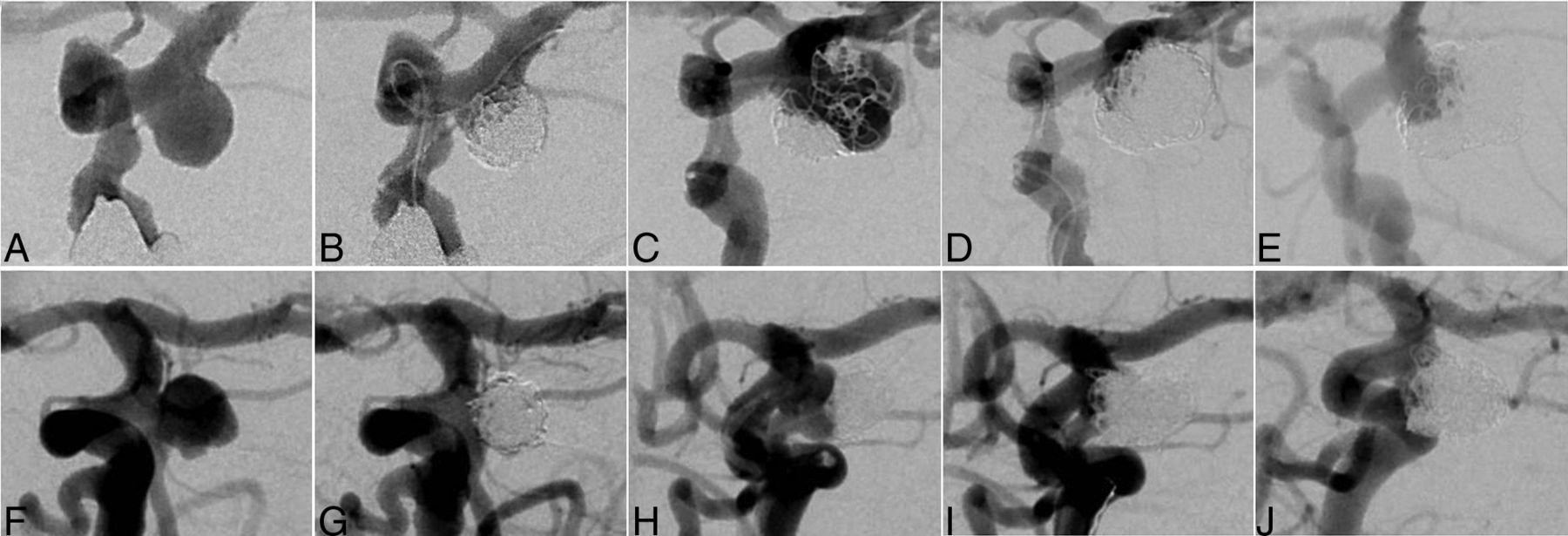
  • FIG 3.
    • Download figure
    • Open in new tab
    • Download powerpoint
    FIG 3.

    Illustrative cases. R group case: A 73-year-old woman who had undergone coil embolization for an incidentally detected unruptured posterior communicating aneurysm with a maximum and neck diameter of 6.7 mm (A). An 8F balloon-guiding catheter (Merci; Stryker Neurovascular) was directed into the extracranial segment of the left ICA. The coils were inserted into the aneurysm using a double-catheter technique; no IMC, balloon, or stent was used, and the embolization result was RROC Class 2 (B). Major recanalization was discovered 27 months after the initial treatment (C). For the second coiling, 2 microcatheters (both Excelsior SL-10; Stryker Neurovascular) were navigated into the aneurysm for coil embolization. The coils were inserted into the aneurysm using a double-catheter technique and no IMC or stent was used. Ultimately, the coil embolization resulted in RROC Class 2 (D). No postoperative complications were noted, but major re-recanalization occurred 1 year after second coiling (E). NR group case: A 63-year-old woman who had undergone coil embolization for an incidentally detected unruptured cerebral aneurysm at the origin of the fetal posterior cerebral artery with a maximum diameter of 6.7 mm and a neck diameter of 5.0 mm (F). The coils were inserted into the aneurysm using a primary coiling; no IMC, balloon, or stent was used, and the embolization result was RROC Class 2 (G). Major recanalization was discovered 104 months after the initial treatment (H). A 6F guiding sheath (Asahi Fubuki; Asahi Intecc) was directed into the extracranial segment of the left ICA. A 3.2F Tactics catheter as the IMC was then guided coaxially to the supraclinoid segment of the left ICA. One microcatheter (Phenom 17; Medtronic) was navigated from the Tactics catheter into the aneurysm for coil embolization, and the other microcatheter (Excelsior SL-10) was directed into the left fetal posterior cerebral artery for stent placement. A Neuroform Atlas (Stryker Neurovascular) stent was placed from the left fetal posterior cerebral artery to the supraclinoid segment of the left ICA. Finally, the coil embolization achieved RROC Class 1 (I). No postoperative complications were noted, and follow-up DSA 1 year after the second coiling showed no re-recanalization (J). Subsequently, at 42 months, the patient remained without re-recanalization (image not shown).

Tables

  • Figures
    • View popup
    Table 1:

    Baseline clinical characteristics in R and NR groups

    CharacteristicsR Group (n=72)NR Group (n=116)P Value
    Age, years65 [56, 71]65 [56, 72].47
    Sex, female47 (65)89 (77).10
    Medical history 
     Hypertension46 (64)52 (45).02b
     Diabetes mellitus4 (5.6)9 (7.8).77
     Hyperlipidemia14 19)27 (23).59
     Polycystic kidney disease1 (1.4)4 (3.4).65
     Prior stroke4 (5.6)2 (1.7).21
     Cerebral small vessel diseasea39 (54)50 (43).18
    Smoking 
     Current10 (14)18 (16).46
     Past smoker18 (25)20 (17) 
     None44 (61)78 (67) 
    Drinking15 (21)18 (16).43
    Family history 
     Cerebral aneurysm10 (14)24 (21).33
     Polycystic kidney1 (1.4)1 (0.9)1
    Aneurysm characteristics 
     Thrombosed aneurysm6 (8.3)5 (4.3).34
     Multiple aneurysms26 (36)43 (37)1
     Aneurysm location 
      ACA/ACoA10 (14)17 15).13
      MCA9 (13)18 (16) 
      ICA37 (51)65 (56) 
      PCA0 (0)1 (0.9) 
      BA/SCA16 (22)11 (9.5) 
      VA/PICA0 (0)4 (3.4) 
      Posterior circulation15 (21)16 (14).23
    • Note:—ACA indicates anterior cerebral artery; ACoA, anterior communicating artery; BA, basilar artery; NR, non-major re-recanalization; PCA, posterior cerebral artery; R, major re-recanalization; SCA, superior cerebellar artery; VA, vertebral artery.

    • a Cerebral small vessel disease was diagnosed if 1 or more of the following radiologic features were seen on MRI: 1) subcortical small, focal infarction, 2) diffuse white matter lesions present as white matter hyperintensities on T2WI, 3) microbleeding in the subcortical region.

    • b P < .05. Unless otherwise indicated, values represent the number of aneurysms (%) or median (IQR). Not all percentage totals reach 100% because of rounding.

    • View popup
    Table 2:

    Multivariate logistic regression analysis and stepwise selection of associated factors for major re-recanalization

    ParameterOR (95% CI)P Value
    A: Variables with P < .05 or previously reported as associated factors for recanalization
     Hypertension1.62 (0.79–3.33).19
     Thrombosed aneurysm1.45 (0.29–7.27).65
     Posterior circulation aneurysm1.13 (0.44–2.94).80
     Aneurysm size at first coiling, mm1.00 (0.75–1.34).98
     Neck size at first coiling, mm1.20 (0.86–1.68).28
     Aspect ratio at first coiling1.27 (0.44–3.69).66
     Aneurysm volume at first coiling, mm31.00 (0.99–1.00).92
     Ruptured status at second coiling2.70 (0.36–20.20).33
     Balloon-assisted coiling at second coiling0.76 (0.22–2.66).67
     Stent-assisted coiling at second coiling0.41 (0.17–1.02).06
     Use of IMC at second coiling0.43 (0.17–1.11).08
     Bioactive coila1.25 (0.56–2.79).59
     Large coilb0.90 (0.25–3.28).87
     Hydrogel coilc2.35 × 10−7 (0 – Inf).99
     RROC class 1 at second coiling0.15 (0.029–0.79).03d
    B: After stepwise selection by using the P value (until P < .05)
     Neck size at first coiling, mm1.18 (1.04–1.33).008d
     Stent-assisted coiling at second coiling0.34 (0.15–0.79).01d
     Use of IMC at second coiling0.35 (0.16–0.80).01d
     RROC class 1 at second coiling0.16 (0.033–0.70).02d
    • Note:—Inf indicates infinitesimal.

    • ↵a Bioactive coils are all the Matrix2 (Stryker Neurovascular).

    • ↵b Large coils with a primary diameter of 0.014 inches or larger are all the Target XL (Stryker Neurovascular).

    • ↵c Hydrogel coils are all part of the HydroCoil Embolic System (MicroVention Terumo).

    • ↵d P < .05.

PreviousNext
Back to top
Advertisement
Print
Download PDF
Email Article

Thank you for your interest in spreading the word on American Journal of Neuroradiology.

NOTE: We only request your email address so that the person you are recommending the page to knows that you wanted them to see it, and that it is not junk mail. We do not capture any email address.

Enter multiple addresses on separate lines or separate them with commas.
Factors Associated with Major Re-Recanalization following Second Coiling for Recanalized Aneurysms: A Multicenter Experience over 20 Years during Long-Term Follow-up
(Your Name) has sent you a message from American Journal of Neuroradiology
(Your Name) thought you would like to see the American Journal of Neuroradiology web site.
CAPTCHA
This question is for testing whether or not you are a human visitor and to prevent automated spam submissions.
Cite this article
Michiyasu Fuga, Toshihiro Ishibashi, Issei Kan, Ken Aoki, Rintaro Tachi, Koreaki Irie, Naoki Kato, Shunsuke Hataoka, Gota Nagayama, Tohru Sano, Toshihide Tanaka, Yuichi Murayama
Factors Associated with Major Re-Recanalization following Second Coiling for Recanalized Aneurysms: A Multicenter Experience over 20 Years during Long-Term Follow-up
American Journal of Neuroradiology May 2025, DOI: 10.3174/ajnr.A8671

Citation Manager Formats

  • BibTeX
  • Bookends
  • EasyBib
  • EndNote (tagged)
  • EndNote 8 (xml)
  • Medlars
  • Mendeley
  • Papers
  • RefWorks Tagged
  • Ref Manager
  • RIS
  • Zotero
0 Responses
Respond to this article
Share
Bookmark this article
Factors Associated with Major Re-Recanalization following Second Coiling for Recanalized Aneurysms: A Multicenter Experience over 20 Years during Long-Term Follow-up
Michiyasu Fuga, Toshihiro Ishibashi, Issei Kan, Ken Aoki, Rintaro Tachi, Koreaki Irie, Naoki Kato, Shunsuke Hataoka, Gota Nagayama, Tohru Sano, Toshihide Tanaka, Yuichi Murayama
American Journal of Neuroradiology May 2025, DOI: 10.3174/ajnr.A8671
del.icio.us logo Twitter logo Facebook logo Mendeley logo
  • Tweet Widget
  • Facebook Like
  • Google Plus One
Purchase

Jump to section

  • Article
    • Graphical Abstract
    • Abstract
    • ABBREVIATIONS:
    • MATERIALS AND METHODS
    • RESULTS
    • DISCUSSION
    • CONCLUSIONS
    • Footnotes
    • References
  • Figures & Data
  • Supplemental
  • Info & Metrics
  • Responses
  • References
  • PDF

Related Articles

  • PubMed
  • Google Scholar

Cited By...

  • No citing articles found.
  • Crossref
  • Google Scholar

This article has not yet been cited by articles in journals that are participating in Crossref Cited-by Linking.

More in this TOC Section

  • Effect of SARS-CoV2 on Endovascular Thrombectomy
  • Neurologic Complication in Transradial Angiography
  • A Key Factor Shapes LS-DAVFs EVT Outcome
Show more NEUROINTERVENTION

Similar Articles

Advertisement

Indexed Content

  • Current Issue
  • Accepted Manuscripts
  • Article Preview
  • Past Issues
  • Editorials
  • Editor's Choice
  • Fellows' Journal Club
  • Letters to the Editor
  • Video Articles

Cases

  • Case Collection
  • Archive - Case of the Week
  • Archive - Case of the Month
  • Archive - Classic Case

More from AJNR

  • Trainee Corner
  • Imaging Protocols
  • MRI Safety Corner
  • Book Reviews

Multimedia

  • AJNR Podcasts
  • AJNR Scantastics

Resources

  • Turnaround Time
  • Submit a Manuscript
  • Submit a Video Article
  • Submit an eLetter to the Editor/Response
  • Manuscript Submission Guidelines
  • Statistical Tips
  • Fast Publishing of Accepted Manuscripts
  • Graphical Abstract Preparation
  • Imaging Protocol Submission
  • Evidence-Based Medicine Level Guide
  • Publishing Checklists
  • Author Policies
  • Become a Reviewer/Academy of Reviewers
  • News and Updates

About Us

  • About AJNR
  • Editorial Board
  • Editorial Board Alumni
  • Alerts
  • Permissions
  • Not an AJNR Subscriber? Join Now
  • Advertise with Us
  • Librarian Resources
  • Feedback
  • Terms and Conditions
  • AJNR Editorial Board Alumni

American Society of Neuroradiology

  • Not an ASNR Member? Join Now

© 2025 by the American Society of Neuroradiology All rights, including for text and data mining, AI training, and similar technologies, are reserved.
Print ISSN: 0195-6108 Online ISSN: 1936-959X

Powered by HighWire