
This article requires a subscription to view the full text. If you have a subscription you may use the login form below to view the article. Access to this article can also be purchased.
Graphical Abstract

Abstract
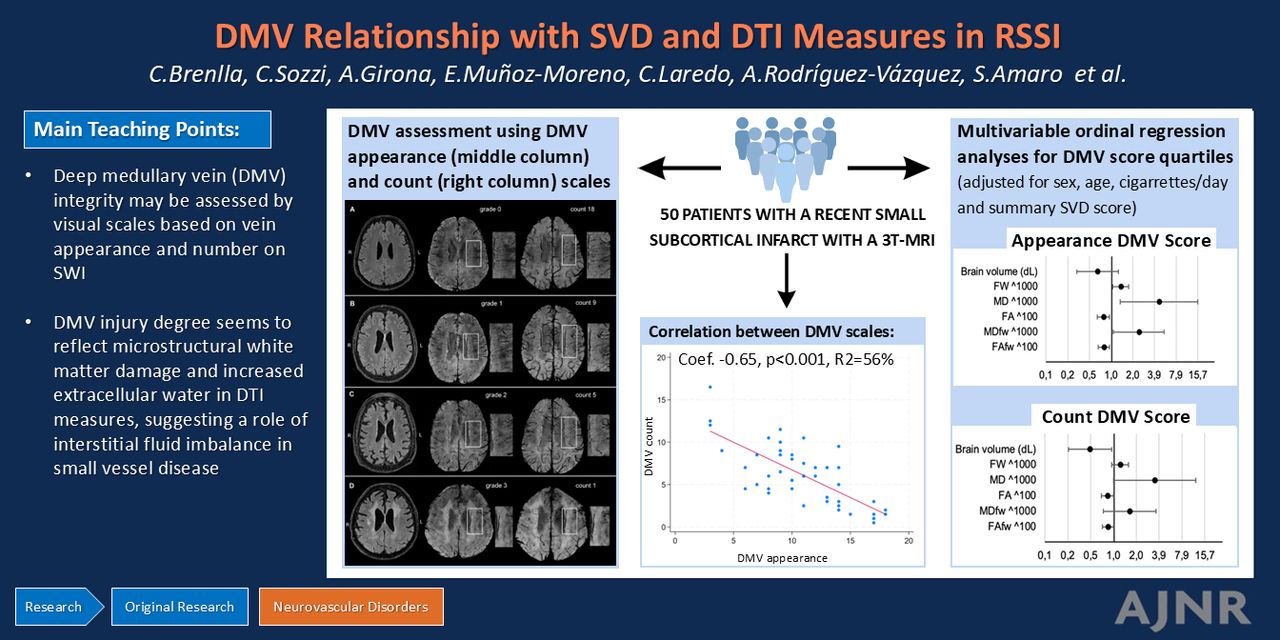
BACKGROUND AND PURPOSE: The role of the venous compartment in cerebral small vessel disease has yet to be fully understood. As such, we evaluated how deep medullary vein (DMV) integrity relates to MRI-based small vessel disease severity markers and glymphatic function assessed by DTI measures in patients with a recent small subcortical infarct.
MATERIALS AND METHODS: We gathered demographic, clinical, and 3T MRI imaging data from 50 patients with a recent small subcortical infarct. We evaluated the venular integrity by using 2 visual scales based on their appearance on SWI. We assessed the number of lacunes and microbleeds, white matter hyperintensities volume, perivascular spaces volume in basal ganglia and white matter, summary small vessel disease score, and brain volume. Diffusivity measures in normal-appearing white matter included free water fraction, mean diffusivity and fractional anisotropy with and without free water correction, and DTI along the perivascular spaces. After categorizing the cohort in quartiles according to both venular scores, we assessed their correlations with small vessel disease markers and diffusivity measures by using multivariable ordinal regression analyses adjusting for age, sex, smoking, and summary small vessel disease score.
RESULTS: In univariate analysis most of the imaging variables, except for microbleeds, perivascular spaces in white matter, and DTI along the perivascular spaces, were associated with 1 or both venular scores. In multivariate analysis, free water (OR, 1.33, 95% CI, 1.03–1.73), mean diffusivity (OR, 4.56, 95% CI, 1.32–15.81), fractional anisotropy (OR, 0.77, 95% CI, 0.63–0.93), free water-corrected mean diffusivity and fractional anisotropy (OR, 2.39, 95% CI, 1.06–5.39; OR 0.78, 95% CI, 0.65–0.94, respectively), associated with vein appearance, while only brain volume (OR, 0.48, 95% CI, 0.25–0.94), fractional anisotropy with and without free water correction (OR, 0.82, 95% CI, 0.86–0.99; OR, 0.83, 95% CI, 0.7–0.99, respectively) remained robust for vein count.
CONCLUSIONS: In patients with a recent small subcortical infarct, disruption of the DMVs, increased extracellular water, and white matter injury appear to be associated.
ABBREVIATIONS:
- BG
- basal ganglia
- DMV
- deep medullary vein
- DTI-ALPS
- DTI along the perivascular spaces
- FA
- fractional anisotropy
- FW
- free water
- IQR
- interquartile range
- MD
- mean diffusivity
- PVS
- perivascular spaces
- SD
- standard deviation
- STRIVE
- STandards for ReportIng Vascular changes on nEuroimaging
- SVD
- small vessel disease
- WMH
- white matter hyperintensities
Footnotes
Carla Brenlla and Caterina Sozzi contributed equally to this article.
Disclosure forms provided by the authors are available with the full text and PDF of this article at www.ajnr.org.
This research has been funded by the Catalan Society of Neurology (2001, https://www.scneurologia.cat/beca-fundacio-societat-catalana-de-neurologia/) and by a grant from the Carlos III Health Institute (ISCIII) co-funded by the European Union (PI22/00771). S.R. receives funding from ISCIII (JR21/00011); A.R. from ISCIII (PI20/00901); X.U. from ISCIII (INT22/00043), and by the European Social Fund “The ESF–Investing in your future.” This work was partially developed at the building Centro Esther Koplowitz, Barcelona, CERCA Programme/Generalitat de Catalunya. This work was performed thanks to the 3T MRI equipment at IDIBAPS (grant IBPS15-EE-3688 cofunded by MINECO and ERDF).
- © 2025 by American Journal of Neuroradiology
Log in using your username and password
Log in through your institution


Jump to section
Related Articles
Cited By...
- No citing articles found.