Advertisement
  • Main menu
  • User menu
  • Search
  • English ▼
    • English
    • Afrikaans
    • Albanian
    • Amharic
    • Arabic
    • Armenian
    • Azerbaijani
    • Basque
    • Belarusian
    • Bengali
    • Bosnian
    • Bulgarian
    • Catalan
    • Cebuano
    • Chichewa
    • Chinese (Simplified)
    • Chinese (Traditional)
    • Corsican
    • Croatian
    • Czech
    • Danish
    • Dutch
    • Esperanto
    • Estonian
    • Filipino
    • Finnish
    • French
    • Frisian
    • Galician
    • Georgian
    • German
    • Greek
    • Gujarati
    • Haitian Creole
    • Hausa
    • Hawaiian
    • Hebrew
    • Hindi
    • Hmong
    • Hungarian
    • Icelandic
    • Igbo
    • Indonesian
    • Irish
    • Italian
    • Japanese
    • Javanese
    • Kannada
    • Kazakh
    • Khmer
    • Korean
    • Kurdish (Kurmanji)
    • Kyrgyz
    • Lao
    • Latin
    • Latvian
    • Lithuanian
    • Luxembourgish
    • Macedonian
    • Malagasy
    • Malay
    • Malayalam
    • Maltese
    • Maori
    • Marathi
    • Mongolian
    • Myanmar (Burmese)
    • Nepali
    • Norwegian
    • Pashto
    • Persian
    • Polish
    • Portuguese
    • Punjabi
    • Romanian
    • Russian
    • Samoan
    • Scottish Gaelic
    • Serbian
    • Sesotho
    • Shona
    • Sindhi
    • Sinhala
    • Slovak
    • Slovenian
    • Somali
    • Spanish
    • Sudanese
    • Swahili
    • Swedish
    • Tajik
    • Tamil
    • Telugu
    • Thai
    • Turkish
    • Ukrainian
    • Urdu
    • Uzbek
    • Vietnamese
    • Welsh
    • Xhosa
    • Yiddish
    • Yoruba
    • Zulu

Main menu

  • Home
  • Content
    • Current Issue
    • Accepted Manuscripts
    • Article Preview
    • Past Issue Archive
    • Video Articles
    • AJNR Case Collection
    • Case of the Week Archive
    • Case of the Month Archive
    • Classic Case Archive
  • Special Collections
    • Advancing NeuroMRI with High-Relaxivity Contrast Agents
    • AJNR Awards
    • Low-Field MRI
    • Alzheimer Disease
    • ASNR Foundation Special Collection
    • Photon-Counting CT
    • View All
  • Multimedia
    • AJNR Podcasts
    • AJNR SCANtastic
    • Trainee Corner
    • MRI Safety Corner
    • Imaging Protocols
  • For Authors
    • Submit a Manuscript
    • Submit a Video Article
    • Submit an eLetter to the Editor/Response
    • Manuscript Submission Guidelines
    • Statistical Tips
    • Fast Publishing of Accepted Manuscripts
    • Graphical Abstract Preparation
    • Imaging Protocol Submission
    • Author Policies
  • About Us
    • About AJNR
    • Editorial Board
    • Editorial Board Alumni
  • More
    • Become a Reviewer/Academy of Reviewers
    • Subscribers
    • Permissions
    • Alerts
    • Feedback
    • Advertisers
    • ASNR Home

User menu

  • Alerts
  • Log in

Search

  • Advanced search
American Journal of Neuroradiology

American Journal of Neuroradiology

ASHNR American Society of Functional Neuroradiology ASHNR American Society of Pediatric Neuroradiology ASSR
  • Alerts
  • Log in

Advanced Search

English ▼
  • English
  • Afrikaans
  • Albanian
  • Amharic
  • Arabic
  • Armenian
  • Azerbaijani
  • Basque
  • Belarusian
  • Bengali
  • Bosnian
  • Bulgarian
  • Catalan
  • Cebuano
  • Chichewa
  • Chinese (Simplified)
  • Chinese (Traditional)
  • Corsican
  • Croatian
  • Czech
  • Danish
  • Dutch
  • Esperanto
  • Estonian
  • Filipino
  • Finnish
  • French
  • Frisian
  • Galician
  • Georgian
  • German
  • Greek
  • Gujarati
  • Haitian Creole
  • Hausa
  • Hawaiian
  • Hebrew
  • Hindi
  • Hmong
  • Hungarian
  • Icelandic
  • Igbo
  • Indonesian
  • Irish
  • Italian
  • Japanese
  • Javanese
  • Kannada
  • Kazakh
  • Khmer
  • Korean
  • Kurdish (Kurmanji)
  • Kyrgyz
  • Lao
  • Latin
  • Latvian
  • Lithuanian
  • Luxembourgish
  • Macedonian
  • Malagasy
  • Malay
  • Malayalam
  • Maltese
  • Maori
  • Marathi
  • Mongolian
  • Myanmar (Burmese)
  • Nepali
  • Norwegian
  • Pashto
  • Persian
  • Polish
  • Portuguese
  • Punjabi
  • Romanian
  • Russian
  • Samoan
  • Scottish Gaelic
  • Serbian
  • Sesotho
  • Shona
  • Sindhi
  • Sinhala
  • Slovak
  • Slovenian
  • Somali
  • Spanish
  • Sudanese
  • Swahili
  • Swedish
  • Tajik
  • Tamil
  • Telugu
  • Thai
  • Turkish
  • Ukrainian
  • Urdu
  • Uzbek
  • Vietnamese
  • Welsh
  • Xhosa
  • Yiddish
  • Yoruba
  • Zulu
  • Home
  • Content
    • Current Issue
    • Accepted Manuscripts
    • Article Preview
    • Past Issue Archive
    • Video Articles
    • AJNR Case Collection
    • Case of the Week Archive
    • Case of the Month Archive
    • Classic Case Archive
  • Special Collections
    • Advancing NeuroMRI with High-Relaxivity Contrast Agents
    • AJNR Awards
    • Low-Field MRI
    • Alzheimer Disease
    • ASNR Foundation Special Collection
    • Photon-Counting CT
    • View All
  • Multimedia
    • AJNR Podcasts
    • AJNR SCANtastic
    • Trainee Corner
    • MRI Safety Corner
    • Imaging Protocols
  • For Authors
    • Submit a Manuscript
    • Submit a Video Article
    • Submit an eLetter to the Editor/Response
    • Manuscript Submission Guidelines
    • Statistical Tips
    • Fast Publishing of Accepted Manuscripts
    • Graphical Abstract Preparation
    • Imaging Protocol Submission
    • Author Policies
  • About Us
    • About AJNR
    • Editorial Board
    • Editorial Board Alumni
  • More
    • Become a Reviewer/Academy of Reviewers
    • Subscribers
    • Permissions
    • Alerts
    • Feedback
    • Advertisers
    • ASNR Home
  • Follow AJNR on Twitter
  • Visit AJNR on Facebook
  • Follow AJNR on Instagram
  • Join AJNR on LinkedIn
  • RSS Feeds

AJNR Awards, New Junior Editors, and more. Read the latest AJNR updates


Improved Turnaround Times | Median time to first decision: 12 days

Research ArticleNeurointervention

Incidence, Risk Factors, and Clinical Implications of Subarachnoid Hyperdensities on Flat-Panel Detector CT following Mechanical Thrombectomy in Patients with Anterior Circulation Acute Ischemic Stroke

Bettina L. Serrallach, Mattia Branca, Adnan Mujanovic, Anna Boronylo, Julie M. Hanke, Arsany Hakim, Sara Pilgram-Pastor, Eike I. Piechowiak, Jan Gralla, Thomas Meinel, Johannes Kaesmacher and Tomas Dobrocky
American Journal of Neuroradiology July 2024, DOI: https://doi.org/10.3174/ajnr.A8277
Bettina L. Serrallach
aFrom the Department of Diagnostic and Interventional Neuroradiology (B.L.S., A.M., A.B., A.H., S.P.-P., E.I.P., J.G., J.K., T.D.), Inselspital, Bern University Hospital and University of Bern, Bern, Switzerland
  • Find this author on Google Scholar
  • Find this author on PubMed
  • Search for this author on this site
  • ORCID record for Bettina L. Serrallach
Mattia Branca
bDepartment of Clinical Research (M.B.), CTU Bern, University of Bern, Bern, Switzerland
  • Find this author on Google Scholar
  • Find this author on PubMed
  • Search for this author on this site
  • ORCID record for Mattia Branca
Adnan Mujanovic
aFrom the Department of Diagnostic and Interventional Neuroradiology (B.L.S., A.M., A.B., A.H., S.P.-P., E.I.P., J.G., J.K., T.D.), Inselspital, Bern University Hospital and University of Bern, Bern, Switzerland
  • Find this author on Google Scholar
  • Find this author on PubMed
  • Search for this author on this site
  • ORCID record for Adnan Mujanovic
Anna Boronylo
aFrom the Department of Diagnostic and Interventional Neuroradiology (B.L.S., A.M., A.B., A.H., S.P.-P., E.I.P., J.G., J.K., T.D.), Inselspital, Bern University Hospital and University of Bern, Bern, Switzerland
  • Find this author on Google Scholar
  • Find this author on PubMed
  • Search for this author on this site
Julie M. Hanke
cDepartment of Neurology (J.M.H., T.M.), Inselspital, Bern University Hospital and University of Bern, Bern, Switzerland
  • Find this author on Google Scholar
  • Find this author on PubMed
  • Search for this author on this site
Arsany Hakim
aFrom the Department of Diagnostic and Interventional Neuroradiology (B.L.S., A.M., A.B., A.H., S.P.-P., E.I.P., J.G., J.K., T.D.), Inselspital, Bern University Hospital and University of Bern, Bern, Switzerland
  • Find this author on Google Scholar
  • Find this author on PubMed
  • Search for this author on this site
  • ORCID record for Arsany Hakim
Sara Pilgram-Pastor
aFrom the Department of Diagnostic and Interventional Neuroradiology (B.L.S., A.M., A.B., A.H., S.P.-P., E.I.P., J.G., J.K., T.D.), Inselspital, Bern University Hospital and University of Bern, Bern, Switzerland
  • Find this author on Google Scholar
  • Find this author on PubMed
  • Search for this author on this site
  • ORCID record for Sara Pilgram-Pastor
Eike I. Piechowiak
aFrom the Department of Diagnostic and Interventional Neuroradiology (B.L.S., A.M., A.B., A.H., S.P.-P., E.I.P., J.G., J.K., T.D.), Inselspital, Bern University Hospital and University of Bern, Bern, Switzerland
  • Find this author on Google Scholar
  • Find this author on PubMed
  • Search for this author on this site
  • ORCID record for Eike I. Piechowiak
Jan Gralla
aFrom the Department of Diagnostic and Interventional Neuroradiology (B.L.S., A.M., A.B., A.H., S.P.-P., E.I.P., J.G., J.K., T.D.), Inselspital, Bern University Hospital and University of Bern, Bern, Switzerland
  • Find this author on Google Scholar
  • Find this author on PubMed
  • Search for this author on this site
  • ORCID record for Jan Gralla
Thomas Meinel
cDepartment of Neurology (J.M.H., T.M.), Inselspital, Bern University Hospital and University of Bern, Bern, Switzerland
  • Find this author on Google Scholar
  • Find this author on PubMed
  • Search for this author on this site
  • ORCID record for Thomas Meinel
Johannes Kaesmacher
aFrom the Department of Diagnostic and Interventional Neuroradiology (B.L.S., A.M., A.B., A.H., S.P.-P., E.I.P., J.G., J.K., T.D.), Inselspital, Bern University Hospital and University of Bern, Bern, Switzerland
  • Find this author on Google Scholar
  • Find this author on PubMed
  • Search for this author on this site
  • ORCID record for Johannes Kaesmacher
Tomas Dobrocky
aFrom the Department of Diagnostic and Interventional Neuroradiology (B.L.S., A.M., A.B., A.H., S.P.-P., E.I.P., J.G., J.K., T.D.), Inselspital, Bern University Hospital and University of Bern, Bern, Switzerland
  • Find this author on Google Scholar
  • Find this author on PubMed
  • Search for this author on this site
  • ORCID record for Tomas Dobrocky
  • Article
  • Figures & Data
  • Supplemental
  • Info & Metrics
  • Responses
  • References
  • PDF
Loading

Article Figures & Data

Figures

  • Tables
  • FIG 1.
    • Download figure
    • Open in new tab
    • Download powerpoint
    FIG 1.

    Examples of SH grades I to IV. A, SH grade I with hyperdensities in 2 neighboring sulci. B, SH grade II with hyperdensities in >2 neighboring sulci but confinement to 1 lobar area. C, SH grade III with diffuse sulcal hyperdensities affecting >2 lobes. D, SH grade IV with diffuse hyperdensities affecting >2 lobes and intraventricular extension.

  • FIG 2.
    • Download figure
    • Open in new tab
    • Download powerpoint
    FIG 2.

    Flow chart depicting patient selection process.
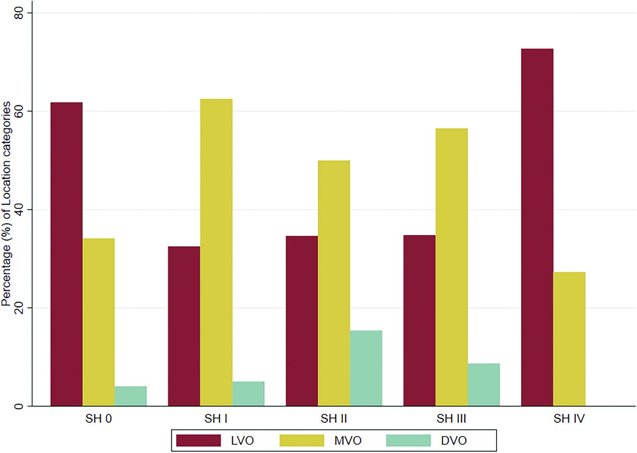
  • FIG 3.
    • Download figure
    • Open in new tab
    • Download powerpoint
    FIG 3.

    Baseline intracranial occlusion site (LVO, MVO, and DVO) stratified by SH.

  • FIG 4.
    • Download figure
    • Open in new tab
    • Download powerpoint
    FIG 4.

    A, Distribution of mRS scores at 90 days for patients without SH and patients with SH I–IV on FDCT. B, The association of different grades of SH 0–IV with mRS at 3 months, mRS dichotomized (0–2 versus 3–6), and mortality (equivalent to mRS 6). Analyses were performed using multivariable ordinal/logistic regression, adjusting for prespecified confounders (see Materials and Methods).

  • FIG 5.
    • Download figure
    • Open in new tab
    • Download powerpoint
    FIG 5.

    Possible mechanism leading to SH. A, Ventrolateral view of the brain and circle of Willis. B, Close-up showing a proximal M2 occlusion with an inserted stent retriever. C, Close-up during the retrieval of the stent retriever. During navigation, smaller vessels (M2 and beyond) tend to straighten more than proximal vessels. The perforators are exposed to excessive forces during thrombectomy due to stretching and may be sheared off, leading to subtle extravasation, which is occult on standard DSA but may be detected on FDCT as SH, in this case, a subarachnoid hemorrhage. © Inselspital, Bern University Hospital, Department of Neuroradiology.

Tables

  • Figures
    • View popup
    Table 1:

    Baseline characteristics stratified by SH seen on FDCT

    Total NaSH 0–IVTotal NaSH 0Total NaSH I–IVP Value
    Baseline characteristics
     Age (median) [lq, uq]22375.5 [63.3–83.1]12377.6 [63.7–85.0]10074.5 [63.3–82.1].24
     Sex (male), No. (%)223110 (49.3%)12365 (52.8%)10045 (45.0%).24
     Hypertension, No. (%)223164 (73.5%)12393 (75.6%)10071 (71.0%).44
     Diabetes mellitus, No. (%)22350 (22.4%)12331 (25.2%)10019 (19.0%).27
     Coronary heart disease, No. (%)22330 (13.5%)12318 (14.6%)10012 (12.0%).57
     Smoking (current), No. (%)22349 (22.0%)12330 (24.4%)10019 (19.0%).33
     Hyperlipidemia, No. (%)223131 (58.7%)12371 (57.7%)10060 (60.0%).73
     Atrial fibrillation, No. (%)22377 (34.5%)12348 (39.0%)10029 (29.0%).12
     NIHSS at baseline (median) [lq, uq]21912.0 [5.0–20.0]12113.0 [6.0–19.0]9811.0 [5.0–20.0].37
     mRS (prestroke) (median) [lq, uq]2120.0 [0.0–1.0]1180.0 [0.0–1.0]940.0 [0.0– 1.0].66
     Baseline intracranial occlusion site, No. (%)223123100<.001
     LVO114 (51.1%)76 (61.8%)38 (38.0%)
     Medium- and distal- vessel occlusion109 (48.9%)47 (38.2%)62 (62.0%)
    ASPECTS (median) [lq, uq]2207.0 [6.0–9.0]1227.0 [5.0–9.0]987.0 [6.0–9.0].45
    IV thrombolysis, No. (%)223101 (45.3%)12349 (39.8%)10052 (52.0%).07
    Time of symptom onset known, No. (%)223123100.44
     No59 (26.5%)34 (27.6%)25 (25.0%)
     Wake up40 (17.9%)25 (20.3%)15 (15.0%)
     Yes124 (55.6%)64 (52.0%)60 (60.0%)
    Onset-to-groin puncture (median) (lq, uq) (min)223192.0 [155.0–266.0]123186.0 [156.5–266.0]100202.5 [150.0–264.0].99
    Medications (prestroke)
     Antihypertensives, No. (%)220130 (59.1%)12076 (63.3%)10054 (54.0%).16
     Lipid-lowering drugs, No. (%)22370 (31.4%)12340 (32.5%)10030 (30.0%).69
     Anticoagulation, No. (%)22344 (19.7%)12328 (22.8%)10016 (16.0%).21
     Antiplatelet, No. (%)22347 (21.1%)12324 (19.5%)10023 (23.0%).53
    • Note:—lq indicates lower quartile; uq, upper quartile.

    • ↵a N indicates number of patients without missing data.

    • View popup
    Table 2:

    Procedural characteristics and outcome measures stratified by SH seen on FDCT

    Total NaSH 0–IVTotal NaSH 0Total NaSH I–IVP Value
    Procedural characteristics
     No. of passes, (median) [lq, uq]2182.0 [1.0–3.0]1211.0 [1.0–2.0]972.0 [1.0–4.0]<.001
     Most distal device position, No. (%)223123100<.001
      Large vessel34 (15.2%)30 (24.4%)4 (4.0%)
      Medium vessel140 (62.8%)72 (58.5%)68 (68.0%)
      Distal vessel49 (22.0%)21 (17.1%)28 (28.0%)
     Amount of contrast medium (mL) (median) [lq, uq]217120 [100–180]119110 [90–160]98133 [100–190].035
     Active extravasation seen on DSA, No. (%)22313 (5.8%)1230 (0.0%)10013 (13.0%)<.001
     Parenchymal hyperdensities on FDCT, No. (%)223110 (49.3%)12367 (54.5%)10043 (43.0%).09
     eTICI score, No. (%)22112398.014
      014 (6.3%)3 (2.4%)11 (11.2%)
      14 (1.8%)1 (0.8%)3 (3.1%)
      2a8 (3.6%)6 (4.9%)2 (2.0%)
      2b5028 (12.7%)13 (10.6%)15 (15.3%)
      2b6736 (16.3%)16 (13.0%)20 (20.4%)
      2c50 (22.6%)32 (26.0%)18 (18.4%)
      381 (36.7%)52 (42.3%)29 (29.6%)
    Outcome measures
     mRS at 3 months, (median) [lq, uq]1963.0 [1.0–6.0]1072.0 [1.0–5.0]893.0 [1.0–6.0].21
     NIHSS at 24 hours (median) [lq, uq]2138.0 [3.0–16.0]1208.0 [3.0–16.0]936.0 [3.0–15.0].76
    • Note:—lq indicates lower quartile; uq, upper quartile.

    • ↵a N indicates number of patients without missing data.

PreviousNext
Back to top
Advertisement
Print
Download PDF
Email Article
Cite this article
0 Responses
Respond to this article
Share
Bookmark this article
Incidence, Risk Factors, and Clinical Implications of Subarachnoid Hyperdensities on Flat-Panel Detector CT following Mechanical Thrombectomy in Patients with Anterior Circulation Acute Ischemic Stroke
Bettina L. Serrallach, Mattia Branca, Adnan Mujanovic, Anna Boronylo, Julie M. Hanke, Arsany Hakim, Sara Pilgram-Pastor, Eike I. Piechowiak, Jan Gralla, Thomas Meinel, Johannes Kaesmacher, Tomas Dobrocky
American Journal of Neuroradiology Jul 2024, DOI: 10.3174/ajnr.A8277
del.icio.us logo Twitter logo Facebook logo Mendeley logo
  • Tweet Widget
Purchase

Jump to section

  • Article
    • Abstract
    • ABBREVIATIONS:
    • MATERIALS AND METHODS
    • RESULTS
    • DISCUSSION
    • CONCLUSIONS
    • Acknowledgments
    • Footnotes
    • References
  • Figures & Data
  • Supplemental
  • Info & Metrics
  • Responses
  • References
  • PDF

Related Articles

  • PubMed
  • Google Scholar

Cited By...

  • REcanalization of Distal Cerebral Vessels In Acute Stroke Using ApeRio (REVISAR)
  • Correspondence on 'Subarachnoid iodine leakage on dual-energy computed tomography after mechanical thrombectomy is associated with malignant brain edema' by Ogata et al
  • Correspondence on 'Subarachnoid iodine leakage on dual-energy computed tomography after mechanical thrombectomy is associated with malignant brain edema' by Ogata et al
  • DOT sign indicates persistent hypoperfusion and poor outcome in patients with incomplete reperfusion following thrombectomy
  • Crossref
  • Google Scholar

This article has not yet been cited by articles in journals that are participating in Crossref Cited-by Linking.

More in this TOC Section

  • Connectivity for remote neurovascular procedures
  • Cavernous dural AVF treated by transfacial route
  • A Retrospective Study in Tentorial DAVFs
Show more NEUROINTERVENTION

Similar Articles

Advertisement

Indexed Content

  • Current Issue
  • Accepted Manuscripts
  • Article Preview
  • Past Issues
  • Editorials
  • Editor's Choice
  • Fellows' Journal Club
  • Letters to the Editor
  • Video Articles

Cases

  • Case Collection
  • Archive - Case of the Week
  • Archive - Case of the Month
  • Archive - Classic Case

More from AJNR

  • Trainee Corner
  • Imaging Protocols
  • MRI Safety Corner
  • Book Reviews

Multimedia

  • AJNR Podcasts
  • AJNR Scantastics

Resources

  • Turnaround Time
  • Submit a Manuscript
  • Submit a Video Article
  • Submit an eLetter to the Editor/Response
  • Manuscript Submission Guidelines
  • Statistical Tips
  • Fast Publishing of Accepted Manuscripts
  • Graphical Abstract Preparation
  • Imaging Protocol Submission
  • Evidence-Based Medicine Level Guide
  • Publishing Checklists
  • Author Policies
  • Become a Reviewer/Academy of Reviewers
  • News and Updates

About Us

  • About AJNR
  • Editorial Board
  • Editorial Board Alumni
  • Alerts
  • Permissions
  • Not an AJNR Subscriber? Join Now
  • Advertise with Us
  • Librarian Resources
  • Feedback
  • Terms and Conditions
  • AJNR Editorial Board Alumni

American Society of Neuroradiology

  • Not an ASNR Member? Join Now

© 2025 by the American Society of Neuroradiology All rights, including for text and data mining, AI training, and similar technologies, are reserved.
Print ISSN: 0195-6108 Online ISSN: 1936-959X

Powered by HighWire
Email this Article

Thank you for your interest in spreading the word on American Journal of Neuroradiology.

NOTE: We only request your email address so that the person you are recommending the page to knows that you wanted them to see it, and that it is not junk mail. We do not capture any email address.

Enter multiple addresses on separate lines or separate them with commas.
Incidence, Risk Factors, and Clinical Implications of Subarachnoid Hyperdensities on Flat-Panel Detector CT following Mechanical Thrombectomy in Patients with Anterior Circulation Acute Ischemic Stroke
(Your Name) has sent you a message from American Journal of Neuroradiology
(Your Name) thought you would like to see the American Journal of Neuroradiology web site.
CAPTCHA
This question is for testing whether or not you are a human visitor and to prevent automated spam submissions.
Cite this article
Bettina L. Serrallach, Mattia Branca, Adnan Mujanovic, Anna Boronylo, Julie M. Hanke, Arsany Hakim, Sara Pilgram-Pastor, Eike I. Piechowiak, Jan Gralla, Thomas Meinel, Johannes Kaesmacher, Tomas Dobrocky
Incidence, Risk Factors, and Clinical Implications of Subarachnoid Hyperdensities on Flat-Panel Detector CT following Mechanical Thrombectomy in Patients with Anterior Circulation Acute Ischemic Stroke
American Journal of Neuroradiology Jul 2024, DOI: 10.3174/ajnr.A8277

Citation Manager Formats

  • BibTeX
  • Bookends
  • EasyBib
  • EndNote (tagged)
  • EndNote 8 (xml)
  • Medlars
  • Mendeley
  • Papers
  • RefWorks Tagged
  • Ref Manager
  • RIS
  • Zotero

We use cookies on this site to enhance your user experience. By clicking any link on this page you are giving your consent for us to set cookies.