
Article Figures & Data
Figures
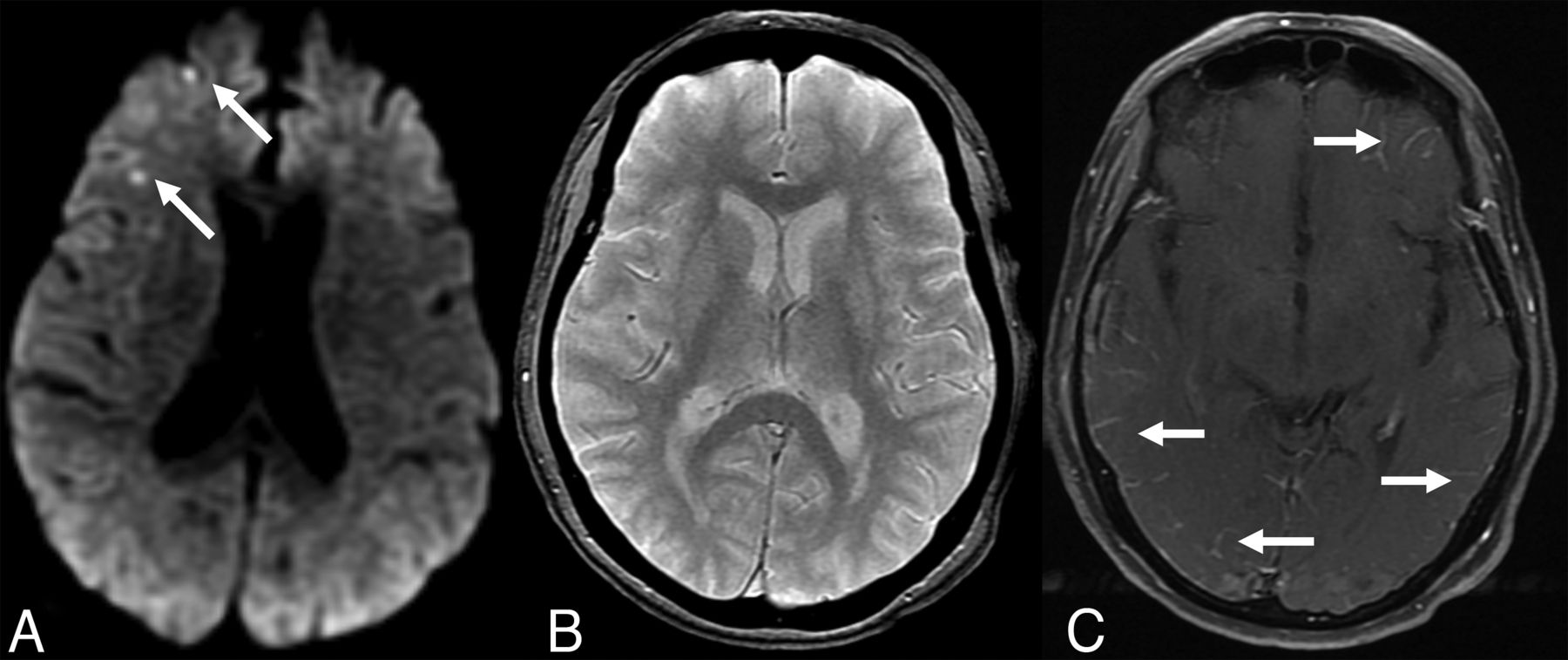
- FIG 1.
MR imaging at 1.5T obtained in the emergency department. Axial DWI (A), T2*-GRE (B), and contrast-enhanced T1-TSE (C) images at identical section levels. A, Arrows show punctuate areas of restricted diffusion in the right frontal cortex. There were a few more similar foci in other regions of the brain (not shown). B, T2*-GRE was negative for any intracranial hemorrhage or cerebral microbleeds. C, Arrows point to bilateral multifocal abnormal leptomeningeal enhancement.
- FIG 2.
Axial precontrast (A) and axial postcontrast (B) black-blood vessel wall MR imaging obtained at 3T. Correlated with precontrast vessel wall MR imaging (A), postcontrast image (B) shows bilateral scattered leptomeningeal disease (arrows) in the supratentorial regions. No definite periarterial or arterial wall enhancement is appreciated.
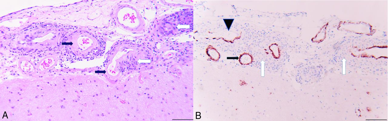
- FIG 3.
A, H&E-stained section from the right frontal lobe biopsy with granulomatous angiitis (white arrows) in the leptomeninges. The affected vessels have hyalinization of the tunica media and loss of smooth muscle nuclei, consistent with amyloid angiopathy (black arrows). B, Immunohistochemical staining for amyloid-β counterstained with hematoxylin. The leptomeningeal vessels involved by granulomatous inflammation (white arrows) have brown immunoreactivity for amyloid-β (black arrow). The vessel in the upper left has partial destruction of the wall with focal loss of amyloid immunoreactivity (black arrowhead). Scale bar =100 µm at (A) and (B).
- FIG 4.
7T brain MR imaging obtained after discharge. Axial T2* images (A and B) show numerous scattered punctate susceptibility artifacts in the cortical/subcortical distribution (solid arrows), consistent with cerebral microbleeds and cortical superficial siderosis in the supratentorial parenchyma (dashed arrows), which are completely obscured at 1.5T GRE (Fig 1). Precontrast (C) and postcontrast (D) black blood vessel wall MR imaging with zoomed smaller images at the center, focusing on larger vessels at the circle of Willis. Compared with vessel wall MR imaging at 3T (Fig 2), the vessel walls have higher resolution and improved appearance. Arrows at (D) show leptomeningeal enhancement in the left frontal regions, which is harder to appreciate compared with 3T. Note that it is harder to suppress the peripheral CSF signal (white oval windows) at 7T, leptomeningeal enhancement at these regions cannot be perceived without a precontrast control study, or these regions could be equivocally called enhancement. No suspicious stenosis, contrast enhancement, or wall thickening is identified at the circle of Willis.
- FIG 5.
Three-month follow-up 7T MR imaging. Axial T2* images are at the upper row (A and B) and corresponding axial SWI (C and D) are at the lower row. After 3 months, the patient is symptom-free, however, images show an increased number of punctate microhemorrhages in comparison with the corresponding images at previous 7T MR imaging axial T2* (Fig 4A, -B). Arrows point out some of the peripheral microhemorrhages and compared with T2* (A and B), SWI provides a clearer and sharper visualization of some of these microhemorrhages (C and D). In closer look, the entire cortex shows a gyriform susceptibility artifact in certain regions on SWI, most significant in the occipital lobes, which is not technically a superficial pial siderosis but actually corresponds to newly described intragyral hemorrhage sign (arrowheads in C and D). This observation was not well appreciated on T2*.


Jump to section
Related Articles
Cited By...
- No citing articles found.