
Article Figures & Data
Figures
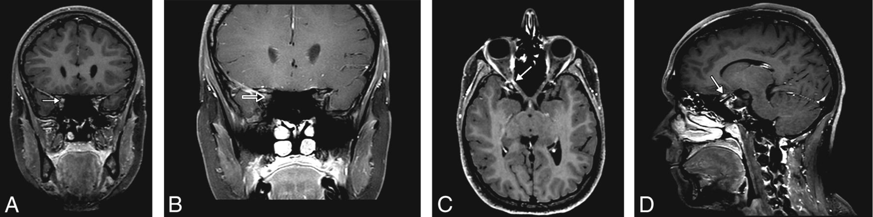
- FIG 1.
A 21-year-old woman presenting with acute vision loss and orbital pain of the right eye. WBCE-3D TSE T1WI reformatted in the coronal plane (A) shows enhancement of the intraorbital segment of the right optic nerve at the orbital apex (white arrow), whereas no optic nerve enhancement was detected on OCE-2D coronal T1WI (black arrow) (B). The WBCE-3D T1WI sequence reformatted in the axial (C) and sagittal (D) planes confirming and precisely localizing the enhancement of the optic nerve.
- FIG 2.
A 27-year-old woman presenting with acute vision loss and orbital pain of the right eye. OCE-2D coronal T1WI (A) shows enhancement of the intraorbital segment of the right optic nerve (arrow), also seen on the WBCE-3D TSE T1WI reformatted in the coronal (B), axial (C), and sagittal (D) planes.
- FIG 3.
A 53-year-old woman presenting with acute vision loss of the right eye. WBCE-3D TSE T1WI reformatted in the axial plane (A) shows enhancement of the right optic disc (arrowhead). The WBCE-3D T1WI sequence reformatted in the sagittal plane (B), confirming the enhancement. No enhancement was detected on OCE-2D coronal T1WI (C).


Tables
- Table 1:
Detailed MR imaging acquisition parameters of OCE-2D coronal T1WI and WBCE-3D TSE T1WI
MR Imaging Sequence OCE-2D T1WI WBCE-3D T1WI Sequence type TSE TSE Acquisition mode 2D 3D Acquisition plane Coronal Sagittal TR (ms) 400 500 TE (ms) 12 26 Section thickness (mm) 3 1 Gap No No No. of excitations 1 1 Echo-train length 1 20 Flip angle 90° 90° Bandwidth (Hz) 218 288 Matrix 232 × 224 252 × 252 × 400 Field of view (mm) 140 × 140 252 × 252 × 200 Acquired voxel size (mm) 0.6 × 0.6 × 3 1 × 1 × 1 Reconstructed voxel size (mm) 0.16 × 0.16 × 3 0.49 × 0.49 × 0.50 Acquisition time (min) 04:22 03:37 OCE-2D T1WI (n = 1023) (%) WBCE-3D T1WI (n = 1023) (%) P Value Optic nerve enhancement 205/1023 (20) 245/1023 (24) <.001a Localization of the enhancement Bilateral 14/1023 (1.4) 23/1023 (2.2) .05 Intraorbital 138/1023 (13.5) 151/1023 (14.8) <.4 Canalicular 90/1023 (8.8) 108/1023 (10.6) .04a Cisternal 38/1023 (3.7) 27/1023 (2.6) .09 Optic chiasm 9/1023 (0.9) 10/1023 (1.0) >.9 Optic disc 0/1023 (0.0) 27/1023 (2.6) NA Artifacts None 562/1023 (55.0) 853/1023 (83.4) P < .001a Moderate 360/1023 (35.2) 102/1023 (10.0) Severe 101/1023 (9.8) 68/1023 (6.6) Reader-reported confidence 1 36/1023 (3.5) 314/1023 (30.7) P < .001a 2 28/1023 (2.7) 194/1023 (19.0) 3 99/1023 (9.7) 236/1023 (23.1) 4 269/1023 (26.3) 188/1023 (18.4) 5 591/1023 (57.8) 91/1023 (8.9) Note:— NA indicates not applicable.
a Statistically significant difference.
- Table 3:
Inter- and intraobserver agreement for a WBCE-3D TSE T1WI and OCE-2D coronal T1WI when detecting optic nerve or optic chiasm enhancement
Interobserver Intraobserver OCE-2D T1WI κ (95% CI) WBCE-3D T1WI κ (95% CI) OCE-2D T1WI κ (95% CI) WBCE-3D T1WI κ (95% CI) Optic nerve enhancement 0.80 (0.75–0.85) 0.65 (0.60–0.71) 0.75 (0.70–0.80) 1 (1–1) Localization of the enhancement Bilateral 0.90 (0.78–1) 0.53 (0.34–0.71) 0.75 (0.58–0.92) 1 (1–1) Intraorbital segment 0.76 (0.70–0.82) 0.63 (0.57–0.70) 0.81 (0.76–0.87) 0.81 (0.76–0.87) Canalicular segment 0.64 (0.56–0.73) 0.72 (0.65–0.79) 0.71 (0.64–0.79) 0.68 (0.58–0.75) Cisternal segment 0.58 (0.45–0.72) 0.54 (0.37–0.72) 0.49 (0.34–0.65) 0.81 (0.76–0.87) Optic chiasm 0.44 (0.15–0.73) 0.73 (0.51–0.96) 0.33 (0.04–0.61) 1 (1–1)


