Advertisement

Main menu

  • Home
  • Content
    • Current Issue
    • Accepted Manuscripts
    • Article Preview
    • Past Issue Archive
    • Video Articles
    • AJNR Case Collection
    • Case of the Week Archive
    • Case of the Month Archive
    • Classic Case Archive
  • Special Collections
    • AJNR Awards
    • Low-Field MRI
    • Alzheimer Disease
    • ASNR Foundation Special Collection
    • Photon-Counting CT
    • View All
  • Multimedia
    • AJNR Podcasts
    • AJNR SCANtastic
    • Trainee Corner
    • MRI Safety Corner
    • Imaging Protocols
  • For Authors
    • Submit a Manuscript
    • Submit a Video Article
    • Submit an eLetter to the Editor/Response
    • Manuscript Submission Guidelines
    • Statistical Tips
    • Fast Publishing of Accepted Manuscripts
    • Graphical Abstract Preparation
    • Imaging Protocol Submission
    • Author Policies
  • About Us
    • About AJNR
    • Editorial Board
    • Editorial Board Alumni
  • More
    • Become a Reviewer/Academy of Reviewers
    • Subscribers
    • Permissions
    • Alerts
    • Feedback
    • Advertisers
    • ASNR Home

User menu

  • Alerts
  • Log in

Search

  • Advanced search
American Journal of Neuroradiology

American Journal of Neuroradiology

ASHNR American Society of Functional Neuroradiology ASHNR American Society of Pediatric Neuroradiology ASSR
  • Alerts
  • Log in

Advanced Search

  • Home
  • Content
    • Current Issue
    • Accepted Manuscripts
    • Article Preview
    • Past Issue Archive
    • Video Articles
    • AJNR Case Collection
    • Case of the Week Archive
    • Case of the Month Archive
    • Classic Case Archive
  • Special Collections
    • AJNR Awards
    • Low-Field MRI
    • Alzheimer Disease
    • ASNR Foundation Special Collection
    • Photon-Counting CT
    • View All
  • Multimedia
    • AJNR Podcasts
    • AJNR SCANtastic
    • Trainee Corner
    • MRI Safety Corner
    • Imaging Protocols
  • For Authors
    • Submit a Manuscript
    • Submit a Video Article
    • Submit an eLetter to the Editor/Response
    • Manuscript Submission Guidelines
    • Statistical Tips
    • Fast Publishing of Accepted Manuscripts
    • Graphical Abstract Preparation
    • Imaging Protocol Submission
    • Author Policies
  • About Us
    • About AJNR
    • Editorial Board
    • Editorial Board Alumni
  • More
    • Become a Reviewer/Academy of Reviewers
    • Subscribers
    • Permissions
    • Alerts
    • Feedback
    • Advertisers
    • ASNR Home
  • Follow AJNR on Twitter
  • Visit AJNR on Facebook
  • Follow AJNR on Instagram
  • Join AJNR on LinkedIn
  • RSS Feeds

AJNR Awards, New Junior Editors, and more. Read the latest AJNR updates

Research ArticleSPINE IMAGING AND SPINE IMAGE-GUIDED INTERVENTION

Lateral Spinal CSF Leaks in Patients with Spontaneous Intracranial Hypotension: Radiologic-Anatomic Study of Different Variants

Wouter I. Schievink, Marcel M. Maya, Angelique Sao-Mai S. Tay, Rachelle B. Taché, Ravi S. Prasad, Vikram Wadhwa and Miriam Nuño
American Journal of Neuroradiology June 2024, DOI: https://doi.org/10.3174/ajnr.A8261
Wouter I. Schievink
aFrom the Department of Neurosurgery (W.I.S., M.M.M., A.S.-M., S.T., R.B.T., R.S.P., V.W.), Cedars-Sinai Medical Center, Los Angeles, California
  • Find this author on Google Scholar
  • Find this author on PubMed
  • Search for this author on this site
  • ORCID record for Wouter I. Schievink
Marcel M. Maya
aFrom the Department of Neurosurgery (W.I.S., M.M.M., A.S.-M., S.T., R.B.T., R.S.P., V.W.), Cedars-Sinai Medical Center, Los Angeles, California
bDepartment of Imaging (M.M.M., R.S.P., V.W.), Cedars-Sinai Medical Center, Los Angeles, California
  • Find this author on Google Scholar
  • Find this author on PubMed
  • Search for this author on this site
  • ORCID record for Marcel M. Maya
Angelique Sao-Mai S. Tay
aFrom the Department of Neurosurgery (W.I.S., M.M.M., A.S.-M., S.T., R.B.T., R.S.P., V.W.), Cedars-Sinai Medical Center, Los Angeles, California
  • Find this author on Google Scholar
  • Find this author on PubMed
  • Search for this author on this site
Rachelle B. Taché
aFrom the Department of Neurosurgery (W.I.S., M.M.M., A.S.-M., S.T., R.B.T., R.S.P., V.W.), Cedars-Sinai Medical Center, Los Angeles, California
  • Find this author on Google Scholar
  • Find this author on PubMed
  • Search for this author on this site
  • ORCID record for Rachelle B. Taché
Ravi S. Prasad
aFrom the Department of Neurosurgery (W.I.S., M.M.M., A.S.-M., S.T., R.B.T., R.S.P., V.W.), Cedars-Sinai Medical Center, Los Angeles, California
bDepartment of Imaging (M.M.M., R.S.P., V.W.), Cedars-Sinai Medical Center, Los Angeles, California
  • Find this author on Google Scholar
  • Find this author on PubMed
  • Search for this author on this site
Vikram Wadhwa
aFrom the Department of Neurosurgery (W.I.S., M.M.M., A.S.-M., S.T., R.B.T., R.S.P., V.W.), Cedars-Sinai Medical Center, Los Angeles, California
bDepartment of Imaging (M.M.M., R.S.P., V.W.), Cedars-Sinai Medical Center, Los Angeles, California
  • Find this author on Google Scholar
  • Find this author on PubMed
  • Search for this author on this site
  • ORCID record for Vikram Wadhwa
Miriam Nuño
cDepartment of Public Health Sciences (M.N.), University of California, Davis, California
  • Find this author on Google Scholar
  • Find this author on PubMed
  • Search for this author on this site
  • Article
  • Figures & Data
  • Supplemental
  • Info & Metrics
  • Responses
  • References
  • PDF
Loading

Article Figures & Data

Figures

  • Tables
  • FIG 1.
    • Download figure
    • Open in new tab
    • Download powerpoint
    FIG 1.

    Illustration depicting the 3 variants of spontaneous lateral spinal CSF leaks. We found spontaneous lateral CSF leaks to be associated with the axilla of the nerve root sleeve in about two-thirds of patients and associated with the shoulder of the nerve root sleeve in about one-fourth of patients. Uncommonly (7.5%), the lateral dural tear was found at the level of the pedicle and was not associated with the nerve root sleeve.
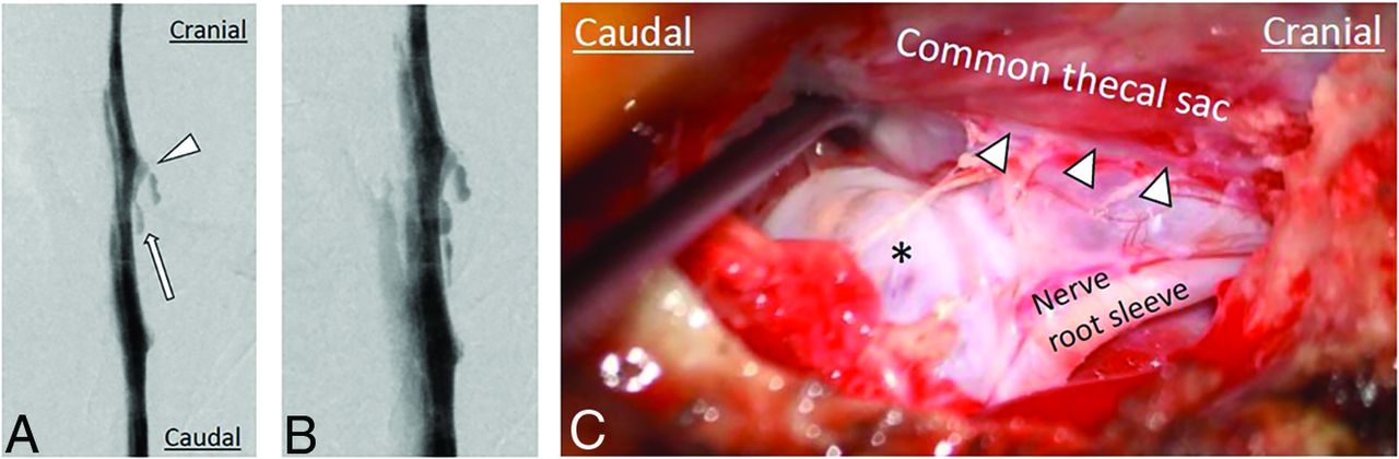
  • FIG 2.
    • Download figure
    • Open in new tab
    • Download powerpoint
    FIG 2.

    Imaging and intraoperative findings of spontaneous lateral spinal CSF leaks arising from the axilla of the nerve root sleeve. DSMs (A and B) show a lateral CSF leak (arrow) arising caudal to the nerve root sleeve (arrowhead). C, Intraoperative photograph shows a lateral dural tear (arrowheads) caudal to the take-off of the nerve root sleeve with arachnoid (asterisk) protruding through the dural tear.
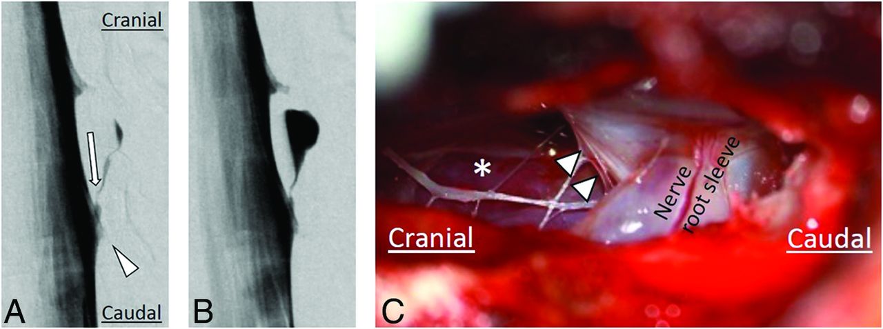
  • FIG 3.
    • Download figure
    • Open in new tab
    • Download powerpoint
    FIG 3.

    Imaging and intraoperative findings of spontaneous lateral spinal CSF leaks arising from the shoulder of the nerve root sleeve. DSMs (A and B) show a lateral CSF leak (arrow) arising cranial to the nerve root sleeve (arrowhead). C, Intraoperative photograph shows a lateral dural tear (arrowheads) cranial to the take-off of the nerve root sleeve after the arachnoid membrane has been resected, leaving a clear view of the extended extradural space (asterisk).

  • FIG 4.
    • Download figure
    • Open in new tab
    • Download powerpoint
    FIG 4.

    Imaging and intraoperative findings of spontaneous lateral spinal CSF leaks arising at the level of the pedicle. DSM (A) shows a small, defined lateral extradural CSF collection (arrow) at the level of the pedicle, not associated with the nerve root sleeve. Axial T2-weighted MRI (B) shows a corresponding small extradural CSF collection (arrow) that was not visible on post-DSM CT (C). The intrathecal contrast on the post-DSM CT is faint, possibly limiting the sensitivity of leak detection. Intraoperative photograph (D) shows a lateral dural tear (arrowheads) with arachnoid billowing out (asterisk) through the dural defect. DSMs (E–G) show a lateral dural tear (arrow) at the level of the pedicle, not associated with the nerve root sleeve, resulting in an extensive CSF leak, with CSF spreading in both cranial and caudal directions (arrowheads). Intraoperative photographs (H and I) show a linear dural tear (arrowheads) without arachnoid protruding through the dural tear. The underlying spinal cord is visible after spreading the dural tear (H).

Tables

  • Figures
    • View popup
    Table 1:

    Cohort demographics and clinical characteristics (N = 53)

    VariablesNo. (%)
    Age at onset of symptoms (yr)
     Mean (SD)35.5 (10.8)
     Range12–76
    Sex
     Male16 (30.2)
     Female37 (69.8)
    Symptom duration (mo)
     Mean (SD)35.8 (49.7)
     Range0–205
    CSF leak type
     Axilla of nerve root sleeve36 (67.9)
     Pedicle4 (7.6)
     Shoulder of nerve root sleeve13 (24.5)
    CSF leak sidea
     Left27 (46.6)
     Right31 (53.4)
    CSF leak levela
     Cervical3 (5.7)
     Thoracic 4–64 (7.8)
     Thoracic 7–913 (24.5)
     Thoracic 10–1231 (58.5)
     Lumbar2 (3.8)
    • ↵a From a total of 58 CSF leak sites.

    • View popup
    Table 2:

    Characteristics per leak type

    VariablesCSF Leak Type
    Axilla 36 (67.9%)Pedicle 4 (7.6%)Shoulder 13 (24.5%) P Value
    Age at onset of symptoms (yr).0295
     Mean (SD)38.1 (10.2)32.8 (12.8)28.9 (9.3)
     Minimum-maximum20–7614–4312–43
    Sex
     Male11 (30.6)1 (25.0)4 (30.8)1.0000
     Female25 (69.4)3 (75.0)9 (69.2)
    Symptom duration (mo).7359
     Mean (SD)34.8 (48.4)47.8 (45.7)35.2 (57.7)
     Range1–2050–1060–195 
PreviousNext
Back to top
Advertisement
Print
Download PDF
Email Article

Thank you for your interest in spreading the word on American Journal of Neuroradiology.

NOTE: We only request your email address so that the person you are recommending the page to knows that you wanted them to see it, and that it is not junk mail. We do not capture any email address.

Enter multiple addresses on separate lines or separate them with commas.
Lateral Spinal CSF Leaks in Patients with Spontaneous Intracranial Hypotension: Radiologic-Anatomic Study of Different Variants
(Your Name) has sent you a message from American Journal of Neuroradiology
(Your Name) thought you would like to see the American Journal of Neuroradiology web site.
CAPTCHA
This question is for testing whether or not you are a human visitor and to prevent automated spam submissions.
Cite this article
Wouter I. Schievink, Marcel M. Maya, Angelique Sao-Mai S. Tay, Rachelle B. Taché, Ravi S. Prasad, Vikram Wadhwa, Miriam Nuño
Lateral Spinal CSF Leaks in Patients with Spontaneous Intracranial Hypotension: Radiologic-Anatomic Study of Different Variants
American Journal of Neuroradiology Jun 2024, DOI: 10.3174/ajnr.A8261

Citation Manager Formats

  • BibTeX
  • Bookends
  • EasyBib
  • EndNote (tagged)
  • EndNote 8 (xml)
  • Medlars
  • Mendeley
  • Papers
  • RefWorks Tagged
  • Ref Manager
  • RIS
  • Zotero
0 Responses
Respond to this article
Share
Bookmark this article
Lateral Spinal CSF Leaks in Patients with Spontaneous Intracranial Hypotension: Radiologic-Anatomic Study of Different Variants
Wouter I. Schievink, Marcel M. Maya, Angelique Sao-Mai S. Tay, Rachelle B. Taché, Ravi S. Prasad, Vikram Wadhwa, Miriam Nuño
American Journal of Neuroradiology Jun 2024, DOI: 10.3174/ajnr.A8261
del.icio.us logo Twitter logo Facebook logo Mendeley logo
  • Tweet Widget
  • Facebook Like
  • Google Plus One
Purchase

Jump to section

  • Article
    • Abstract
    • ABBREVIATIONS:
    • MATERIALS AND METHODS
    • RESULTS
    • DISCUSSION
    • CONCLUSIONS
    • Footnotes
    • References
  • Figures & Data
  • Supplemental
  • Info & Metrics
  • Responses
  • References
  • PDF

Related Articles

  • PubMed
  • Google Scholar

Cited By...

  • Spontaneous Intracranial Hypotension Associated with Vascular Malformations
  • Spinal CSF Leaks: The Neuroradiologist Transforming Care
  • Expounding on the Distinction between Lateral Dural Tears and Leaking Meningeal Diverticula in Spontaneous Intracranial Hypotension
  • Crossref (5)
  • Google Scholar

This article has been cited by the following articles in journals that are participating in Crossref Cited-by Linking.

  • Expounding on the Distinction between Lateral Dural Tears and Leaking Meningeal Diverticula in Spontaneous Intracranial Hypotension
    Ajay A. Madhavan, Richard I. Farb, Waleed Brinjikji, Jeremy K. Cutsforth-Gregory, Wouter I. Schievink
    American Journal of Neuroradiology 2024 45 8
  • Spinal CSF Leaks: The Neuroradiologist Transforming Care
    Mark D. Mamlouk, Andrew L. Callen, Ajay A. Madhavan, Niklas Lützen, Lalani Carlton Jones, Ian T. Mark, Waleed Brinjikji, John C. Benson, Jared T. Verdoorn, D.K. Kim, Timothy J. Amrhein, Linda Gray, William P. Dillon, Marcel M. Maya, Thien J. Huynh, Vinil N. Shah, Tomas Dobrocky, Eike I. Piechowiak, Joseph Levi Chazen, Michael D. Malinzak, Jessica L. Houk, Peter G. Kranz
    American Journal of Neuroradiology 2024 45 11
  • Commentary: Surgical Treatment of Spontaneous Intracranial Hypotension From Spinal CSF Leak: Single Institution Case Series
    Catherine M. Garcia, Wouter I. Schievink, Julie L. Chan
    Neurosurgery 2025
  • Spinal CSF leaks in spontaneous intracranial hypotension: A single-institution analysis of incidence, typology and treatment outcomes
    Bala McRae-Posani, Andrew Kim, David Edasery, Sara Strauss, Michelle Roytman, John K. Park, Gayle Salama
    Clinical Neurology and Neurosurgery 2025 255
  • Spontaneous Intracranial Hypotension Associated with Vascular Malformations
    Mark D. Mamlouk, Adriana Gutierrez, William P. Dillon
    American Journal of Neuroradiology 2025 46 2

Similar Articles

Advertisement

Indexed Content

  • Current Issue
  • Accepted Manuscripts
  • Article Preview
  • Past Issues
  • Editorials
  • Editor's Choice
  • Fellows' Journal Club
  • Letters to the Editor
  • Video Articles

Cases

  • Case Collection
  • Archive - Case of the Week
  • Archive - Case of the Month
  • Archive - Classic Case

More from AJNR

  • Trainee Corner
  • Imaging Protocols
  • MRI Safety Corner
  • Book Reviews

Multimedia

  • AJNR Podcasts
  • AJNR Scantastics

Resources

  • Turnaround Time
  • Submit a Manuscript
  • Submit a Video Article
  • Submit an eLetter to the Editor/Response
  • Manuscript Submission Guidelines
  • Statistical Tips
  • Fast Publishing of Accepted Manuscripts
  • Graphical Abstract Preparation
  • Imaging Protocol Submission
  • Evidence-Based Medicine Level Guide
  • Publishing Checklists
  • Author Policies
  • Become a Reviewer/Academy of Reviewers
  • News and Updates

About Us

  • About AJNR
  • Editorial Board
  • Editorial Board Alumni
  • Alerts
  • Permissions
  • Not an AJNR Subscriber? Join Now
  • Advertise with Us
  • Librarian Resources
  • Feedback
  • Terms and Conditions
  • AJNR Editorial Board Alumni

American Society of Neuroradiology

  • Not an ASNR Member? Join Now

© 2025 by the American Society of Neuroradiology All rights, including for text and data mining, AI training, and similar technologies, are reserved.
Print ISSN: 0195-6108 Online ISSN: 1936-959X

Powered by HighWire