
Article Figures & Data
Figures
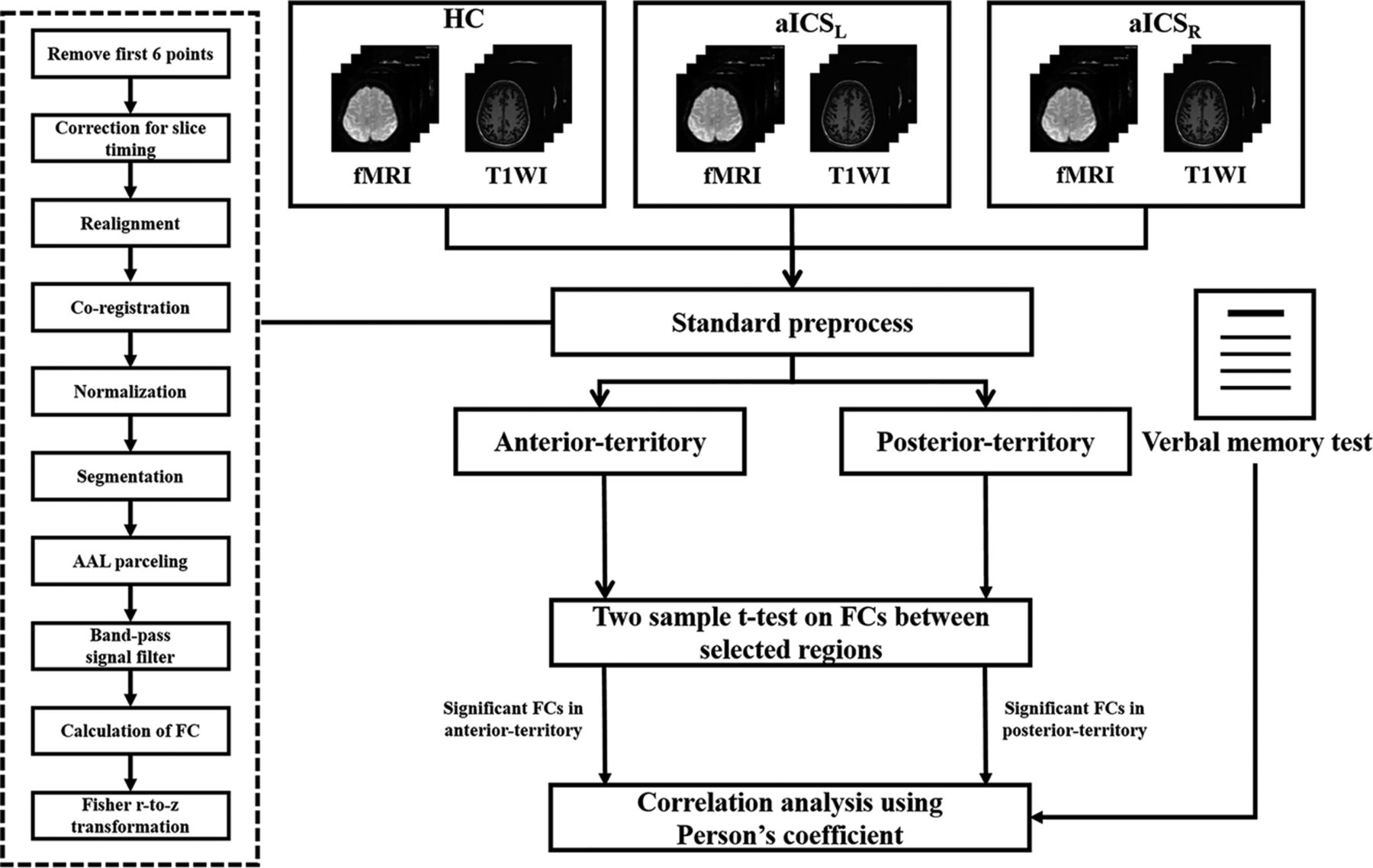
- FIG 1.
Flow chart for the study.
- FIG 2.
The 9 brain regions were parceled based on the automated anatomical labeling atlas with 116 areas (AAL 116) and highlighted in the following analyses. The green nodes indicate brain regions in the anterior circulation territory; the wine-colored nodes indicate brain regions in the posterior circulation territory.
- FIG 3.
The t maps from the comparisons of FC among the aICSL, aICSR, and HC groups. A, The matrices show the comparison between the aICS and HC groups in regions in the anterior territory. The boxplot presents the distribution of altered FC in the anterior circulation territory. B, The matrices show the comparison between the aICS and HC groups in regions in the posterior territory. The boxplot presents the distribution of altered FC in the posterior circulation territory. Red or blue nodes indicate that patients with stenosis in the right internal carotid artery show a significant increase or reduction in FC, respectively. Fron_Sup_Med: superior medial frontal gyrus, Tem_Mid_pole: middle temporal pole, R: right, and L: left. *P value < .05 (FDR corrected).
- FIG 4.
Scatterplots showing correlations between the delayed recall of verbal memory and altered FC in the anterior and posterior vascular territories after aICSL and aICSR. The dots represent the strength of the altered FC and delayed recall of verbal memory for patients. The fitted lines are also displayed in the scatterplots. A, In the anterior circulation territory FC matrix, the FC between the right superior medial frontal gyrus and left lingual gyrus was significantly correlated with delayed recall of verbal memory after aICSL. B, In the posterior circulation territory FC matrix, the FC between the right middle temporal pole and cerebellum VIII was significantly correlated with delayed recall of verbal memory after aICSR.
- FIG 5.
Diagram summarizing the underlying compensatory mechanism (neuroplasticity) after aICSL and aICSR. A, Anterior circulation territory compensation for verbal memory recall after aICSL. B, Patients with aICSR showed posterior circulation territory compensation for recall verbal memory. The green nodes indicate brain regions in the anterior circulation territory; the wine-colored nodes represent brain regions in the posterior circulation territory. The dashed double arrows indicate altered FC after aICS. The altered connections related to verbal memory recall are presented with double arrows.
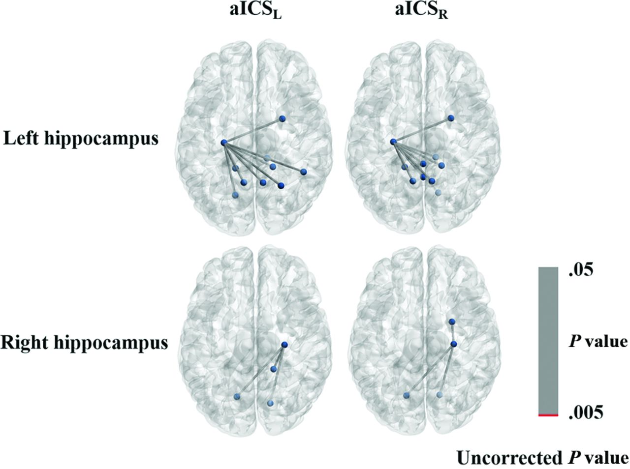
- FIG 6.
Graph displaying the FC of the left or right hippocampus gyrus with uncorrected P values less than .05. The connections with P values less than .005 are shown in red; those connections with P values between .005 and .05 are shown in gray.


Tables
HC (n = 15) aICS (n = 55) P Values (Power) aICSL (n = 22) aICSR (n = 33) aICSL vs HC aICSR vs HC aICSL vs aICSR Age 70.67 ± 3.09 64.50 ± 11.68 68.82 ± 9.03 .06 .45 .13 Sex (M:F) 8:7 16:6 25:8 .23 .06 .85 Stenosis (%) – 80.22 ± 12.68 78.91 ± 12.75 – – .71 Handedness (L/R) 0/15 0/22 2/31 1 .33 .24 Mini-Mental State Examination 29.33 ± 0.82 27.32 ± 2.21 28.67 ± 1.31 .002b(0.94) .08 .01b(0.76) Digit Span Test Forward 8.40 ± 0.74 7.95 ± 1.50 8.21 ± 0.99 .29 .52 .45 Reverse 5.73 ± 1.03 4.45 ± 1.65 5.03 ± 1.67 .01a(0.77) .14 .21 Verbal Learning Test Immediate recall 53.93 ± 8.37 45.95 ± 12.43 49.64 ± 8.37 .04a (0.81) .15 .23 Delayed recall 10.93 ± 1.44 8.14 ± 3.44 9.48 ± 2.00 .01b (0.92) .02a (0.87) .07 - Table 2:
The distribution of the severity of white matter hyperintensities for aICSL and aICSR groups. The total Fazekas score is the summation of scores for periventricular white matter and deep white matter
HC (n = 15) aICSL (n = 22) aICSR (n = 33) P Value aICSL vs HC aICSR vs HC aICSL vs aICSR Fazekas score for periventricular white matter .501 .586 .637 0 1 (6.7%) 3 (13.6%) 4 (12.1%) 1 11 (73.3%) 9 (50.0%) 19 (60.6%) 2 3 (20.0%) 6 (31.8%) 6 (18.2%) 3 0 (0.0%) 1 (4.5%) 3 (9.1%) Fazekas score for deep white matter .705 .693 .693 0 3 (20.0%) 3 (13.6%) 6 (18.2%) 1 10 (66.7%) 10 (59.1%) 20 (60.6%) 2 2 (13.3%) 5 (22.7%) 3 (12.1%) 3 0 (0.0%) 1 (4.5%) 3 (9.1%) Total Fazekas score .413 .801 .157 0 1 (6.7%) 3 (13.6%) 1 (3.0%) 1 2 (13.3%) 0 (0.0%) 8 (24.2%) 2 8 (53.3%) 8 (45.5%) 14 (42.4%) 3 3 (20.0%) 3 (18.2%) 3 (12.1%) 4 1 (6.7%) 4 (18.2%) 3 (9.1%) 5 0 (0.0%) 0 (0.0%) 1 (3.0%) 6 0 (0.0%) 1 (4.5%) 2 (6.1%) Note:—T2 FLAIR images are not available in 10 HCs, 3 aICSL and 1 aICSR patients. The Fazekas scores for those cases were assessed with diffusion tensor imaging.
- Table 3:
List of primary language regions with altered FC. The first 3 columns show the distribution of FC in the HC, aICSL, and aICSR groups. The fourth and fifth columns list FDR-corrected P values of comparison between the HC and aICSL groups and between the HC and aICSR groups, respectively
HC aICSL aICSR aICSL vs HC aICSR vs HC Homopotic Broca_L-Broca_R 0.22 ± 0.16 0.18 ± 0.13 0.22 ± 0.17 .73 .99 Geschwind_L-Geschwind_R 0.89 ± 0.25 0.95 ± 0.32 0.85 ± 0.33 .73 .93 Wernicke_L-Wernicke_R 1.21 ± 0.29 0.98 ± 0.36 1.04 ± 0.32 .60 .71 Left hemisphere Broca-Geschwind 0.21 ± 0.15 0.23 ± 0.16 0.21 ± 0.20 .73 .99 Broca-Wernicke 0.22 ± 0.14 0.19 ± 0.14 0.26 ± 0.22 .73 .99 Geschwind-Wernicke 0.18 ± 0.15 0.29 ± 0.33 0.22 ± 0.19 .73 .93 Right hemisphere Broca-Geschwind 0.15 ± 0.11 0.18 ± 0.13 0.20 ± 0.20 .73 .75 Broca-Wernicke 0.57 ± 0.39 0.61 ± 0.28 0.48 ± 0.25 .73 .75 Wernicke-Geschwind 0.18 ± 0.12 0.20 ± 0.14 0.26 ± 0.23 .53 .75 Note:—FDR-corrected P value. L: left, R: right.


