
Article Figures & Data
Figures
- FIG 1.
The time-density curve and color-coded QDSA. A, Lateral view of the color-coded QDSA. Selected ROIs are the following: 1) cavernous sinus segment of the ICA; 2) distal segment of the feeding artery; 3) AVM nidus; 4) proximal segment of the main draining vein; and 5) junction area of the draining vein and the venous sinus. B, Quantitative parameters in the time-density curve from QDSA. TTP indicates the time required for the bolus to reach peak attenuation,
 .
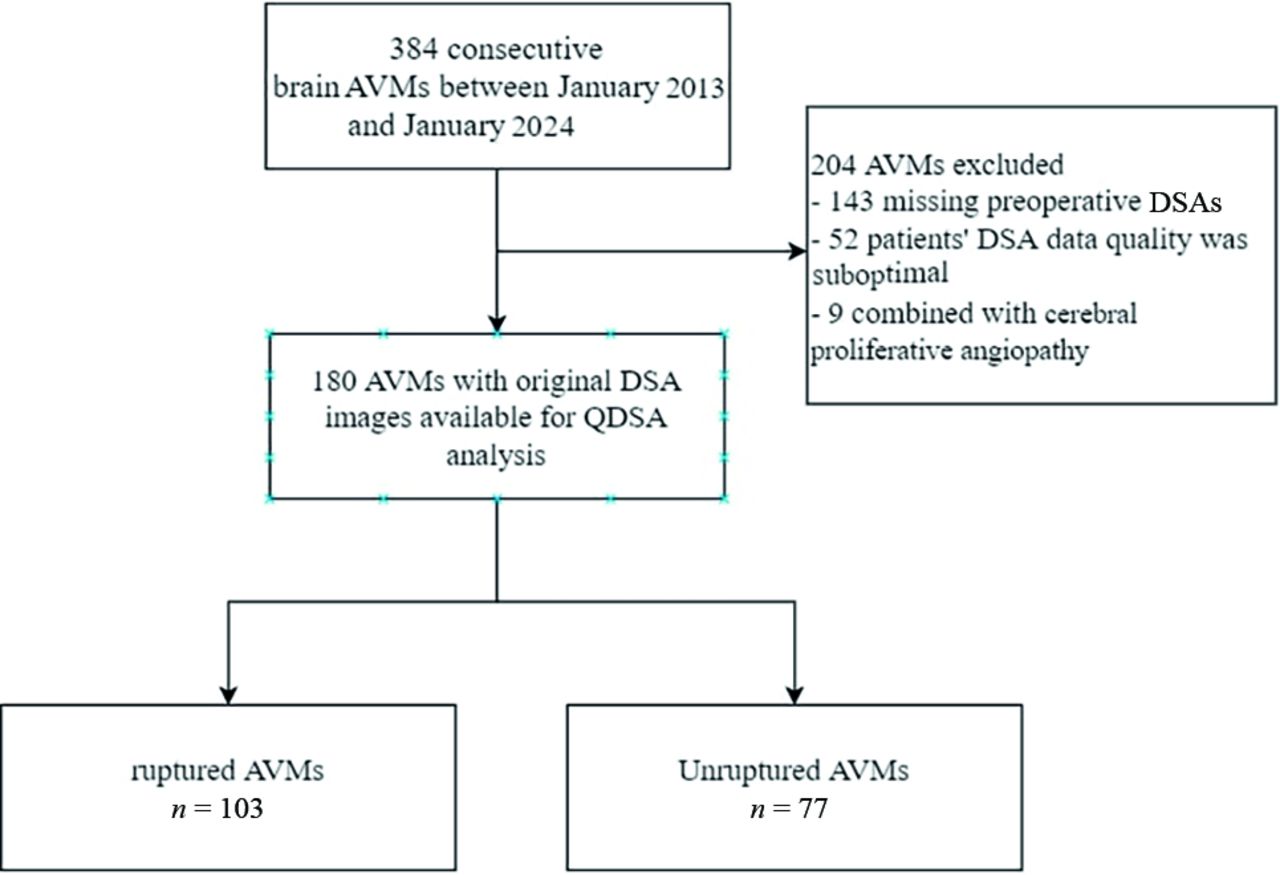
. - FIG 2.
Flow diagram of the enrolled patients.
- FIG 3.
Comparison of 2 AVM rupture-prediction models before and after the inclusion of the LFI. A. Comparing R2ED and R2ED+LFI reveals that the original R2ED AUC value is 0.755. After the inclusion of LFI, the AUC value increases to 0.791. Based on the DeLong' test, the significance level is P = .03. B, Comparing VALE and VALE+LFI reveals that the original VALE AUC value is 0.760. After the inclusion of LFI, the AUC value increases to 0.823. Based on DeLong test, the significance level is P = .005.


Tables
Characteristics All Cases Unruptured Ruptured P Value No. of patients 180 77 103 Age (mean) (yr) 24.6 (SD, 12.7) 25.2 (SD, 11.9) 24.2 (SD, 13.3) .60 Sex (male) 110 (61.1%) 50 (64.9%) 60 (58.3%) .22 Admission mRs score (mean) 1.0 (SD, 1.1) 0.8 (SD, 0.6) 1.2 (SD, 1.3) .002a Clinical presentation Seizure 49 (27.2%) 32 (41.6%) 17 (16.5%) <.001a Headache (nonruptured) 25 (13.9%) 24 (31.2%) 1 (1.0%) <.001a Focal neurologic deficit 20 (11.1%) 12 (15.6%) 8 (7.8%) .08 Spetzler-Martin grade .07 I 30 (16.7%) 8 (10.4%) 22 (21.4%) II 61 (33.9%) 26 (33.8%) 35 (34.0%) III 53 (29.4%) 25 (32.5%) 28 (27.2%) IV 28 (15.6%) 15 (19.5%) 13 (12.6%) V 8 (4.4%) 3 (3.9%) 5 (4.9%) ↵a Statistical significance (P < .05).
- Table 2:
Comparison of angioarchitecture and hemodynamics between the unruptured and ruptured AVMs
Characteristics Unruptured Ruptured P Value No. of patients 77 103 Mean nidus volume (mean) (mL) 40.2 (SD, 46.4) 13.6 (SD, 21.0) <.001a Angioarchitecture Feeding artery dilation 57 (74.0%) 51 (49.5%) .001a Single feeding artery 12 (15.6%) 35 (34.0%) .003a Deep venous drainage 17 (22.1%) 38 (36.9%) .035a Single draining vein 33 (42.9%) 65 (63.1%) .07 Drainage vein stenosis 11 (14.3%) 27 (26.2%) .07 Diffuse nidus 15 (19.5%) 38 (36.9%) .01a Flow-related aneurysm 14 (18.2%) 43 (41.7%) .001a Hemodynamics Feeding artery TTP (mean) (sec) 2.88 (SD, 0.80) 3.14 (SD, 1.01) .07 FWHM (mean) (sec) 2.83 (SD, 1.02) 3.33 (SD, 1.29) .006a Inflow gradient (mean) 1265.61 (SD, 912.53) 1317.85 (SD, 1018.43) .72 Outflow gradient (mean) 636.89 (SD, 505.72) 600.35 (SD, 511.84) .63 Stasis index (mean) 2.42 (SD, 0.99) 2.63 (SD, 1.04) .16 Drainage vein TTP (mean) (sec) 3.62 (SD, 1.07) 3.76 (SD, 1.30) .42 FWHM (mean) (sec) 3.00 (SD, 0.75) 3.56 (SD, 1.29) <.001a Inflow gradient (mean) 1854.28 (SD, 2794.89) 1289.84 (SD, 975.82) .06 Outflow gradient (mean) 741.99 (SD, 568.85) 610.58 (SD, 522.51) .11 Stasis index (mean) 2.38 (SD, 0.65) 2.52 (SD, 0.97) .28 MTT (ICA-sinus) (mean) (sec) 1.48 (SD, 1.03) 1.97 (SD, 1.70) .02a MTT (feeding-draining) (mean) (sec) 0.73 (SD, 0.97) 0.62 (SD, 1.04) .47 ADW (mean) 695.02 (SD, 607.38) 742.75 (SD, 720.42) .64 TRV (mean) 225.35 (SD, 211.26) 115.93 (SD, 157.39) <.001a LFI (mean) 49.40 (SD, 98.25) 390.27 (SD, 919.81) <.001a ↵a Statistical significance (P < .05).
- Table 3:
Univariate and multivariate logistic regression analyses of LFI and 3 LFI-combined models of factors associated with rupture in AVMs
Univariable Analysis Angioarchitecture Model Hemodynamic Model Combined Model OR (95% CI) P Value OR (95% CI) P Value OR (95% CI) P Value OR (95% CI) P Value LFI 1.007 (1.003–1.010) .001a 1.004 (1.001–1.008) .02a 1.005 (1.001–1.009) .009a 1.004 (1.000–1.007) .03a Mean nidus volume 0.979 (0.964–0.994) .007a 0.959 (0.931–0.988) .005a Angioarchitecture Feeding artery dilation 1.047 (0.443–2.472) .92 1.028 (0.429–2.461) .95 Single feeding artery 0.783 (0.299–2.049) .62 1.112 (0.768–1.608) .57 Deep venous drainage 2.315 (1.018–6.947) .05a 2.684 (1.132–6.361) .03a Diffuse nidus 2.863 (1.180–6.947) .02a 2.990 (1.114–8.022) .03a Flow-related aneurysm 3.417 (1.521–7.678) .003a 3.617 (1.544–8.474) .003a Hemodynamic Feeding artery FWHM (sec) 1.067 (0.703–1.619) .76 0.949 (0.590–1.524) .83 Drainage vein FWHM (sec) 1.218 (0.750–1.978) .43 1.17 1 (0.695–1.974) .55 MTT (ICA-sinus) 1.084 (0.811–1.448) .59 1.297 (0.933–1.803) .12 TRV 0.999 (0.997–1.001) .26 1.004 (0.999–1.009) .14 ↵a Statistical significance (P < .05).
- Table 5:
10-fold cross-validation of performance with 2 previous models after incorporating LFI
Model AUC (95%CI) P Value Accuracy Precision Sensitivity Specificity R2eD 0.601 (0.515–0.687) Reference 0.611 0.674 0.621 0.597 R2eD + LFI 0.624 (0.541–0.707) .15 0.639 0.702 0.641 0.636 VALE 0.603 (0.518–0.688) Reference 0.556 0.610 0.621 0.468 VALE + LFI 0.706 (0.629–0.783) <.001a 0.683 0.717 0.738 0.610 ↵a Statistical significance (P < .05).


