Advertisement

Main menu

  • Home
  • Content
    • Current Issue
    • Accepted Manuscripts
    • Article Preview
    • Past Issue Archive
    • Video Articles
    • AJNR Case Collection
    • Case of the Week Archive
    • Case of the Month Archive
    • Classic Case Archive
  • Special Collections
    • AJNR Awards
    • Low-Field MRI
    • Alzheimer Disease
    • ASNR Foundation Special Collection
    • Photon-Counting CT
    • View All
  • Multimedia
    • AJNR Podcasts
    • AJNR SCANtastic
    • Trainee Corner
    • MRI Safety Corner
    • Imaging Protocols
  • For Authors
    • Submit a Manuscript
    • Submit a Video Article
    • Submit an eLetter to the Editor/Response
    • Manuscript Submission Guidelines
    • Statistical Tips
    • Fast Publishing of Accepted Manuscripts
    • Graphical Abstract Preparation
    • Imaging Protocol Submission
    • Author Policies
  • About Us
    • About AJNR
    • Editorial Board
    • Editorial Board Alumni
  • More
    • Become a Reviewer/Academy of Reviewers
    • Subscribers
    • Permissions
    • Alerts
    • Feedback
    • Advertisers
    • ASNR Home

User menu

  • Alerts
  • Log in

Search

  • Advanced search
American Journal of Neuroradiology

American Journal of Neuroradiology

ASHNR American Society of Functional Neuroradiology ASHNR American Society of Pediatric Neuroradiology ASSR
  • Alerts
  • Log in

Advanced Search

  • Home
  • Content
    • Current Issue
    • Accepted Manuscripts
    • Article Preview
    • Past Issue Archive
    • Video Articles
    • AJNR Case Collection
    • Case of the Week Archive
    • Case of the Month Archive
    • Classic Case Archive
  • Special Collections
    • AJNR Awards
    • Low-Field MRI
    • Alzheimer Disease
    • ASNR Foundation Special Collection
    • Photon-Counting CT
    • View All
  • Multimedia
    • AJNR Podcasts
    • AJNR SCANtastic
    • Trainee Corner
    • MRI Safety Corner
    • Imaging Protocols
  • For Authors
    • Submit a Manuscript
    • Submit a Video Article
    • Submit an eLetter to the Editor/Response
    • Manuscript Submission Guidelines
    • Statistical Tips
    • Fast Publishing of Accepted Manuscripts
    • Graphical Abstract Preparation
    • Imaging Protocol Submission
    • Author Policies
  • About Us
    • About AJNR
    • Editorial Board
    • Editorial Board Alumni
  • More
    • Become a Reviewer/Academy of Reviewers
    • Subscribers
    • Permissions
    • Alerts
    • Feedback
    • Advertisers
    • ASNR Home
  • Follow AJNR on Twitter
  • Visit AJNR on Facebook
  • Follow AJNR on Instagram
  • Join AJNR on LinkedIn
  • RSS Feeds

AJNR Awards, New Junior Editors, and more. Read the latest AJNR updates

Research ArticleNEUROVASCULAR/STROKE IMAGING

Concurrent Moyamoya-like Intracranial Steno-Occlusive Disease and Dural Arteriovenous Fistulas

Vaishnavi L. Rao, Jee Won Shin, James P. Klaas and Giuseppe Lanzino
American Journal of Neuroradiology May 2024, DOI: https://doi.org/10.3174/ajnr.A8197
Vaishnavi L. Rao
aFrom the Department of Neurosurgery (V.L.R., G.L.), Mayo Clinic, Rochester, Minnesota
  • Find this author on Google Scholar
  • Find this author on PubMed
  • Search for this author on this site
  • ORCID record for Vaishnavi L. Rao
Jee Won Shin
bMayo Clinic Alix School of Medicine (J.W.S.), Rochester, Minnesota
  • Find this author on Google Scholar
  • Find this author on PubMed
  • Search for this author on this site
James P. Klaas
cDepartment of Neurology (J.P.K.), Mayo Clinic, Rochester, Minnesota
  • Find this author on Google Scholar
  • Find this author on PubMed
  • Search for this author on this site
  • ORCID record for James P. Klaas
Giuseppe Lanzino
aFrom the Department of Neurosurgery (V.L.R., G.L.), Mayo Clinic, Rochester, Minnesota
  • Find this author on Google Scholar
  • Find this author on PubMed
  • Search for this author on this site
  • ORCID record for Giuseppe Lanzino
  • Article
  • Figures & Data
  • Supplemental
  • Info & Metrics
  • Responses
  • References
  • PDF
Loading

SUMMARY:

The simultaneous presentation of intracranial steno-occlusive disease, Moyamoya disease, or Moyamoya-like vasculopathy and dural arteriovenous fistulas (DAVFs) has been documented in very few case reports worldwide. We aimed to better characterize this association by reviewing the clinical and radiologic findings of 4 patients with concurrent intracranial steno-occlusive disease or Moyamoya-like vasculopathy and DAVFs evaluated in our institution. All 4 patients were of Asian descent. One patient presented with ischemic stroke secondary to intracranial stenosis, 2 presented with symptoms related to the DAVF, and the diagnosis was incidental in the fourth patient. Three patients underwent embolization of the DAVF, which was followed by surgical ligation in 2. One patient underwent extracranial-intracranial bypass for Moyamoya-like intracranial steno-occlusive disease. One patient is being managed conservatively with close follow-up. Our case series details findings in 4 patients with associated intracranial steno-occlusive disease and DAVFs. Further studies and reporting of similar cases are necessary to establish whether this is pure coincidence or if there is indeed a relationship between these 2 conditions, especially in certain ethnic groups.

ABBREVIATIONS:

DAVF
dural arteriovenous fistula
MMD
Moyamoya disease

Moyamoya disease (MMD), Moyamoya-like vasculopathy, and intracranial steno-occlusive disease are cerebrovascular conditions with a higher incidence in East Asian countries than in the Western Hemisphere.1 Dural arteriovenous fistulas (DAVFs) are relatively uncommon cerebrovascular entities characterized by abnormal shunting between the meningeal arteries and the dural venous sinuses or subarachnoid veins,2 accounting for 10%–15% of intracranial vascular malformations,3 with an incidence as high as 0.29 per 100,000 person-years in Japan.4 There have been very few case reports worldwide documenting the co-occurrence of these 2 disease entities before surgical intervention or head trauma. Here, we describe an institutional series of 4 patients presenting with concurrent Moyamoya-like vascular changes or intracranial steno-occlusive disease and DAVFs.

CASE SERIES

We identified all patients with intracranial steno-occlusive disease, Moyamoya, Moyamoya-like vasculopathy, and DAVFs from a prospectively maintained database of cases evaluated and/or treated by the senior author (G.L.) at our institution between January 2008 and 2023. The diagnosis of idiopathic Moyamoya or Moyamoya-like vasculopathy was made on the basis of the presence of both stenosis or occlusion of the terminal ICA or the proximal middle and/or anterior cerebral artery and an abnormal network of blood vessels on cerebral angiography, according to the 2021 diagnostic criteria for MMD published by the Ministry of Health, Labor and Welfare in Japan.5 More recently, patients underwent high-resolution MR imaging with vessel wall enhancement to distinguish eccentric (more consistent with intracranial atherosclerosis) from concentric (more consistent with idiopathic vasculopathy) enhancing patterns.6,7 The Cognard system was used to classify the type of DAVF, because it is useful for predicting the associated risk and guiding management decisions.8

This retrospective study was reviewed and approved by our institutional review board (19–001663), and all protocols were followed in accordance with institutional review board guidelines. A common denominator of these patients was their Asian ethnicity. Demographics, clinical presentation, and treatment are summarized in the Online Supplemental Data. Angiographic findings are presented in Figs 1–4. MR vessel wall imaging was available for cases 2 and 4, which presented more recently after vessel wall imaging was incorporated into practice at our institution. For these patients, vessel wall imaging demonstrated circumferential vessel wall enhancement involving the distal left ICA in case 2 and lack of any vessel wall enhancement whatsoever in case 4, suggesting a steno-occlusive picture outside intracranial atherosclerosis. For cases 1 and 3, we elected to treat the DAVFs because they were symptomatic, while in case 2, we attempted treatment of the DAVF because of the retrograde cortical venous drainage/venous hypertension and its potential role in aggravating perfusion issues related to the steno-occlusive disease. For case 4, we elected for observation of the DAVF because of its asymptomatic status/incidental finding and potential risks associated with embolization (ie, compromise of retinal blood flow supply) or surgical therapy (brain manipulation in the setting of steno-occlusive disease and marginal perfusion). The steno-occlusive disease in each patient was medically managed with aspirin monotherapy, while patient 2 underwent surgical revascularization for recurrent left-hemispheric infarcts.

FIG 1.
  • Download figure
  • Open in new tab
  • Download powerpoint
FIG 1.

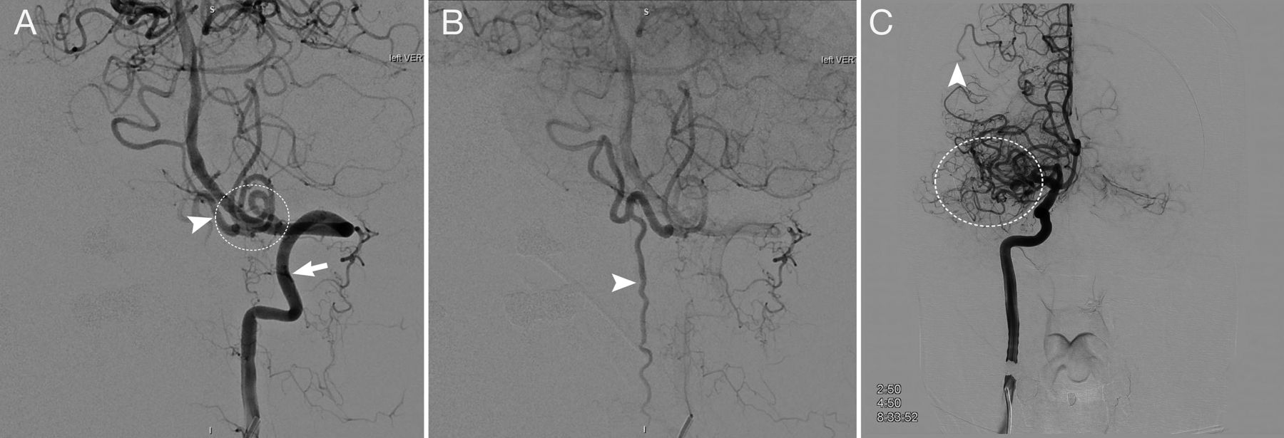
Case 1. A 57-year-old Asian man who presented with several weeks of progressive myelopathy. A, Conventional angiography shows a craniocervical junction AVF (white circle) with a large arterialized draining vein (arrowhead) arising from the dura adjacent to the point where the vertebral artery pierces the dura, supplied by branches of the left vertebral artery (arrow). B, The same injection demonstrates the venous drainage of the fistula down the anterior and posterior spinal veins, which are filled early (arrowhead). C, A right ICA injection, anterior-posterior view shows incidental occlusion of the M1 segment with Moyamoya-like collaterals (white circle) reconstituting the distal MCA territory in addition to leptomeningeal collaterals (arrowhead) from the distal anterior cerebral artery. He underwent surgical ligation of the fistula after failed embolization. The asymptomatic MCA occlusion was treated medically.

FIG 2.
  • Download figure
  • Open in new tab
  • Download powerpoint
FIG 2.

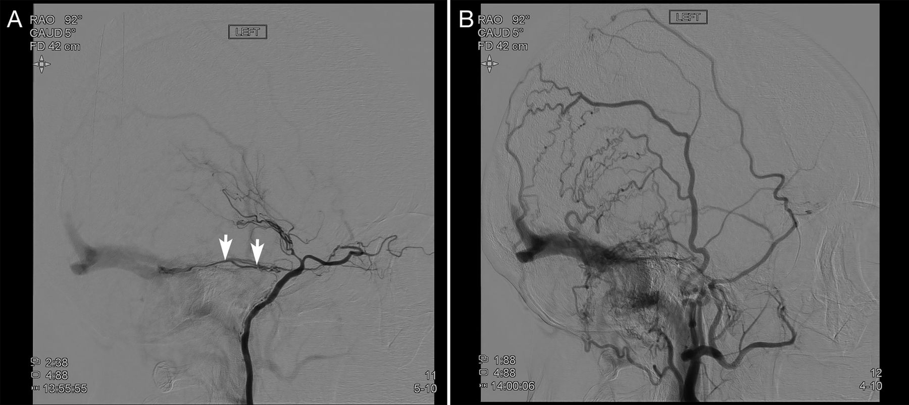
Case 2. A 63-year-old Asian man who presented with multiple left-hemisphere infarcts during 1 year. A, A left common carotid artery injection shows a left sphenoparietal DAVF (circle) supplied by the left middle meningeal artery with exclusive retrograde cortical venous drainage (arrowheads). B, There is delayed filling of the left ICA (arrow), which is occluded distal to the origin of the ophthalmic artery (arrowheads). C, Selective right ICA injection, lateral view, shows severe stenosis of the right supraclinoid ICA (arrows). The patient underwent partial embolization of the DAVF followed by surgical ligation and left superficial temporal artery–to-MCA bypass.

FIG 3.
  • Download figure
  • Open in new tab
  • Download powerpoint
FIG 3.

Case 3, A 74-year-old Asian woman who presented with episodic vision loss, papilledema, and cognitive decline. A, Conventional angiography, selective left ICA injection, shows an incidental left supraclinoid ICA occlusion as well as a left transverse-sigmoid junction DAVF with arterial feeders from the left meningohypophyseal artery (arrows). B, External carotid artery injection, lateral view, shows a high-flow transverse-sigmoid sinus DAVF. The DAVF was occluded via a transvenous approach with resolution of symptoms at follow up. Low-dose aspirin was prescribed for the incidental carotid occlusion.

FIG 4.
  • Download figure
  • Open in new tab
  • Download powerpoint
FIG 4.

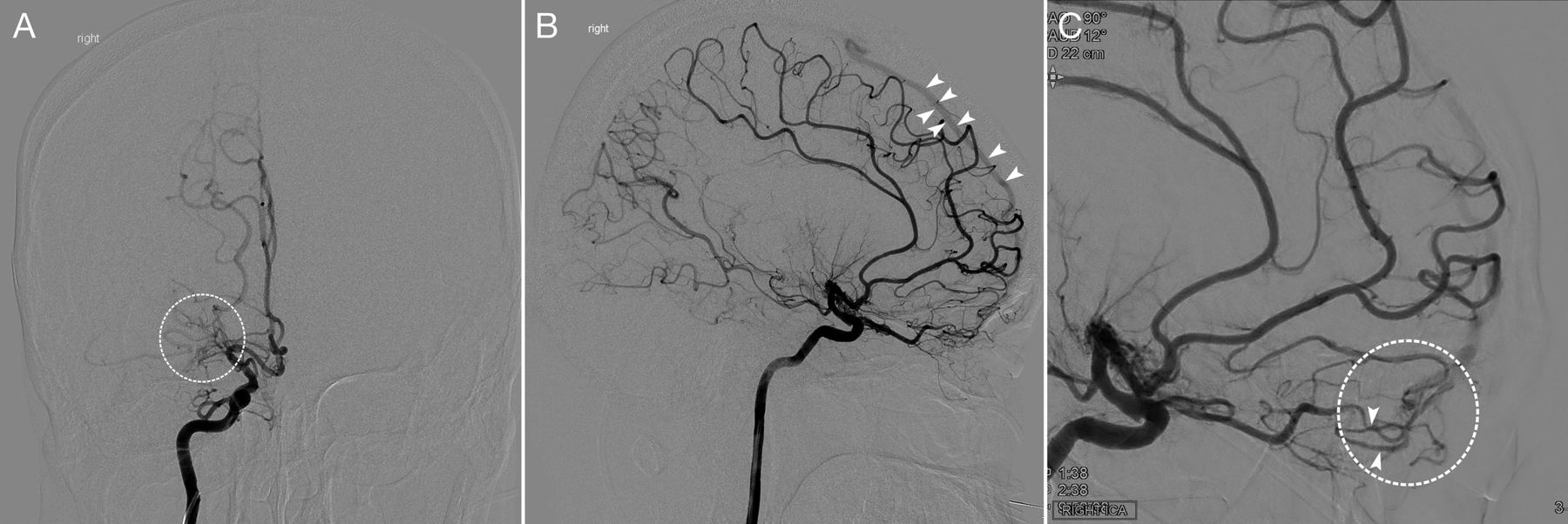
Case 4. A 42-year-old Asian man with right-sided headache. A, Selective right ICA injection, anterior-posterior view, early arterial phase, shows occlusion of the MCA with a Moyamoya network (white circle). B, Selective right ICA injection, lateral view, early arterial phase, shows a lack of opacification in the MCA territory and early filling of the superior sagittal sinus (arrowheads). C, Right ICA selective injection, magnified oblique view, shows enlarged and tortuous distal ophthalmic artery branches (arrowheads) feeding a dural/pial AVF (white circle), with early opacification of a cortical vein draining into the superior sagittal sinus.

DISCUSSION

We describe 4 patients of Asian ethnicity with concomitant idiopathic Moyamoya disease, Moyamoya-like vasculopathy, or steno-occlusive disease and intracranial DAVFs. The rare concurrent appearance of intracranial steno-occlusive disease and DAVFs as seen in our 4 cases has been previously documented in only a few single case reports and usually after surgical therapy or trauma. De novo formation of DAVFs in patients with Moyamoya-like vasculopathy has been observed in a delayed fashion following direct bypass,9,10 indirect bypass,11 and head trauma;12 however, there have been a very few cases of simultaneous presentation in the absence of prior surgery or trauma. These include a patient with unilateral MMD who presented with intraventricular hemorrhage and was found to have an ipsilateral transverse-sigmoid DAVF, which was ultimately treated with endovascular embolization,13 similar to case 3 in our series, as well as ipsilateral cavernous and tentorial DAVFs observed in patients with MMD in China14 and Spain,15 respectively.

A proposed mechanism for the formation of DAVFs in these patients is that aberrant flow dynamics in intracranial steno-occlusive disease leads to an increased demand and decreased perfusion, with the subsequent ischemia triggering an angiogenic response in the dural vasculature, resulting in arteriovenous shunting.16 Indeed, increased expression of proangiogenic factors has been reported in the dura of patients with MMD, including vascular endothelial growth factor17 and fibroblast growth factor.18 These factors are also involved in the formation of DAVFs.19

Of note, all 4 patients in our series are of East Asian ethnicity, suggesting that a genetic association may potentially exist in cases of concurrent intracranial steno-occlusive disease and DAVF. None of the previous case reports we came across specified the patients’ ethnicity, so we could not confirm such an association on the basis of prior literature. Nevertheless, variants of the HLA genes and the RNF213 susceptibility gene have been found to be associated with East Asian patients with MMD.20,21 Unfortunately, the patients included in our study did not undergo genetic testing to evaluate these pathogenic variants; however, this association would be an interesting area of future study. Overall, intracranial stenosis is more prevalent in Asians,22 often with a younger mean age of onset and independent of vascular risk factors compared with Caucasians.23

In our small institutional case series, we describe the simultaneous presentation of the two entities of Moyamoya pattern/steno-occlusive intracranial disease and DAVF in 4 patients of East Asian descent. While our series does not establish causality, future reporting and study of similar cases may further determine whether a potential relationship exists between ethnicity and the concurrence of these cerebrovascular phenomena. As more patients like these are identified, the pathophysiology of these two conditions can be more clearly elucidated and the presence of an underlying genetic association can be investigated.

Acknowledgments

We acknowledge Peggy Chihak and Arielle Davis for their help in formatting the figures and manuscript.

Footnotes

  • Disclosure forms provided by the authors are available with the full text and PDF of this article at www.ajnr.org.

References

  1. 1.↵
    1. Zhang H,
    2. Zheng L,
    3. Feng L
    . Epidemiology, diagnosis, and treatment of moyamoya disease. Exp Ther Med 2019;17:1977–84 doi:10.3892/etm.2019.7198 pmid:30867689
    CrossRefPubMed
  2. 2.↵
    1. Reynolds MR,
    2. Lanzino G,
    3. Zipfel GJ
    . Intracranial dural arteriovenous fistulae. Stroke 2017;48:1424–31 doi:10.1161/STROKEAHA.116.012784 pmid:28432263
    FREE Full Text
  3. 3.↵
    1. Newton TH,
    2. Cronqvist S
    . Involvement of dural arteries in intracranial arteriovenous malformations. Radiology 1969;93:1071–78 doi:10.1148/93.5.1071 pmid:5350675
    CrossRefPubMed
  4. 4.↵
    1. Satomi J,
    2. Satoh K
    . Epidemiology and etiology of dural arteriovenous fistula. Brain Nerve 2008;60:883–86 pmid:18717191
    PubMed
  5. 5.↵
    1. Kuroda S,
    2. Fujimura M,
    3. Takahashi J, et al
    ; Research Committee on Moyamoya Disease (Spontaneous Occlusion of Circle of Willis) of the Ministry of Health, Labor, and Welfare, Japan. Diagnostic criteria for moyamoya disease: 2021 revised version. Neurol Med Chir (Tokyo) 2022;62:307–12 doi:10.2176/jns-nmc.2022-0072 pmid:35613882
    CrossRefPubMed
  6. 6.↵
    1. Mossa-Basha M,
    2. de Havenon A,
    3. Becker KJ, et al
    . Added value of vessel wall magnetic resonance imaging in the differentiation of moyamoya vasculopathies in a non-Asian cohort. Stroke 2016;47:1782–88 doi:10.1161/STROKEAHA.116.013320 pmid:27272486
    Abstract/FREE Full Text
  7. 7.↵
    1. Liu S,
    2. Lu M,
    3. Gao X, et al
    . The diagnostic performance of high-resolution magnetic resonance-vessel wall imaging in differentiating atherosclerosis-associated moyamoya vasculopathy from moyamoya disease. Eur Radiol 2023;33:6918–26 doi:10.1007/s00330-023-09951-z pmid:37453985
    CrossRefPubMed
  8. 8.↵
    1. Cognard C,
    2. Gobin YP,
    3. Pierot L, et al
    . Cerebral dural arteriovenous fistulas: clinical and angiographic correlation with a revised classification of venous drainage. Radiology 1995;194:671–80 doi:10.1148/radiology.194.3.7862961 pmid:7862961
    CrossRefPubMed
  9. 9.↵
    1. Feroze AH,
    2. Kushkuley J,
    3. Choudhri O, et al
    . Development of arteriovenous fistula after revascularization bypass for Moyamoya disease: case report. Operative Neurosurgery 2015;11(Suppl 2):E202–06 doi:10.1227/NEU.0000000000000558 pmid:25251198
    CrossRefPubMed
  10. 10.↵
    1. Hanaoka M,
    2. Matsubara S,
    3. Satoh K, et al
    . Dural arteriovenous fistulae after cerebral infarction: report of two cases. Neurosurgery 2011;68:E575–79; discussion E580 doi:10.1227/NEU.0b013e3182039803 pmid:21135717
    CrossRefPubMed
  11. 11.↵
    1. Koduri S,
    2. Wilkinson DA,
    3. Griauzde JM, et al
    . Development of bilateral dural arteriovenous fistulae following pial synangiosis for moyamoya syndrome: case report. J Neurosurg Pediatr 2019;24:9–13 doi:10.3171/2019.2.PEDS18603 pmid:30978680
    CrossRefPubMed
  12. 12.↵
    1. Zaletel M,
    2. Surlan-Popović K,
    3. Pretnar-Oblak J, et al
    . Moyamoya syndrome with arteriovenous dural fistula after head trauma. Acta Clini Croat 2011;50:115–19 pmid:22034792
    PubMed
  13. 13.↵
    1. Killory BD,
    2. Gonzalez LF,
    3. Wait SD, et al
    . Simultaneous unilateral moyamoya disease and ipsilateral dural arteriovenous fistula: case report. Neurosurgery 2008;62:E1375–76; discussion E1376 doi:10.1227/01.neu.0000333311.87554.9c pmid:18824958
    CrossRefPubMed
  14. 14.↵
    1. Liu P,
    2. Xu Y,
    3. Lv X, et al
    . Progression of unilateral moyamoya disease resulted in spontaneous occlusion of ipsilateral cavernous dural arteriovenous fistula: case report. Interv Neuroradiol 2016;22:362–64 doi:10.1177/1591019916632371 pmid:26916656
    CrossRefPubMed
  15. 15.↵
    1. Cajal Campo B,
    2. Delgado Acosta F
    . Enfermedad moyamoya y fístula arteriovenosa dural simultánea. Revista de Neurologia 2010;50:317–18 doi:10.33588/rn.5005.2009707 pmid:20217652
    CrossRefPubMed
  16. 16.↵
    1. Lawton MT,
    2. Jacobowitz R,
    3. Spetzler RF
    . Redefined role of angiogenesis in the pathogenesis of dural arteriovenous malformations. J Neurosurg 1997;87:267–74 doi:10.3171/jns.1997.87.2.0267 pmid:9254092
    CrossRefPubMed
  17. 17.↵
    1. Sakamoto S,
    2. Kiura Y,
    3. Yamasaki F, et al
    . Expression of vascular endothelial growth factor in dura mater of patients with moyamoya disease. Neurosurg Rev 2008;31:77–81; discussion 81 doi:10.1007/s10143-007-0102-8 pmid:17912564
    CrossRefPubMed
  18. 18.↵
    1. Hoshimaru M,
    2. Takahashi JA,
    3. Kikuchi H, et al
    . Possible roles of basic fibroblast growth factor in the pathogenesis of moyamoya disease: an immunohistochemical study. J Neurosurg 1991;75:267–70 doi:10.3171/jns.1991.75.2.0267 pmid:2072165
    CrossRefPubMed
  19. 19.↵
    1. Uranishi R,
    2. Nakase H,
    3. Sakaki T
    . Expression of angiogenic growth factors in dural arteriovenous fistula. J Neurosurg 1999;91:781–86 doi:10.3171/jns.1999.91.5.0781 pmid:10541235
    CrossRefPubMed
  20. 20.↵
    1. Mertens R,
    2. Graupera M,
    3. Gerhardt H, et al
    . The genetic basis of moyamoya disease. Transl Stroke Res 2021;13:25–45 doi:10.1007/s12975-021-00940-2 pmid:34529262
    CrossRefPubMed
  21. 21.↵
    1. Hiraide T,
    2. Suzuki H,
    3. Momoi M, et al
    . RNF213-associated vascular disease: a concept unifying various vasculopathies. Life (Basel) 2022;12:555 doi:10.3390/life12040555 pmid:35455046
    CrossRefPubMed
  22. 22.↵
    1. Suri MF,
    2. Johnston SC
    . Epidemiology of intracranial stenosis. J Neuroimaging 2009;19(Suppl 1)11S–6S doi:10.1111/j.1552-6569.2009.00415.x pmid:19807851
    CrossRefPubMed
  23. 23.↵
    1. Leng X,
    2. Hurford R,
    3. Feng X, et al
    . Intracranial arterial stenosis in Caucasian versus Chinese patients with TIA and minor stroke: two contemporaneous cohorts and a systematic review. J Neurol Neurosurg Psychiatry 2021 Mar 30 [Epub ahead of print] doi:10.1136/jnnp-2020-325630 pmid:33785575
    Abstract/FREE Full Text
  • Received November 15, 2023.
  • Accepted after revision January 15, 2024.
  • © 2024 by American Journal of Neuroradiology
PreviousNext
Back to top
Advertisement
Print
Download PDF
Email Article

Thank you for your interest in spreading the word on American Journal of Neuroradiology.

NOTE: We only request your email address so that the person you are recommending the page to knows that you wanted them to see it, and that it is not junk mail. We do not capture any email address.

Enter multiple addresses on separate lines or separate them with commas.
Concurrent Moyamoya-like Intracranial Steno-Occlusive Disease and Dural Arteriovenous Fistulas
(Your Name) has sent you a message from American Journal of Neuroradiology
(Your Name) thought you would like to see the American Journal of Neuroradiology web site.
CAPTCHA
This question is for testing whether or not you are a human visitor and to prevent automated spam submissions.
Cite this article
Vaishnavi L. Rao, Jee Won Shin, James P. Klaas, Giuseppe Lanzino
Concurrent Moyamoya-like Intracranial Steno-Occlusive Disease and Dural Arteriovenous Fistulas
American Journal of Neuroradiology May 2024, DOI: 10.3174/ajnr.A8197

Citation Manager Formats

  • BibTeX
  • Bookends
  • EasyBib
  • EndNote (tagged)
  • EndNote 8 (xml)
  • Medlars
  • Mendeley
  • Papers
  • RefWorks Tagged
  • Ref Manager
  • RIS
  • Zotero
0 Responses
Respond to this article
Share
Bookmark this article
Concurrent Moyamoya-like Intracranial Steno-Occlusive Disease and Dural Arteriovenous Fistulas
Vaishnavi L. Rao, Jee Won Shin, James P. Klaas, Giuseppe Lanzino
American Journal of Neuroradiology May 2024, DOI: 10.3174/ajnr.A8197
del.icio.us logo Twitter logo Facebook logo Mendeley logo
  • Tweet Widget
  • Facebook Like
  • Google Plus One
Purchase

Jump to section

  • Article
    • SUMMARY:
    • ABBREVIATIONS:
    • CASE SERIES
    • DISCUSSION
    • Acknowledgments
    • Footnotes
    • References
  • Figures & Data
  • Supplemental
  • Info & Metrics
  • Responses
  • References
  • PDF

Related Articles

  • PubMed
  • Google Scholar

Cited By...

  • No citing articles found.
  • Crossref
  • Google Scholar

This article has not yet been cited by articles in journals that are participating in Crossref Cited-by Linking.

More in this TOC Section

  • DMV relationship with SVD and DTI measures in RSSI
  • Role of Hypoperfusion Intensity Ratio in Vessel Occlusions: A Review on Safety and Clinical Outcomes
  • DMV, IFS and CSVD burden
Show more Neurovascular/Stroke Imaging

Similar Articles

Advertisement

Indexed Content

  • Current Issue
  • Accepted Manuscripts
  • Article Preview
  • Past Issues
  • Editorials
  • Editor's Choice
  • Fellows' Journal Club
  • Letters to the Editor
  • Video Articles

Cases

  • Case Collection
  • Archive - Case of the Week
  • Archive - Case of the Month
  • Archive - Classic Case

More from AJNR

  • Trainee Corner
  • Imaging Protocols
  • MRI Safety Corner

Multimedia

  • AJNR Podcasts
  • AJNR Scantastics

Resources

  • Turnaround Time
  • Submit a Manuscript
  • Submit a Video Article
  • Submit an eLetter to the Editor/Response
  • Manuscript Submission Guidelines
  • Statistical Tips
  • Fast Publishing of Accepted Manuscripts
  • Graphical Abstract Preparation
  • Imaging Protocol Submission
  • Evidence-Based Medicine Level Guide
  • Publishing Checklists
  • Author Policies
  • Become a Reviewer/Academy of Reviewers
  • News and Updates

About Us

  • About AJNR
  • Editorial Board
  • Editorial Board Alumni
  • Alerts
  • Permissions
  • Not an AJNR Subscriber? Join Now
  • Advertise with Us
  • Librarian Resources
  • Feedback
  • Terms and Conditions
  • AJNR Editorial Board Alumni

American Society of Neuroradiology

  • Not an ASNR Member? Join Now

© 2025 by the American Society of Neuroradiology All rights, including for text and data mining, AI training, and similar technologies, are reserved.
Print ISSN: 0195-6108 Online ISSN: 1936-959X

Powered by HighWire